|
Hudson–Stahli Line
The Hudson–Stahli line is a line of iron deposition lying roughly on the border between the middle and lower thirds of the cornea.Ophthalmology Myron Yanoff, Jay S. Duker Edition 3, illustrated Elsevier Health Sciences, 2008 , It lies in the corneal epithelium. Usually it has about 0.5 mm in thickness and is 1–2 mm long. It is generally horizontal, with possible mild downward trend in the middle. It is present normally in people over the age of 50, but seems to dissipate to some degree by the age of 70. The Hudson–Stahli line is not associated with any pathology calling for clinical intervention. Formation of the line may depend upon the rate of tear secretion. However, the Hudson–Stahli line can be enhanced in hydroxychloroquine toxicity. See also *Fleischer ring – corneal iron depositions in keratoconus Keratoconus (KC) is a disorder of the eye that results in progressive thinning of the cornea. This may result in blurry vision, double vision, nearsigh ... [...More Info...] [...Related Items...] OR: [Wikipedia] [Google] [Baidu] |
Iron
Iron () is a chemical element with symbol Fe (from la, ferrum) and atomic number 26. It is a metal that belongs to the first transition series and group 8 of the periodic table. It is, by mass, the most common element on Earth, right in front of oxygen (32.1% and 30.1%, respectively), forming much of Earth's outer and inner core. It is the fourth most common element in the Earth's crust. In its metallic state, iron is rare in the Earth's crust, limited mainly to deposition by meteorites. Iron ores, by contrast, are among the most abundant in the Earth's crust, although extracting usable metal from them requires kilns or furnaces capable of reaching or higher, about higher than that required to smelt copper. Humans started to master that process in Eurasia during the 2nd millennium BCE and the use of iron tools and weapons began to displace copper alloys, in some regions, only around 1200 BCE. That event is considered the transition from the Bronze Age to the Iron A ... [...More Info...] [...Related Items...] OR: [Wikipedia] [Google] [Baidu] |
Corneal Epithelium
The corneal epithelium (epithelium corneæ anterior layer) is made up of epithelial tissue and covers the front of the cornea. It acts as a barrier to protect the cornea, resisting the free flow of fluids from the tears, and prevents bacteria from entering the epithelium and corneal stroma. Anatomy The corneal epithelium consists of several layers of cells. The cells of the deepest layer are columnar, known as basal cells which are attached by multiprotein complexes known as hemidesmosomes to an underlying basement membrane. Then follow two or three layers of polyhedral cells, commonly known as wing cells. The majority of these are prickle cells, similar to those found in the stratum mucosum of the cuticle. Lastly, there are three or four layers of squamous cells, with flattened nuclei. The layers of the epithelium are constantly undergoing mitosis. Basal and wing cells migrate to the anterior of the cornea, while squamous cells age and slough off into the tear film. Central ... [...More Info...] [...Related Items...] OR: [Wikipedia] [Google] [Baidu] |
Tears
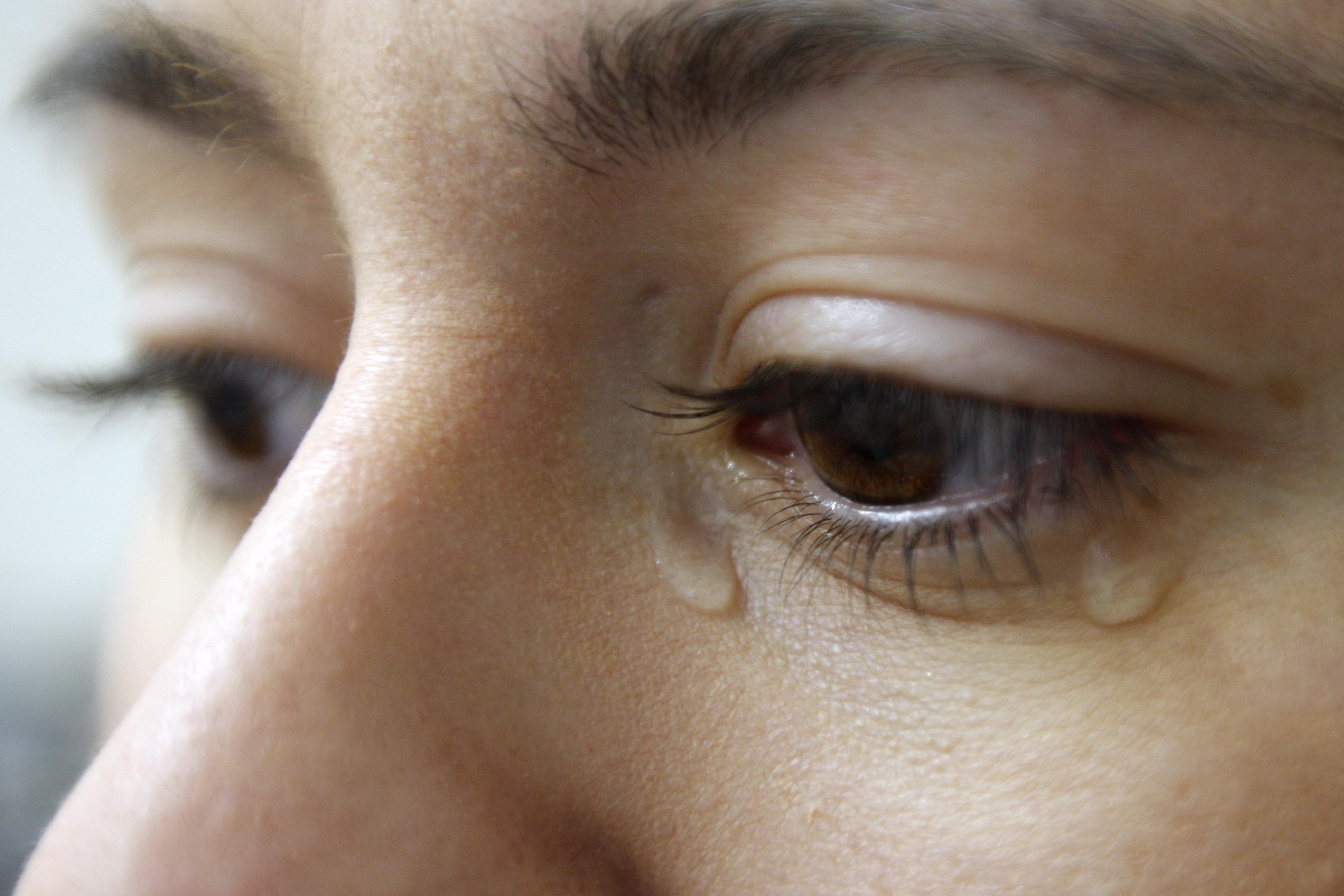
Tears are a clear liquid secreted by the lacrimal glands (tear gland) found in the eyes of all land mammals. Tears are made up of water, electrolytes, proteins, lipids, and mucins that form layers on the surface of eyes. The different types of tears—basal, reflex, and emotional—vary significantly in composition. The functions of tears include lubricating the eyes (basal tears), removing irritants (reflex tears), and also aiding the immune system. Tears also occur as a part of the body's natural pain response. Emotional secretion of tears may serve a biological function by excreting stress-inducing hormones built up through times of emotional distress. Tears have symbolic significance among humans. Physiology Chemical composition Tears are made up of three layers: lipid, aqueous, and mucous. Tears are composed of water, salts, antibodies, and lysozymes (antibacterial enzymes); though composition varies among different tear types. The composition of tears caused by an ... [...More Info...] [...Related Items...] OR: [Wikipedia] [Google] [Baidu] |
Hydroxychloroquine
Hydroxychloroquine, sold under the brand name Plaquenil among others, is a medication used to prevent and treat malaria in areas where malaria remains sensitive to chloroquine. Other uses include treatment of rheumatoid arthritis, lupus, and porphyria cutanea tarda. It is taken by mouth, often in the form of hydroxychloroquine sulfate. Common side effects may include vomiting, headache, changes in vision, and muscle weakness. Severe side effects may include allergic reactions, vision problems, and heart problems. Although all risk cannot be excluded, it remains a treatment for rheumatic disease during pregnancy. Hydroxychloroquine is in the antimalarial and 4-aminoquinoline families of medication. Hydroxychloroquine was approved for medical use in the United States in 1955. It is on the World Health Organization's List of Essential Medicines. In 2020, it was the 126th most commonly prescribed medication in the United States, with more than 4million prescriptions. Hydrox ... [...More Info...] [...Related Items...] OR: [Wikipedia] [Google] [Baidu] |
Fleischer Ring
Fleischer rings are pigmented rings in the peripheral cornea, resulting from iron deposition in basal epithelial cells, in the form of hemosiderin. They are usually yellowish to dark-brown, and may be complete or broken. The rings are best seen using the slit lamp under cobalt blue filter. They are named for Bruno Fleischer. Fleischer rings are indicative of keratoconus, a degenerative corneal condition that causes the cornea to thin and change to a conic shape. Confusion with Kayser-Fleischer rings Some confusion exists between Fleischer rings and Kayser-Fleischer rings. Kayser-Fleischer rings are caused by copper deposits in descemet's membrane of cornea, and are indicative of Wilson's disease, whereas Fleischer rings are caused by iron deposits in basal epithelial cells. One example of a medical condition that can present with Fleischer rings is keratoconus. See also * Hudson–Stahli line * Limbal ring References Eye diseases Eye color {{med-sign-stub ... [...More Info...] [...Related Items...] OR: [Wikipedia] [Google] [Baidu] |
Keratoconus
Keratoconus (KC) is a disorder of the eye that results in progressive thinning of the cornea. This may result in blurry vision, double vision, nearsightedness, irregular astigmatism, and light sensitivity leading to poor quality-of-life. Usually both eyes are affected. In more severe cases a scarring or a circle may be seen within the cornea. While the cause is unknown, it is believed to occur due to a combination of genetic, environmental, and hormonal factors. Patients with a parent, sibling, or child who has keratoconus have 15 to 67 times higher risk in developing corneal ectasia compared to patients with no affected relatives. Proposed environmental factors include rubbing the eyes and allergies. The underlying mechanism involves changes of the cornea to a cone shape. Diagnosis is most often by topography. Topography measures the curvature of the cornea and creates a colored "map" of the cornea. Keratoconus causes very distinctive changes in the appearance of these ma ... [...More Info...] [...Related Items...] OR: [Wikipedia] [Google] [Baidu] |
Ophthalmology
Ophthalmology ( ) is a surgical subspecialty within medicine that deals with the diagnosis and treatment of eye disorders. An ophthalmologist is a physician who undergoes subspecialty training in medical and surgical eye care. Following a medical degree, a doctor specialising in ophthalmology must pursue additional postgraduate residency training specific to that field. This may include a one-year integrated internship that involves more general medical training in other fields such as internal medicine or general surgery. Following residency, additional specialty training (or fellowship) may be sought in a particular aspect of eye pathology. Ophthalmologists prescribe medications to treat eye diseases, implement laser therapy, and perform surgery when needed. Ophthalmologists provide both primary and specialty eye care - medical and surgical. Most ophthalmologists participate in academic research on eye diseases at some point in their training and many include research as part ... [...More Info...] [...Related Items...] OR: [Wikipedia] [Google] [Baidu] |


