|
Fleischer Ring
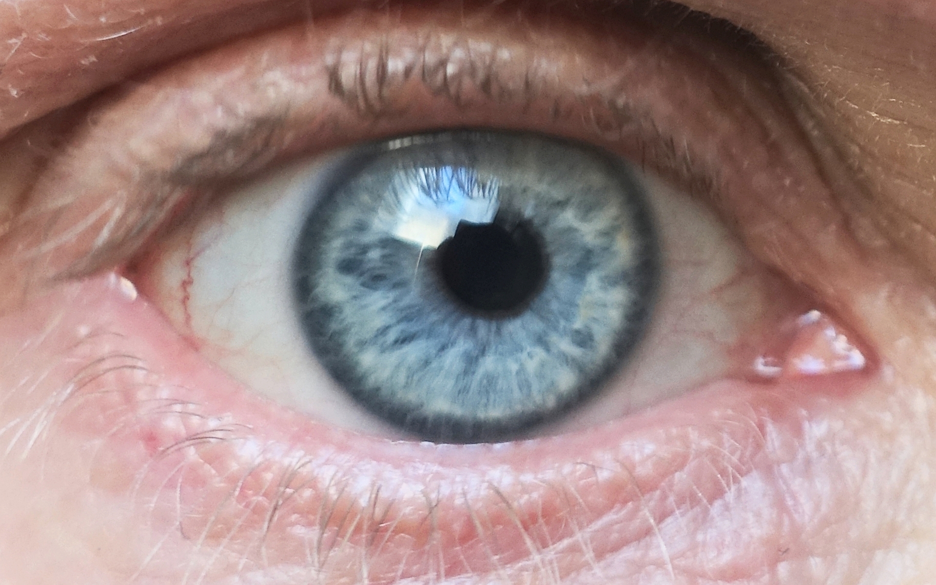
Fleischer rings are pigmented rings in the peripheral cornea, resulting from iron deposition in basal epithelial cells, in the form of hemosiderin. They are usually yellowish to dark-brown, and may be complete or broken. The rings are best seen using the slit lamp under cobalt blue filter. They are named for Bruno Fleischer. Fleischer rings are indicative of keratoconus, a degenerative corneal condition that causes the cornea to thin and change to a conic shape. Confusion with Kayser-Fleischer rings Some confusion exists between Fleischer rings and Kayser-Fleischer rings. Kayser-Fleischer rings are caused by copper deposits in descemet's membrane of cornea, and are indicative of Wilson's disease, whereas Fleischer rings are caused by iron deposits in basal epithelial cells. One example of a medical condition that can present with Fleischer rings is keratoconus. See also * Hudson–Stahli line * Limbal ring References Eye diseases Eye color {{med-sign-stub ... [...More Info...] [...Related Items...] OR: [Wikipedia] [Google] [Baidu] |
Cornea
The cornea is the transparent front part of the eye that covers the iris, pupil, and anterior chamber. Along with the anterior chamber and lens, the cornea refracts light, accounting for approximately two-thirds of the eye's total optical power. In humans, the refractive power of the cornea is approximately 43 dioptres. The cornea can be reshaped by surgical procedures such as LASIK. While the cornea contributes most of the eye's focusing power, its focus is fixed. Accommodation (the refocusing of light to better view near objects) is accomplished by changing the geometry of the lens. Medical terms related to the cornea often start with the prefix "'' kerat-''" from the Greek word κέρας, ''horn''. Structure The cornea has unmyelinated nerve endings sensitive to touch, temperature and chemicals; a touch of the cornea causes an involuntary reflex to close the eyelid. Because transparency is of prime importance, the healthy cornea does not have or need blood vessels with ... [...More Info...] [...Related Items...] OR: [Wikipedia] [Google] [Baidu] |
Iron
Iron () is a chemical element with symbol Fe (from la, ferrum) and atomic number 26. It is a metal that belongs to the first transition series and group 8 of the periodic table. It is, by mass, the most common element on Earth, right in front of oxygen (32.1% and 30.1%, respectively), forming much of Earth's outer and inner core. It is the fourth most common element in the Earth's crust. In its metallic state, iron is rare in the Earth's crust, limited mainly to deposition by meteorites. Iron ores, by contrast, are among the most abundant in the Earth's crust, although extracting usable metal from them requires kilns or furnaces capable of reaching or higher, about higher than that required to smelt copper. Humans started to master that process in Eurasia during the 2nd millennium BCE and the use of iron tools and weapons began to displace copper alloys, in some regions, only around 1200 BCE. That event is considered the transition from the Bronze Age to the Iron A ... [...More Info...] [...Related Items...] OR: [Wikipedia] [Google] [Baidu] |
Hemosiderin
Hemosiderin image of a kidney viewed under a microscope. The brown areas represent hemosiderin Hemosiderin or haemosiderin is an iron-storage complex that is composed of partially digested ferritin and lysosomes. The breakdown of heme gives rise to biliverdin and iron. The body then traps the released iron and stores it as hemosiderin in tissues. Hemosiderin is also generated from the abnormal metabolic pathway of ferritin. It is only found within cells (as opposed to circulating in blood) and appears to be a complex of ferritin, denatured ferritin and other material. The iron within deposits of hemosiderin is very poorly available to supply iron when needed. Hemosiderin can be identified histologically with ''Perls' Prussian blue stain''; iron in hemosiderin turns blue to black when exposed to potassium ferrocyanide. In normal animals, hemosiderin deposits are small and commonly inapparent without special stains. Excessive accumulation of hemosiderin is usually detected within ... [...More Info...] [...Related Items...] OR: [Wikipedia] [Google] [Baidu] |
Slit Lamp
A slit lamp is an instrument consisting of a high-intensity light source that can be focused to shine a thin sheet of light into the eye. It is used in conjunction with a biomicroscope. The lamp facilitates an examination of the anterior segment and posterior segment of the human eye, which includes the eyelid, sclera, conjunctiva, iris, natural crystalline lens, and cornea. The binocular slit-lamp examination provides a stereoscopic magnified view of the eye structures in detail, enabling anatomical diagnoses to be made for a variety of eye conditions. A second, hand-held lens is used to examine the retina. History Two conflicting trends emerged in the development of the slit lamp. One trend originated from clinical research and aimed to apply the increasingly complex and advanced technology of the time. [...More Info...] [...Related Items...] OR: [Wikipedia] [Google] [Baidu] |
Bruno Fleischer
Bruno Otto Fleischer (2 May 1874 – 26 March 1965) was a German ophthalmologist. Kayser-Fleischer rings and Fleischer ring Fleischer rings are pigmented rings in the peripheral cornea, resulting from iron deposition in basal epithelial cells, in the form of hemosiderin. They are usually yellowish to dark-brown, and may be complete or broken. The rings are best seen usi ...s are named for him. References Further reading * German ophthalmologists 1965 deaths 1874 births Eye color {{germany-med-bio-stub ... [...More Info...] [...Related Items...] OR: [Wikipedia] [Google] [Baidu] |
Keratoconus
Keratoconus (KC) is a disorder of the eye that results in progressive thinning of the cornea. This may result in blurry vision, double vision, nearsightedness, irregular astigmatism, and light sensitivity leading to poor quality-of-life. Usually both eyes are affected. In more severe cases a scarring or a circle may be seen within the cornea. While the cause is unknown, it is believed to occur due to a combination of genetic, environmental, and hormonal factors. Patients with a parent, sibling, or child who has keratoconus have 15 to 67 times higher risk in developing corneal ectasia compared to patients with no affected relatives. Proposed environmental factors include rubbing the eyes and allergies. The underlying mechanism involves changes of the cornea to a cone shape. Diagnosis is most often by topography. Topography measures the curvature of the cornea and creates a colored "map" of the cornea. Keratoconus causes very distinctive changes in the appearance of these ma ... [...More Info...] [...Related Items...] OR: [Wikipedia] [Google] [Baidu] |
Copper
Copper is a chemical element with the symbol Cu (from la, cuprum) and atomic number 29. It is a soft, malleable, and ductile metal with very high thermal and electrical conductivity. A freshly exposed surface of pure copper has a pinkish-orange color. Copper is used as a conductor of heat and electricity, as a building material, and as a constituent of various metal alloys, such as sterling silver used in jewelry, cupronickel used to make marine hardware and coins, and constantan used in strain gauges and thermocouples for temperature measurement. Copper is one of the few metals that can occur in nature in a directly usable metallic form ( native metals). This led to very early human use in several regions, from circa 8000 BC. Thousands of years later, it was the first metal to be smelted from sulfide ores, circa 5000 BC; the first metal to be cast into a shape in a mold, c. 4000 BC; and the first metal to be purposely alloyed with another metal, tin, to create ... [...More Info...] [...Related Items...] OR: [Wikipedia] [Google] [Baidu] |
Wilson's Disease
Wilson's disease is a genetic disorder in which excess copper builds up in the body. Symptoms are typically related to the brain and liver. Liver-related symptoms include vomiting, weakness, fluid build up in the abdomen, swelling of the legs, yellowish skin and itchiness. Brain-related symptoms include tremors, muscle stiffness, trouble in speaking, personality changes, anxiety, and psychosis. Wilson's disease is caused by a mutation in the Wilson disease protein (''ATP7B'') gene. This protein transports excess copper into bile, where it is excreted in waste products. The condition is autosomal recessive; for a person to be affected, they must inherit a mutated copy of the gene from both parents. Diagnosis may be difficult and often involves a combination of blood tests, urine tests and a liver biopsy. Genetic testing may be used to screen family members of those affected. Wilson's disease is typically treated with dietary changes and medication. Dietary changes involve e ... [...More Info...] [...Related Items...] OR: [Wikipedia] [Google] [Baidu] |
Hudson–Stahli Line
The Hudson–Stahli line is a line of iron deposition lying roughly on the border between the middle and lower thirds of the cornea.Ophthalmology Myron Yanoff, Jay S. Duker Edition 3, illustrated Elsevier Health Sciences, 2008 , It lies in the corneal epithelium. Usually it has about 0.5 mm in thickness and is 1–2 mm long. It is generally horizontal, with possible mild downward trend in the middle. It is present normally in people over the age of 50, but seems to dissipate to some degree by the age of 70. The Hudson–Stahli line is not associated with any pathology calling for clinical intervention. Formation of the line may depend upon the rate of tear secretion. However, the Hudson–Stahli line can be enhanced in hydroxychloroquine toxicity. See also *Fleischer ring – corneal iron depositions in keratoconus Keratoconus (KC) is a disorder of the eye that results in progressive thinning of the cornea. This may result in blurry vision, double vision, nearsigh ... [...More Info...] [...Related Items...] OR: [Wikipedia] [Google] [Baidu] |
Limbal Ring
A limbal ring is a dark ring around the iris of the eye, where the sclera meets the cornea.Johnson and Johnson Vision Care Inc. Tinted contact lenses with combined limbal ring and iris patterns. US7246903B2. United States Patent and Trademark Office, July 24, 2007. http://patft.uspto.gov/netacgi/nph-Parser?Sect1=PTO1&Sect2=HITOFF&p=1&u=/netahtml/PTO/srchnum.html&r=1&f=G&l=50&d=PALL&s1=7246903.PN. It is a dark-colored manifestation of the corneal limbus resulting from optical properties of the region. The appearance and visibility of the limbal ring can be negatively affected by a variety of medical conditions concerning the peripheral cornea. It has been suggested that limbal ring thickness may correlate with health or youthfulness and may contribute to facial attractiveness. Some contact lenses are colored to simulate limbal rings. Youth, health, and attractiveness Both health and age are positively correlated with a prominent limbal ring. For instance, a darker limbal ring tends ... [...More Info...] [...Related Items...] OR: [Wikipedia] [Google] [Baidu] |
Eye Diseases
This is a partial list of human eye diseases and disorders. The World Health Organization publishes a classification of known diseases and injuries, the International Statistical Classification of Diseases and Related Health Problems, or ICD-10. This list uses that classification. H00-H06 Disorders of eyelid, lacrimal system and orbit * (H02.1) Ectropion * (H02.2) Lagophthalmos * (H02.3) Blepharochalasis * (H02.4) Ptosis * (H02.5) Stye, an acne type infection of the sebaceous glands on or near the eyelid. * (H02.6) Xanthelasma of eyelid * (H03.0*) Parasitic infestation of eyelid in diseases classified elsewhere ** Dermatitis of eyelid due to Demodex species ( B88.0+ ) ** Parasitic infestation of eyelid in: *** leishmaniasis ( B55.-+ ) *** loiasis ( B74.3+ ) *** onchocerciasis ( B73+ ) *** phthiriasis ( B85.3+ ) * (H03.1*) Involvement of eyelid in other infectious diseases classified elsewhere ** Involvement of eyelid in: *** herpesviral (herpes simplex) infection ( B00.5+ ) * ... [...More Info...] [...Related Items...] OR: [Wikipedia] [Google] [Baidu] |


