|
Hirschberg Test
In the fields of optometry and ophthalmology, the Hirschberg test, also Hirschberg corneal reflex test, is a screening test that can be used to assess whether a person has strabismus (ocular misalignment). A photographic version of the Hirschberg is used to quantify strabismus. Technique It is performed by shining a light in the person's eyes and observing where the light reflects off the corneas. In a person with normal ocular alignment the light reflex lies slightly nasal from the center of the cornea (approximately 11 prism diopters—or 0.5mm from the pupillary axis), as a result of the cornea acting as a temporally-turned convex mirror to the observer. When doing the test, the light reflexes of both eyes are compared, and will be symmetrical in an individual with normal fixation. For an abnormal result, based on where the light lands on the cornea, the examiner can detect if there is an exotropia (abnormal eye is turned out), esotropia (abnormal eye is turned in), hypertro ... [...More Info...] [...Related Items...] OR: [Wikipedia] [Google] [Baidu] |
Anatomical Terms Of Location
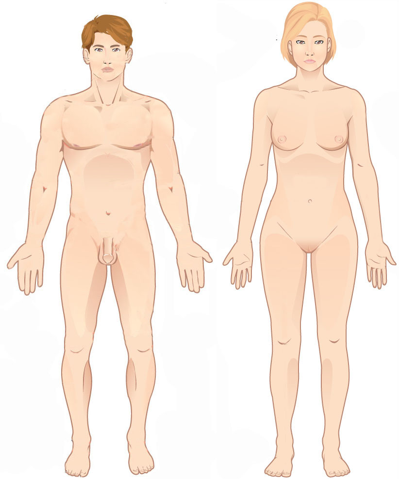
Standard anatomical terms of location are used to unambiguously describe the anatomy of animals, including humans. The terms, typically derived from Latin or Greek roots, describe something in its standard anatomical position. This position provides a definition of what is at the front ("anterior"), behind ("posterior") and so on. As part of defining and describing terms, the body is described through the use of anatomical planes and anatomical axes. The meaning of terms that are used can change depending on whether an organism is bipedal or quadrupedal. Additionally, for some animals such as invertebrates, some terms may not have any meaning at all; for example, an animal that is radially symmetrical will have no anterior surface, but can still have a description that a part is close to the middle ("proximal") or further from the middle ("distal"). International organisations have determined vocabularies that are often used as standard vocabularies for subdisciplines o ... [...More Info...] [...Related Items...] OR: [Wikipedia] [Google] [Baidu] |
Graves Ophthalmopathy
Graves’ ophthalmopathy, also known as thyroid eye disease (TED), is an autoimmune inflammatory disorder of the orbit and periorbital tissues, characterized by upper eyelid retraction, lid lag, swelling, redness (erythema), conjunctivitis, and bulging eyes (exophthalmos). It occurs most commonly in individuals with Graves' disease, and less commonly in individuals with Hashimoto's thyroiditis, or in those who are euthyroid. It is part of a systemic process with variable expression in the eyes, thyroid, and skin, caused by autoantibodies that bind to tissues in those organs. The autoantibodies target the fibroblasts in the eye muscles, and those fibroblasts can differentiate into fat cells (adipocytes). Fat cells and muscles expand and become inflamed. Veins become compressed and are unable to drain fluid, causing edema. Annual incidence is 16/100,000 in women, 3/100,000 in men. About 3–5% have severe disease with intense pain, and sight-threatening corneal ulceration or compr ... [...More Info...] [...Related Items...] OR: [Wikipedia] [Google] [Baidu] |
Medical Signs
Signs and symptoms are the observed or detectable signs, and experienced symptoms of an illness, injury, or condition. A sign for example may be a higher or lower temperature than normal, raised or lowered blood pressure or an abnormality showing on a medical scan. A symptom is something out of the ordinary that is experienced by an individual such as feeling feverish, a headache or other pain or pains in the body. Signs and symptoms Signs A medical sign is an objective observable indication of a disease, injury, or abnormal physiological state that may be detected during a physical examination, examining the patient history, or diagnostic procedure. These signs are visible or otherwise detectable such as a rash or bruise. Medical signs, along with symptoms, assist in formulating diagnostic hypothesis. Examples of signs include elevated blood pressure, nail clubbing of the fingernails or toenails, staggering gait, and arcus senilis and arcus juvenilis of the eyes. Indicat ... [...More Info...] [...Related Items...] OR: [Wikipedia] [Google] [Baidu] |
Terms For Anatomical Location
Standard anatomical terms of location are used to unambiguously describe the anatomy of animals, including humans. The terms, typically derived from Latin or Greek roots, describe something in its standard anatomical position. This position provides a definition of what is at the front ("anterior"), behind ("posterior") and so on. As part of defining and describing terms, the body is described through the use of anatomical planes and anatomical axes. The meaning of terms that are used can change depending on whether an organism is bipedal or quadrupedal. Additionally, for some animals such as invertebrates, some terms may not have any meaning at all; for example, an animal that is radially symmetrical will have no anterior surface, but can still have a description that a part is close to the middle ("proximal") or further from the middle ("distal"). International organisations have determined vocabularies that are often used as standard vocabularies for subdisciplines of ana ... [...More Info...] [...Related Items...] OR: [Wikipedia] [Google] [Baidu] |
Eye Examination
An eye examination is a series of tests performed to assess vision and ability to focus on and discern objects. It also includes other tests and examinations pertaining to the eyes. Eye examinations are primarily performed by an optometrist, ophthalmologist, or an orthoptist. Health care professionals often recommend that all people should have periodic and thorough eye examinations as part of routine primary care, especially since many eye diseases are asymptomatic. Eye examinations may detect potentially treatable blinding eye diseases, ocular manifestations of systemic disease, or signs of tumours or other anomalies of the brain. A full eye examination consists of an external examination, followed by specific tests for visual acuity, pupil function, extraocular muscle motility, visual fields, intraocular pressure and ophthalmoscopy through a dilated pupil. A minimal eye examination consists of tests for visual acuity, pupil function, and extraocular muscle motilit ... [...More Info...] [...Related Items...] OR: [Wikipedia] [Google] [Baidu] |
Cover Test
A cover test or ''cover-uncover test'' is an objective determination of the presence and amount of ocular deviation. It is typically performed by orthoptists, ophthalmologists and optometrists during eye examinations. The two primary types of cover tests are: * the alternating cover test * the unilateral cover test (or the cover-uncover test). The test involves having the patient focusing on both a distance as well as near object at different times during the examination. A cover is placed over an eye for a short moment then removed while observing both eyes for movement. The misaligned eye will deviate inwards or outwards. The process is repeated on both eyes and then with the child focusing on a distant object. The cover test is used to determine both the type of ocular deviation and measure the amount of deviation. The two primary types of ocular deviations are the tropia and the phoria. A tropia is a misalignment of the two eyes when a patient is looking with both eyes uncov ... [...More Info...] [...Related Items...] OR: [Wikipedia] [Google] [Baidu] |
Candle
A candle is an ignitable wick embedded in wax, or another flammable solid substance such as tallow, that provides light, and in some cases, a fragrance. A candle can also provide heat or a method of keeping time. A person who makes candles is traditionally known as a chandler. Various devices have been invented to hold candles, from simple tabletop candlesticks, also known as candle holders, to elaborate candelabra and chandeliers. For a candle to burn, a heat source (commonly a naked flame from a match or lighter) is used to light the candle's wick, which melts and vaporizes a small amount of fuel (the wax). Once vaporized, the fuel combines with oxygen in the atmosphere to ignite and form a constant flame. This flame provides sufficient heat to keep the candle burning via a self-sustaining chain of events: the heat of the flame melts the top of the mass of solid fuel; the liquefied fuel then moves upward through the wick via capillary action; the liquefied fuel fina ... [...More Info...] [...Related Items...] OR: [Wikipedia] [Google] [Baidu] |
Julius Hirschberg
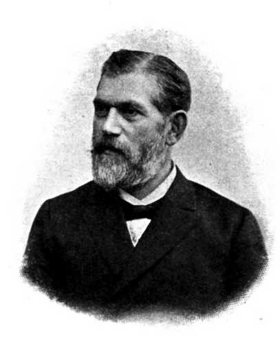
Julius Hirschberg (18 September 1843 – 17 February 1925) was a German ophthalmologist and medical historian. He was of Jewish ancestry. In 1875, Hirschberg coined the term "campimetry" for the measurement of the visual field on a flat surface (tangent screen test) and in 1879 he became the first to use an electromagnet to remove metallic foreign bodies from the eye.Manage Account - Modern Medicine at www.ophthalmologytimes.com In 1886, he developed the for measuring strabismus
[...More Info...] [...Related Items...] OR: [Wikipedia] [Google] [Baidu] |
Inferior Rectus Muscle
The inferior rectus muscle is a muscle in the orbit near the eye. It is one of the four recti muscles in the group of extraocular muscles. It originates from the common tendinous ring, and inserts into the anteroinferior surface of the eye. It depresses the eye (downwards). Structure The inferior rectus muscle originates from the common tendinous ring (annulus of Zinn). It inserts into the anteroinferior surface of the eye. This insertion has a width of around 10.5 mm. It is around 7 mm from the corneal limbus. Blood supply The inferior rectus muscle is supplied by an inferior muscular branch of the ophthalmic artery. It may also be supplied by a branch of the infraorbital artery. It is drained by the corresponding veins: the inferior muscular branch of the ophthalmic vein, and sometimes a branch of the infraorbital vein. Nerve supply The inferior rectus muscle is supplied by the inferior division of the oculomotor nerve (III). Development The inferior rectus muscle deve ... [...More Info...] [...Related Items...] OR: [Wikipedia] [Google] [Baidu] |
Co-morbid
In medicine, comorbidity - from Latin morbus ("sickness"), co ("together"), -ity (as if - several sicknesses together) - is the presence of one or more additional conditions often co-occurring (that is, concomitant or concurrent) with a primary condition. Comorbidity describes the effect of all other conditions an individual patient might have other than the primary condition of interest, and can be physiological or psychological. In the context of mental health, comorbidity often refers to disorders that are often coexistent with each other, such as depression and anxiety disorders. The concept of multimorbidity is related to comorbidity but presents a different meaning and approach. Definition The term "comorbid" has three definitions: # to indicate a medical condition existing simultaneously but independently with another condition in a patient. # to indicate a medical condition in a patient that causes, is caused by, or is otherwise related to another condition in the s ... [...More Info...] [...Related Items...] OR: [Wikipedia] [Google] [Baidu] |
Medial Rectus Muscle
The medial rectus muscle is a muscle in the orbit near the eye. It is one of the extraocular muscles. It originates from the common tendinous ring, and inserts into the anteromedial surface of the eye. It is supplied by the inferior division of the oculomotor nerve (III). It rotates the eye medially (adduction). Structure The medial rectus muscle shares an origin with several other extrinsic eye muscles, the common tendinous ring. It inserts into the anteromedial surface of the eye. This insertion has a width of around 11 mm. Nerve supply The medial rectus muscle is supplied by the inferior division of the oculomotor nerve (III). A branch of it enters the muscle around two fifths along its length. It usually divides into 2 smaller branches, occasionally 3. These further subdivide, becoming smaller down the length of the muscle until they become imperceptible to standard staining around 17 mm from the insertion of the muscle. Relations The insertion of the medial rectus mu ... [...More Info...] [...Related Items...] OR: [Wikipedia] [Google] [Baidu] |
Cover Test
A cover test or ''cover-uncover test'' is an objective determination of the presence and amount of ocular deviation. It is typically performed by orthoptists, ophthalmologists and optometrists during eye examinations. The two primary types of cover tests are: * the alternating cover test * the unilateral cover test (or the cover-uncover test). The test involves having the patient focusing on both a distance as well as near object at different times during the examination. A cover is placed over an eye for a short moment then removed while observing both eyes for movement. The misaligned eye will deviate inwards or outwards. The process is repeated on both eyes and then with the child focusing on a distant object. The cover test is used to determine both the type of ocular deviation and measure the amount of deviation. The two primary types of ocular deviations are the tropia and the phoria. A tropia is a misalignment of the two eyes when a patient is looking with both eyes uncov ... [...More Info...] [...Related Items...] OR: [Wikipedia] [Google] [Baidu] |


