|
Hannington-Kiff Sign
The Hannington-Kiff sign is a clinical sign in which there is an absent adductor reflex in the thigh in the presence of a positive patellar reflex. It occurs in patients with an obturator hernia, due to compression of the obturator nerve. The adductor reflex is elicited by tapping over either the medial epicondyle of the femur or the medial condyle of the tibia, which should cause the adductor muscles of the hip to contract, moving the leg inwards. The sign was described by John G Hannington-Kiff in 1980. See also * Howship–Romberg sign The Howship–Romberg sign is inner thigh pain on internal rotation of the hip. It can be caused by an obturator hernia. It is named for John Howship and Moritz Heinrich Romberg.M. H. von Romberg. Pathologie und Therapie der Senisbilitäts- und Mo ... References {{Digestive system and abdomen symptoms and signs Medical signs ... [...More Info...] [...Related Items...] OR: [Wikipedia] [Google] [Baidu] |
Medical Sign
Signs and symptoms are the observed or detectable signs, and experienced symptoms of an illness, injury, or condition. A sign for example may be a higher or lower temperature than normal, raised or lowered blood pressure or an abnormality showing on a medical scan. A symptom is something out of the ordinary that is experienced by an individual such as feeling feverish, a headache or other pain or pains in the body. Signs and symptoms Signs A medical sign is an objective observable indication of a disease, injury, or abnormal physiological state that may be detected during a physical examination, examining the patient history, or diagnostic procedure. These signs are visible or otherwise detectable such as a rash or bruise. Medical signs, along with symptoms, assist in formulating diagnostic hypothesis. Examples of signs include elevated blood pressure, nail clubbing of the fingernails or toenails, staggering gait, and arcus senilis and arcus juvenilis of the eyes. Indicati ... [...More Info...] [...Related Items...] OR: [Wikipedia] [Google] [Baidu] |
Patellar Reflex
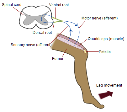
The patellar reflex, also called the knee reflex or knee-jerk, is a stretch reflex which tests the L2, L3, and L4 segments of the spinal cord. Mechanism Striking of the patellar tendon with a reflex hammer just below the patella stretches the muscle spindle in the quadriceps muscle. This produces a signal which travels back to the spinal cord and synapses (without interneurons) at the level of L3 or L4 in the spinal cord, completely independent of higher centres. From there, an alpha motor neuron conducts an efferent impulse back to the quadriceps femoris muscle, triggering contraction. This contraction, coordinated with the relaxation of the antagonistic flexor hamstring muscle causes the leg to kick. There is a latency of around 18 ms between stretch of the patellar tendon and the beginning of contraction of the quadriceps femoris muscle. This is a reflex of proprioception which helps maintain posture and balance, allowing to keep one's balance with little effort or conscious th ... [...More Info...] [...Related Items...] OR: [Wikipedia] [Google] [Baidu] |
Obturator Hernia
An obturator hernia is a rare type of hernia of the pelvic floor in which pelvic or abdominal contents protrudes through the obturator foramen. Because of differences in anatomy, it is much more common in women, especially multiparous and older women who have recently lost much weight. The diagnosis is often made intraoperatively after presenting with bowel obstruction. The Howship–Romberg sign is suggestive of an obturator hernia, exacerbated by thigh extension, medial rotation and abduction. It is characterized by lancinating pain in the medial thigh/obturator distribution, extending to the knee; caused by hernia compression of the obturator nerve The obturator nerve in human anatomy arises from the ventral divisions of the second, third, and fourth lumbar nerves in the lumbar plexus; the branch from the third is the largest, while that from the second is often very small. Structure The ob .... References External links Hernias {{disease-stub ... [...More Info...] [...Related Items...] OR: [Wikipedia] [Google] [Baidu] |
Obturator Nerve
The obturator nerve in human anatomy arises from the ventral divisions of the second, third, and fourth lumbar nerves in the lumbar plexus; the branch from the third is the largest, while that from the second is often very small. Structure The obturator nerve originates from the anterior divisions of the L2, L3, and L4 spinal nerve roots. It descends through the fibers of the psoas major, and emerges from its medial border near the brim of the pelvis. It then passes behind the common iliac arteries, and on the lateral side of the internal iliac artery and vein, and runs along the lateral wall of the lesser pelvis, above and in front of the obturator vessels, to the upper part of the obturator foramen. Here it enters the thigh, through the obturator canal, and divides into an anterior and a posterior branch, which are separated at first by some of the fibers of the obturator externus, and lower down by the adductor brevis. An accessory obturator nerve may be present in approx ... [...More Info...] [...Related Items...] OR: [Wikipedia] [Google] [Baidu] |
Medial Epicondyle Of The Femur of the are attached to it.Platzer (2004), 9 206
Behind it, and proximal to the medial condyl ...
The medial epicondyle of the femur is an epicondyle, a bony protrusion, located on the medial side of the femur at its distal end. Located above the medial condyle, it bears an elevation, the adductor tubercle,Platzer (2004), p 192 which serves for the attachment of the superficial part, or "tendinous insertion", of the adductor magnus.''Thieme Atlas of Anatomy'' (2006), p 426 This tendinous part here forms an intermuscular septum which forms the medial separation between the thigh's flexors and extensors.tibial collateral ligament [...More Info...] [...Related Items...] OR: [Wikipedia] [Google] [Baidu] |
Medial Condyle Of The Tibia
The medial condyle is the medial (or inner) portion of the upper extremity of tibia. It is the site of insertion for the semimembranosus muscle The semimembranosus muscle () is the most medial of the three hamstring muscles in the thigh. It is so named because it has a flat tendon of origin. It lies posteromedially in the thigh, deep to the semitendinosus muscle. It extends the hip joint .... See also * Lateral condyle of tibia * Medial collateral ligament Additional images File:Gray258.png, Bones of the right leg. Anterior surface. File:Gray259.png, Bones of the right leg. Posterior surface. File:Slide2bib.JPG, Right knee in extension. Deep dissection. Posterior view. File:Slide2cocc.JPG, Right knee in extension. Deep dissection. Posterior view. References External links * * * () Bones of the lower limb Tibia {{musculoskeletal-stub ... [...More Info...] [...Related Items...] OR: [Wikipedia] [Google] [Baidu] |
Adductor Muscles Of The Hip
The adductor muscles of the hip are a group of muscles mostly used for bringing the thighs together (called adduction). Structure The adductor group is made up of: *Adductor brevis *Adductor longus *Adductor magnus * Adductor minimus This is often considered to be a part of adductor magnus. * pectineus * gracilis *Obturator externusPlatzer, Werner (2004), Color Atlas of Human Anatomy, Vol. 1, Locomotor System', Thieme, 5th ed, p 240 and are also part of the medial compartment of thigh The adductors originate on the pubis and ischium bones and insert mainly on the medial posterior surface of the femur. Nerve supply The pectineus is the only adductor muscle that is innervated by the femoral nerve. The other adductor muscles are innervated by the obturator nerve with the exception of a small part of the adductor magnus which is innervated by the tibial nerve. Variation In 33% of people a supernumerary muscle is found between the adductor brevis and adductor minimus. When ... [...More Info...] [...Related Items...] OR: [Wikipedia] [Google] [Baidu] |
Howship–Romberg Sign
The Howship–Romberg sign is inner thigh pain on internal rotation of the hip. It can be caused by an obturator hernia. It is named for John Howship and Moritz Heinrich Romberg.M. H. von Romberg. Pathologie und Therapie der Senisbilitäts- und Motilitätsneurosen. 1857. 3rd edition (unfinished) of Romberg’s Lehrbuch der Nervenkrankheiten des Menschen, page 89. References Medical signs {{med-sign-stub ... [...More Info...] [...Related Items...] OR: [Wikipedia] [Google] [Baidu] |
