|
Globus Pallidus
The globus pallidus (GP), also known as paleostriatum or dorsal pallidum, is a subcortical structure of the brain. It consists of two adjacent segments, one external, known in rodents simply as the globus pallidus, and one internal, known in rodents as the entopeduncular nucleus. It is part of the telencephalon, but retains close functional ties with the subthalamus in the diencephalon – both of which are part of the extrapyramidal motor system. The globus pallidus is a major component of the basal ganglia, with principal inputs from the striatum, and principal direct outputs to the thalamus and the substantia nigra. The latter is made up of similar neuronal elements, has similar afferents from the striatum, similar projections to the thalamus, and has a similar synaptology. Neither receives direct cortical afferents, and both receive substantial additional inputs from the intralaminar thalamus. Globus pallidus is Latin for "pale globe". Structure Pallidal nuclei are made u ... [...More Info...] [...Related Items...] OR: [Wikipedia] [Google] [Baidu] |
Basal Ganglia
The basal ganglia (BG), or basal nuclei, are a group of subcortical nuclei, of varied origin, in the brains of vertebrates. In humans, and some primates, there are some differences, mainly in the division of the globus pallidus into an external and internal region, and in the division of the striatum. The basal ganglia are situated at the base of the forebrain and top of the midbrain. Basal ganglia are strongly interconnected with the cerebral cortex, thalamus, and brainstem, as well as several other brain areas. The basal ganglia are associated with a variety of functions, including control of voluntary motor movements, procedural learning, habit learning, conditional learning, eye movements, cognition, and emotion. The main components of the basal ganglia – as defined functionally – are the striatum, consisting of both the dorsal striatum ( caudate nucleus and putamen) and the ventral striatum ( nucleus accumbens and olfactory tubercle), the globus ... [...More Info...] [...Related Items...] OR: [Wikipedia] [Google] [Baidu] |
Striatopallidal
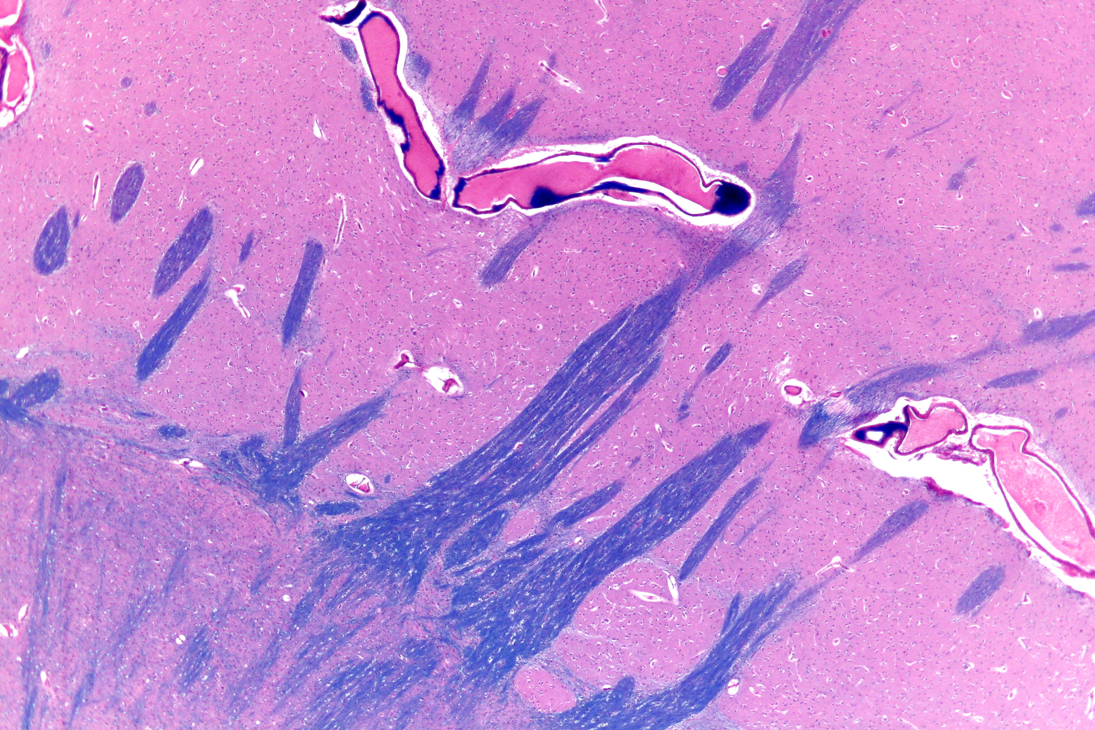
The striatopallidal fibres, also Wilson's pencils, pencil fibres of Wilson, and pencils of Wilson, are prominent myelinated fibres that connect the striatum to the globus pallidus. Their distinctive appearance allows the putamen to be identified on light microscopy. See also *Lentiform nucleus The lentiform nucleus, or lenticular nucleus, comprises the putamen and the globus pallidus within the basal ganglia. With the caudate nucleus, it forms the dorsal striatum. It is a large, lens-shaped mass of gray matter just lateral to the inte ... References {{Reflist, 2 Basal ganglia ... [...More Info...] [...Related Items...] OR: [Wikipedia] [Google] [Baidu] |
Pedunculopontine Nucleus
The pedunculopontine nucleus (PPN) or pedunculopontine tegmental nucleus (PPT or PPTg) is a collection of neurons located in the upper pons in the brainstem. It lies caudal to the substantia nigra and adjacent to the superior cerebellar peduncle. It has two divisions of subnuclei; the pars compacta containing mainly cholinergic neurons, and the pars dissipata containing mainly glutamatergic neurons and some non-cholinergic neurons. The pedunculopontine nucleus is one of the main components of the reticular activating system. It was first described in 1909 by Louis Jacobsohn-Lask, a German neuroanatomist. Projections Pedunculopontine nucleus neurons project axons to a wide range of areas in the brain, particularly parts of the basal ganglia such as the subthalamic nucleus, substantia nigra pars compacta, and globus pallidus internus. It also sends them to targets in the thalamus, cerebellum, basal forebrain, and lower brainstem, and in the cerebral cortex, the suppleme ... [...More Info...] [...Related Items...] OR: [Wikipedia] [Google] [Baidu] |
Prefrontal Cortex
In mammalian brain anatomy, the prefrontal cortex (PFC) covers the front part of the frontal lobe of the cerebral cortex. The PFC contains the Brodmann areas BA8, BA9, BA10, BA11, BA12, BA13, BA14, BA24, BA25, BA32, BA44, BA45, BA46, and BA47. The basic activity of this brain region is considered to be orchestration of thoughts and actions in accordance with internal goals. Many authors have indicated an integral link between a person's will to live, personality, and the functions of the prefrontal cortex. This brain region has been implicated in executive functions, such as planning, decision making, short-term memory, personality expression, moderating social behavior and controlling certain aspects of speech and language. Executive function relates to abilities to differentiate among conflicting thoughts, determine good and bad, better and best, same and different, future consequences of current activities, working toward a defined goal, prediction of outcom ... [...More Info...] [...Related Items...] OR: [Wikipedia] [Google] [Baidu] |
Olfactory Tubercle
The olfactory tubercle (OT), also known as the tuberculum olfactorium, is a multi-sensory processing center that is contained within the olfactory cortex and ventral striatum and plays a role in reward cognition. The OT has also been shown to play a role in locomotor and attentional behaviors, particularly in relation to social and sensory responsiveness, and it may be necessary for behavioral flexibility. The OT is interconnected with numerous brain regions, especially the sensory, arousal, and reward centers, thus making it a potentially critical interface between processing of sensory information and the subsequent behavioral responses. The OT is a composite structure that receives direct input from the olfactory bulb and contains the morphological and histochemical characteristics of the ventral pallidum and the striatum of the forebrain. The dopaminergic neurons of the mesolimbic pathway project onto the GABAergic medium spiny neurons of the nucleus accumbens and ol ... [...More Info...] [...Related Items...] OR: [Wikipedia] [Google] [Baidu] |
Nucleus Accumbens
The nucleus accumbens (NAc or NAcc; also known as the accumbens nucleus, or formerly as the ''nucleus accumbens septi'', Latin for " nucleus adjacent to the septum") is a region in the basal forebrain rostral to the preoptic area of the hypothalamus. The nucleus accumbens and the olfactory tubercle collectively form the ventral striatum. The ventral striatum and dorsal striatum collectively form the striatum, which is the main component of the basal ganglia. The dopaminergic neurons of the mesolimbic pathway project onto the GABAergic medium spiny neurons of the nucleus accumbens and olfactory tubercle. Each cerebral hemisphere has its own nucleus accumbens, which can be divided into two structures: the nucleus accumbens core and the nucleus accumbens shell. These substructures have different morphology and functions. Different NAcc subregions (core vs shell) and neuron subpopulations within each region ( D1-type vs D2-type medium spiny neurons) are responsible for diff ... [...More Info...] [...Related Items...] OR: [Wikipedia] [Google] [Baidu] |
Substantia Innominata
The substantia innominata also innominate substance, or substantia innominata of Meynert (Latin for unnamed substance) is a series of layers in the human brain consisting partly of gray and partly of white matter, which lies below the anterior part of the thalamus and lentiform nucleus. It is included as part of the anterior perforated substance (as it appears to be perforated by many holes which are actually blood vessels). It is part of the basal forebrain structures and includes the nucleus basalis. A portion of the substantia innominata, below the globus pallidus is considered as part of the extended amygdala. Layers It consists of three layers, superior, middle, and inferior. * The ''superior layer'' is named the ansa lenticularis, and its fibers, derived from the medullary lamina of the lentiform nucleus, pass medially to end in the thalamus and subthalamic region, while others are said to end in the tegmentum and red nucleus. * The ''middle layer'' consists of nerve c ... [...More Info...] [...Related Items...] OR: [Wikipedia] [Google] [Baidu] |
Ventral Pallidum
The ventral pallidum (VP) is a structure within the basal ganglia of the brain. It is an output nucleus whose fibres project to thalamic nuclei, such as the ventral anterior nucleus, the ventral lateral nucleus, and the medial dorsal nucleus. The VP is a core component of the reward system which forms part of the limbic loop of the basal ganglia, a pathway involved in the regulation of motivational salience, behavior, and emotions. It is involved in addiction. The VP contains one of the brain's pleasure centers, which mediates the subjective perception of pleasure that results from "consuming" certain rewarding stimuli (e.g., palatable food). Anatomy The ventral pallidum lies within the basal ganglia, a group of subcortical nuclei. Along with the external globus pallidus, it is separated from other basal ganglia nuclei by the anterior commissure. Limbic loop The limbic loop is a functional pathway of the basal ganglia, in which the ventral pallidum is involved. It (and the in ... [...More Info...] [...Related Items...] OR: [Wikipedia] [Google] [Baidu] |
External Globus Pallidus
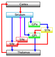
The external globus pallidus (GPe or lateral globus pallidus) combines with the internal globus pallidus (GPi) to form the globus pallidus, an anatomical subset of the basal ganglia. Globus pallidus means "pale globe" in Latin, indicating its appearance. The external globus pallidus is the segment of the globus pallidus that is relatively further (lateral) from the midline of the brain. The GPe GABAergic neurons, allow for its inhibitory function and projects axons to the subthalamic nucleus (in the diencephalon), the striatum, internal globus pallidus (GPi) and substantia nigra pars reticulata. The GPe is particular in comparison to the other elements of the set by the fact that it does not work as an output base of the basal ganglia (not sending axons to the thalamus) but as the main regulator of the basal ganglia system. It is sometimes used as a target for deep brain stimulation as a treatment for Parkinson's disease. Function Indirect pathway The basal ganglia funct ... [...More Info...] [...Related Items...] OR: [Wikipedia] [Google] [Baidu] |
Internal Globus Pallidus
The internal globus pallidus (GPi or medial globus pallidus; in rodents its homologue is known as the entopeduncular nucleus) and the external globus pallidus (GPe) make up the globus pallidus. The GPi is one of the output nuclei of the basal ganglia (the other being the substantia nigra pars reticulata). The GABAergic neurons of the GPi send their axons to the ventral anterior nucleus (VA) and the ventral lateral nucleus (VL) in the dorsal thalamus, to the centromedian complex, and to the pedunculopontine complex. The efferent bundle is constituted first of the ansa and lenticular fasciculus, then crosses the internal capsule within and in parallel to the Edinger's comb system then arrives at the laterosuperior corner of the subthalamic nucleus and constitutes the field H2 of Forel, then H, and suddenly changes its direction to form field H1 that goes to the inferior part of the thalamus. The distribution of axonal islands is widespread in the lateral region of the thalamus. T ... [...More Info...] [...Related Items...] OR: [Wikipedia] [Google] [Baidu] |
Medial Medullary Lamina
Medial may refer to: Mathematics * Medial magma, a mathematical identity in algebra Geometry * Medial axis, in geometry the set of all points having more than one closest point on an object's boundary * Medial graph, another graph that represents the adjacencies between edges in the faces of a plane graph * Medial triangle, the triangle whose vertices lie at the midpoints of an enclosing triangle's sides * Polyhedra: ** Medial deltoidal hexecontahedron ** Medial disdyakis triacontahedron ** Medial hexagonal hexecontahedron ** Medial icosacronic hexecontahedron ** Medial inverted pentagonal hexecontahedron ** Medial pentagonal hexecontahedron ** Medial rhombic triacontahedron Linguistics * A medial sound or letter is one that is found in the middle of a larger unit (like a word) ** Syllable medial, a segment located between the onset and the rime of a syllable * In the older literature, a term for the voiced stops (like ''b'', ''d'', ''g'') * Medial or second person demon ... [...More Info...] [...Related Items...] OR: [Wikipedia] [Google] [Baidu] |
Primate Basal Ganglia
The basal ganglia form a major brain system in all species of vertebrates, but in primates (including humans) there are special features that justify a separate consideration. As in other vertebrates, the primate basal ganglia can be divided into striatal, pallidal, nigral, and subthalamic components. In primates, however, there are two pallidal subdivisions called the external globus pallidus (GPe) and internal globus pallidus (GPi). Also in primates, the dorsal striatum is divided by a large tract called the internal capsule into two masses named the caudate nucleus and the putamen—in most other species no such division exists, and only the striatum as a whole is recognized. Beyond this, there is a complex circuitry of connections between the striatum and cortex that is specific to primates. This complexity reflects the difference in functioning of different cortical areas in the primate brain. Functional imaging studies have been performed mainly using human subjects. ... [...More Info...] [...Related Items...] OR: [Wikipedia] [Google] [Baidu] |


