|
Substantia Innominata
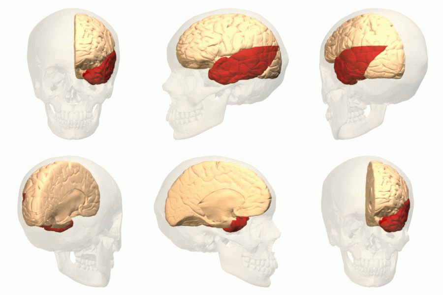
The substantia innominata also innominate substance, or substantia innominata of Meynert (Latin for unnamed substance) is a series of layers in the human brain consisting partly of gray and partly of white matter, which lies below the anterior part of the thalamus and lentiform nucleus. It is included as part of the anterior perforated substance (as it appears to be perforated by many holes which are actually blood vessels). It is part of the basal forebrain structures and includes the nucleus basalis. A portion of the substantia innominata, below the globus pallidus is considered as part of the extended amygdala. Layers It consists of three layers, superior, middle, and inferior. * The ''superior layer'' is named the ansa lenticularis, and its fibers, derived from the medullary lamina of the lentiform nucleus, pass medially to end in the thalamus and subthalamic region, while others are said to end in the tegmentum and red nucleus. * The ''middle layer'' consists of nerve cells ... [...More Info...] [...Related Items...] OR: [Wikipedia] [Google] [Baidu] |
Human Brain
The human brain is the central organ of the human nervous system, and with the spinal cord makes up the central nervous system. The brain consists of the cerebrum, the brainstem and the cerebellum. It controls most of the activities of the body, processing, integrating, and coordinating the information it receives from the sense organs, and making decisions as to the instructions sent to the rest of the body. The brain is contained in, and protected by, the skull bones of the head. The cerebrum, the largest part of the human brain, consists of two cerebral hemispheres. Each hemisphere has an inner core composed of white matter, and an outer surface – the cerebral cortex – composed of grey matter. The cortex has an outer layer, the neocortex, and an inner allocortex. The neocortex is made up of six neuronal layers, while the allocortex has three or four. Each hemisphere is conventionally divided into four lobes – the frontal, temporal, parietal, and occipital lo ... [...More Info...] [...Related Items...] OR: [Wikipedia] [Google] [Baidu] |
Ansa Lenticularis
The ansa lenticularis (''ansa lentiformis'' in older texts) is a part of the brain, making up the superior layer of the substantia innominata. Its fibers, derived from the medullary lamina of the lentiform nucleus, pass medially to end in the thalamus and subthalamic region, while others are said to end in the tegmentum and red nucleus. It is classified by NeuroNames as part of the subthalamus The subthalamus or prethalamus is a part of the diencephalon. Its most prominent structure is the subthalamic nucleus. The subthalamus connects to the globus pallidus, a basal nucleus of the telencephalon. Structure The subthalamus is locate .... References External links * {{Authority control Thalamic connections Basal ganglia connections ... [...More Info...] [...Related Items...] OR: [Wikipedia] [Google] [Baidu] |
Staining
Staining is a technique used to enhance contrast in samples, generally at the microscopic level. Stains and dyes are frequently used in histology (microscopic study of biological tissues), in cytology (microscopic study of cells), and in the medical fields of histopathology, hematology, and cytopathology that focus on the study and diagnoses of diseases at the microscopic level. Stains may be used to define biological tissues (highlighting, for example, muscle fibers or connective tissue), cell populations (classifying different blood cells), or organelles within individual cells. In biochemistry, it involves adding a class-specific ( DNA, proteins, lipids, carbohydrates) dye to a substrate to qualify or quantify the presence of a specific compound. Staining and fluorescent tagging can serve similar purposes. Biological staining is also used to mark cells in flow cytometry, and to flag proteins or nucleic acids in gel electrophoresis. Light microscopes are used for viewin ... [...More Info...] [...Related Items...] OR: [Wikipedia] [Google] [Baidu] |
Insular Cortex
The insular cortex (also insula and insular lobe) is a portion of the cerebral cortex folded deep within the lateral sulcus (the fissure separating the temporal lobe from the parietal and frontal lobes) within each hemisphere of the mammalian brain. The insulae are believed to be involved in consciousness and play a role in diverse functions usually linked to emotion or the regulation of the body's homeostasis. These functions include compassion, empathy, taste, perception, motor control, self-awareness, cognitive functioning, interpersonal experience, and awareness of homeostatic emotions such as hunger, pain and fatigue. In relation to these, it is involved in psychopathology. The insular cortex is divided into two parts: the anterior insula and the posterior insula in which more than a dozen field areas have been identified. The cortical area overlying the insula toward the lateral surface of the brain is the operculum (meaning ''lid''). The opercula are formed from parts o ... [...More Info...] [...Related Items...] OR: [Wikipedia] [Google] [Baidu] |
Temporal Lobe
The temporal lobe is one of the four Lobes of the brain, major lobes of the cerebral cortex in the brain of mammals. The temporal lobe is located beneath the lateral fissure on both cerebral hemispheres of the mammalian brain. The temporal lobe is involved in processing sensory input into derived meanings for the appropriate retention of visual memory, language comprehension, and emotion association. ''Temporal'' refers to the head's Temple (anatomy), temples. Structure The Temple (anatomy)#Etymology, temporal Lobe (anatomy), lobe consists of structures that are vital for declarative or long-term memory. Declarative memory, Declarative (denotative) or Explicit memory, explicit memory is conscious memory divided into semantic memory (facts) and episodic memory (events). Medial temporal lobe structures that are critical for long-term memory include the hippocampus, along with the surrounding Hippocampal formation, hippocampal region consisting of the Perirhinal cortex, perirhinal, ... [...More Info...] [...Related Items...] OR: [Wikipedia] [Google] [Baidu] |
Medial Longitudinal Fasciculus
The medial longitudinal fasciculus (MLF) is an area of crossed over tracts, on each side of the brainstem. These bundles of axons are situated near the midline of the brainstem. They are made up of both ascending and descending fibers that arise from a number of sources and terminate in different areas, including the superior colliculus, the vestibular nuclei, and the cerebellum. It contains the interstitial nucleus of Cajal, responsible for oculomotor control, head posture, and vertical eye movement. The medial longitudinal fasciculus is the main central connection for the oculomotor nerve, trochlear nerve, and abducens nerve. It carries information about the direction that the eyes should move. Lesions of the medial longitudinal fasciculus can cause nystagmus and diplopia, which may be associated with multiple sclerosis, a neoplasm, or a stroke. Structure The medial longitudinal fasciculus is an area of crossed over tracts, on each side of the brainstem. It is medial, ... [...More Info...] [...Related Items...] OR: [Wikipedia] [Google] [Baidu] |
External Capsule
The external capsule is a series of white matter fiber tracts in the brain. These fibers run between the most lateral (toward the side of the head) segment of the lentiform nucleus (more specifically the putamen) and the claustrum. The white matter of the external capsule contains fibers known as corticocortical association fibers. These fibers are responsible for connecting the cerebral cortex to another cortical area. The capsule itself appears as a thin white sheet of white matter. The external capsule is a route for cholinergic fibers from the basal forebrain to the cerebral cortex. The putamen separates the external capsule from the internal capsule medially and the claustrum separates it from the extreme capsule laterally. But the external capsule eventually joins the internal capsule The internal capsule is a white matter structure situated in the inferomedial part of each cerebral hemisphere of the brain. It carries information past the basal ganglia, separating the ... [...More Info...] [...Related Items...] OR: [Wikipedia] [Google] [Baidu] |
Red Nucleus
The red nucleus or nucleus ruber is a structure in the rostral midbrain involved in motor coordination. The red nucleus is pale pink, which is believed to be due to the presence of iron in at least two different forms: hemoglobin and ferritin. The structure is located in the tegmentum of the midbrain next to the substantia nigra and comprises caudal magnocellular and rostral parvocellular components. The red nucleus and substantia nigra are subcortical centers of the extrapyramidal motor system. Function In a vertebrate without a significant corticospinal tract, gait is mainly controlled by the red nucleus. However, in primates, where the corticospinal tract is dominant, the rubrospinal tract may be regarded as vestigial in motor function. Therefore, the red nucleus is less important in primates than in many other mammals. Nevertheless, the crawling of babies is controlled by the red nucleus, as is arm swinging in typical walking. The red nucleus may play an additional role ... [...More Info...] [...Related Items...] OR: [Wikipedia] [Google] [Baidu] |
Tegmentum
The tegmentum (from Latin for "covering") is a general area within the brainstem. The tegmentum is the ventral part of the midbrain and the tectum is the dorsal part of the midbrain. It is located between the ventricular system and distinctive basal or ventral structures at each level. It forms the floor of the midbrain (mesencephalon) whereas the tectum forms the ceiling. It is a multisynaptic network of neurons that is involved in many subconscious homeostatic and reflexive pathways. It is a motor center that relays inhibitory signals to the thalamus and basal nuclei preventing unwanted body movement. The tegmentum area includes various different structures, such as the rostral end of the reticular formation, several nuclei controlling eye movements, the periaqueductal gray matter, the red nucleus, the substantia nigra, and the ventral tegmental area. The tegmentum is the location of several cranial nerve (CN) nuclei. The nuclei of CN III and IV are located in the tegmentu ... [...More Info...] [...Related Items...] OR: [Wikipedia] [Google] [Baidu] |
Nucleus Basalis Of Meynert -l- Very Low Mag
Nucleus ( : nuclei) is a Latin word for the seed inside a fruit. It most often refers to: *Atomic nucleus, the very dense central region of an atom *Cell nucleus, a central organelle of a eukaryotic cell, containing most of the cell's DNA Nucleus may also refer to: Science, technology, and mathematics Astronomy *Active galactic nucleus in astronomy *Comet nucleus, the solid, central part of a comet Biology *Cell nucleus, a central organelle of a eukaryotic cell, containing most of the cell's DNA *Nucleus (neuroanatomy), a cluster of cell bodies of neurons in the central nervous system *Nucleus that forms in the eye in nuclear sclerosis (early cataracts) *''Nucleus'', a scientific journal concerned with the cell nucleus; published by Taylor & Francis *Nucleus, a small colony of honeybees, induced to raise a new queen by the beekeeper Computer systems * Nucleus (operating system), sometimes a synonym for kernel * Nucleus CMS, a weblog system * Nucleus RTOS, a real-time operat ... [...More Info...] [...Related Items...] OR: [Wikipedia] [Google] [Baidu] |
Grey Matter
Grey matter is a major component of the central nervous system, consisting of neuronal cell bodies, neuropil (dendrites and unmyelinated axons), glial cells (astrocytes and oligodendrocytes), synapses, and capillaries. Grey matter is distinguished from white matter in that it contains numerous cell bodies and relatively few myelinated axons, while white matter contains relatively few cell bodies and is composed chiefly of long-range myelinated axons. The colour difference arises mainly from the whiteness of myelin. In living tissue, grey matter actually has a very light grey colour with yellowish or pinkish hues, which come from capillary blood vessels and neuronal cell bodies. Structure Grey matter refers to unmyelinated neurons and other cells of the central nervous system. It is present in the brain, brainstem and cerebellum, and present throughout the spinal cord. Grey matter is distributed at the surface of the cerebral hemispheres (cerebral cortex) and of the cerebellu ... [...More Info...] [...Related Items...] OR: [Wikipedia] [Google] [Baidu] |
Extended Amygdala
The extended amygdala is a macrostructure in the brain that is involved in reward cognition and defined by connectivity and neurochemical staining. It includes the central medial amygdala, sublenticular substantia innominata, and the bed nucleus of the stria terminalis The stria terminalis (or terminal stria) is a structure in the brain consisting of a band of fibers running along the lateral margin of the ventricular surface of the thalamus. Serving as a major output pathway of the amygdala, the stria termina .... The boundaries are indistinct in Nissl stained sections. References External links * More information aBrainInfo Amygdala {{neuroanatomy-stub ... [...More Info...] [...Related Items...] OR: [Wikipedia] [Google] [Baidu] |


