|
Ganglion
A ganglion is a group of neuron cell bodies in the peripheral nervous system. In the somatic nervous system this includes dorsal root ganglia and trigeminal ganglia among a few others. In the autonomic nervous system there are both sympathetic and parasympathetic ganglia which contain the cell bodies of postganglionic sympathetic and parasympathetic neurons respectively. A pseudoganglion looks like a ganglion, but only has nerve fibers and has no nerve cell bodies. Structure Ganglia are primarily made up of somata and dendritic structures which are bundled or connected. Ganglia often interconnect with other ganglia to form a complex system of ganglia known as a plexus. Ganglia provide relay points and intermediary connections between different neurological structures in the body, such as the peripheral and central nervous systems. Among vertebrates there are three major groups of ganglia: *Dorsal root ganglia (also known as the spinal ganglia) contain the cell bodies of se ... [...More Info...] [...Related Items...] OR: [Wikipedia] [Google] [Baidu] |
Ganglion Cyst
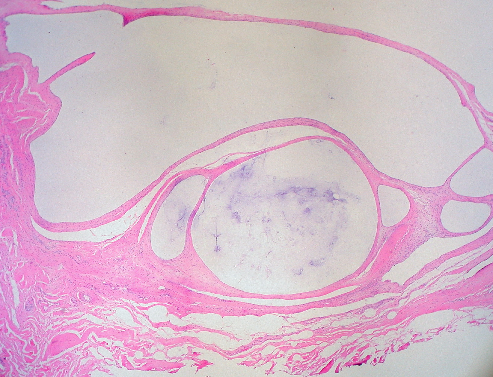
A ganglion cyst is a fluid-filled bump associated with a joint or tendon sheath. It most often occurs at the back of the wrist, followed by the front of the wrist. Onset is often over several months, typically with no further symptoms. Occasionally, pain or numbness may occur. Complications may include carpal tunnel syndrome. The cause is unknown. The underlying mechanism is believed to involve an outpouching of the synovial membrane. Risk factors include gymnastics activity. Diagnosis is typically based on examination with light shining through the lesion being supportive. Medical imaging may be done to rule out other potential causes. Treatment options include watchful waiting, splinting the affected joint, needle aspiration, or surgery. About half the time, they resolve on their own. About three per 10,000 people newly develop ganglion of the wrist or hand a year. They most commonly occur in young and middle-aged females. Presentation The average size of these cysts is 2.0 ... [...More Info...] [...Related Items...] OR: [Wikipedia] [Google] [Baidu] |
Autonomic Nervous System
The autonomic nervous system (ANS), formerly referred to as the vegetative nervous system, is a division of the peripheral nervous system that supplies viscera, internal organs, smooth muscle and glands. The autonomic nervous system is a control system that acts largely unconsciously and regulates bodily functions, such as the heart rate, its force of contraction, digestion, respiratory rate, pupillary dilation, pupillary response, Micturition, urination, and sexual arousal. This system is the primary mechanism in control of the fight-or-flight response. The autonomic nervous system is regulated by integrated reflexes through the brainstem to the spinal cord and organ (anatomy), organs. Autonomic functions include control of respiration, heart rate, cardiac regulation (the cardiac control center), vasomotor activity (the vasomotor center), and certain reflex, reflex actions such as coughing, sneezing, swallowing and vomiting. Those are then subdivided into other areas and are also ... [...More Info...] [...Related Items...] OR: [Wikipedia] [Google] [Baidu] |
Trigeminal Ganglion
A trigeminal ganglion (or Gasserian ganglion, or semilunar ganglion, or Gasser's ganglion) is the sensory ganglion at the base of each of the two trigeminal nerves (CN V), occupying a cavity (Meckel's cave) in the dura mater, covering the trigeminal impression near the apex of the petrous part of the temporal bone. Structure It is somewhat crescent-shaped, with its convexity directed forward: Medially, it is in relation with the internal carotid artery and the posterior part of the cavernous sinus. The motor root runs in front of and medial to the sensory root, and passes beneath the ganglion; it leaves the skull through the foramen ovale, and, immediately below this foramen, joins the mandibular nerve. The greater superficial petrosal nerve lies also underneath the ganglion. The ganglion receives, on its medial side, filaments from the carotid plexus of the sympathetic. It gives off minute branches to the tentorium cerebelli, and to the dura mater in the middle fossa of th ... [...More Info...] [...Related Items...] OR: [Wikipedia] [Google] [Baidu] |
Dorsal Root Ganglion
A dorsal root ganglion (or spinal ganglion; also known as a posterior root ganglion) is a cluster of neurons (a ganglion) in a dorsal root of a spinal nerve. The cell bodies of sensory neurons known as first-order neurons are located in the dorsal root ganglia. The axons of dorsal root ganglion neurons are known as afferents. In the peripheral nervous system, afferents refer to the axons that relay sensory information into the central nervous system (i.e. the brain and the spinal cord). Structure The neurons comprising the dorsal root ganglion are of the pseudo-unipolar type, meaning they have a cell body (soma) with two branches that act as a single axon, often referred to as a ''distal process'' and a ''proximal process''. Unlike the majority of neurons found in the central nervous system, an action potential in posterior root ganglion neuron may initiate in the ''distal process'' in the periphery, bypass the cell body, and continue to propagate along the ''proximal process ... [...More Info...] [...Related Items...] OR: [Wikipedia] [Google] [Baidu] |
Parasympathetic Ganglion
Parasympathetic ganglia are the autonomic ganglia of the parasympathetic nervous system. Most are small terminal ganglia or intramural ganglia, so named because they lie near or within (respectively) the organs they innervate. The exceptions are the four paired parasympathetic ganglia of the head and neck. Of the head and neck These paired ganglia supply all parasympathetic innervation to the head and neck. *ciliary ganglion (sphincter pupillae, ciliary muscle) *pterygopalatine ganglion (lacrimal gland, glands of nasal cavity) *submandibular ganglion (submandibular and sublingual glands) *otic ganglion (parotid gland) Roots Each has three roots entering the ganglion and a variable number of exiting branches. * The motor root carries presynaptic parasympathetic nerve fibers ( GVE) that terminate in the ganglion and synapse with the postsynaptic fibers that, in turn, project to target organs. * The sympathetic root carries postsynaptic sympathetic fibers ( GVE) that traverse the g ... [...More Info...] [...Related Items...] OR: [Wikipedia] [Google] [Baidu] |
Cranial Nerve Ganglia
In neuroanatomy, the cranial nerve ganglia are ganglia of certain cranial nerves. They can be parasympathetic or sensory. All cranial nerve ganglia are bilateral. Parasympathetic The four cranial parasympathetic ganglia are: *ciliary ganglion *pterygopalatine ganglion *otic ganglion *submandibular ganglion Sensory * trigeminal ganglion (CN V) * geniculate ganglion (CN VII) * spiral ganglion (CN VIII) * vestibular ganglion aka Scarpa's ganglion (CN VIII) * superior ganglion of glossopharyngeal nerve * inferior ganglion of glossopharyngeal nerve * superior ganglion of vagus nerve * inferior ganglion of vagus nerve The inferior ganglion of the vagus nerve, (nodose ganglion) is a sensory ganglion of the peripheral nervous system. It is located within the jugular foramen where the vagus nerve exits the skull. It is larger than and below the superior ganglion ... References Nervous ganglia {{neuroanatomy-stub ... [...More Info...] [...Related Items...] OR: [Wikipedia] [Google] [Baidu] |
Nervous System
In biology, the nervous system is the highly complex part of an animal that coordinates its actions and sensory information by transmitting signals to and from different parts of its body. The nervous system detects environmental changes that impact the body, then works in tandem with the endocrine system to respond to such events. Nervous tissue first arose in wormlike organisms about 550 to 600 million years ago. In vertebrates it consists of two main parts, the central nervous system (CNS) and the peripheral nervous system (PNS). The CNS consists of the brain and spinal cord. The PNS consists mainly of nerves, which are enclosed bundles of the long fibers or axons, that connect the CNS to every other part of the body. Nerves that transmit signals from the brain are called motor nerves or '' efferent'' nerves, while those nerves that transmit information from the body to the CNS are called sensory nerves or '' afferent''. Spinal nerves are mixed nerves that serve both fu ... [...More Info...] [...Related Items...] OR: [Wikipedia] [Google] [Baidu] |
Postganglionic Fibers
In the autonomic nervous system, fibers from the ganglion to the effector organ are called postganglionic fibers. Neurotransmitters The neurotransmitters of postganglionic fibers differ: * In the parasympathetic division, neurons are ''cholinergic''. That is to say acetylcholine is the primary neurotransmitter responsible for the communication between neurons on the parasympathetic pathway. * In the sympathetic division, neurons are mostly ''adrenergic'' (that is, epinephrine and norepinephrine function as the primary neurotransmitters). Notable exceptions to this rule include the sympathetic innervation of sweat glands and arrectores pilorum muscles where the neurotransmitter at both pre and post ganglionic synapses is acetylcholine. Another notable structure is the medulla of the adrenal gland, where chromaffin cells function as modified post-ganglionic nerves. Instead of releasing epinephrine and norepinephrine into a synaptic cleft, these cells of the adrenal medulla r ... [...More Info...] [...Related Items...] OR: [Wikipedia] [Google] [Baidu] |
Preganglionic Fibers
In the autonomic nervous system, fibers from the CNS to the ganglion are known as preganglionic fibers. All preganglionic fibers, whether they are in the sympathetic division or in the parasympathetic division, are cholinergic (that is, these fibers use acetylcholine as their neurotransmitter) and they are myelinated. Sympathetic preganglionic fibers tend to be shorter than parasympathetic preganglionic fibers because sympathetic ganglia are often closer to the spinal cord than are the parasympathetic ganglia. Another major difference between the two ANS (autonomic nervous systems) is divergence. Whereas in the parasympathetic division there is a divergence factor of roughly 1:4, in the sympathetic division there can be a divergence of up to 1:20. This is due to the number of synapses formed by the preganglionic fibers with ganglionic neurons. See also * Postganglionic fibers In the autonomic nervous system, fibers from the ganglion to the effector organ are called p ... [...More Info...] [...Related Items...] OR: [Wikipedia] [Google] [Baidu] |
Sympathetic Ganglion
The sympathetic ganglia, or paravertebral ganglia are autonomic ganglia, of the sympathetic nervous system. Ganglia are 20,000 to 30,000 afferent and efferent nerve cell bodies that run along on either side of the spinal cord. Afferent nerve cell bodies bring information from the body to the brain and spinal cord, while efferent nerve cell bodies bring information from the brain and spinal cord to the rest of the body. The cell bodies create long sympathetic chains that are on either side of the spinal cord. They also form para- or pre-vertebral ganglia of gross anatomy. The efferent nerve cell bodies bring information from the brain to the body regarding perceptions of danger. This perception of danger can instigate the fight-or-flight response associated with the sympathetic nervous system. The fight-or-flight response is adaptive when there is a real and present danger which can be avoided or diminished through increased sympathetic activity. Sympathetic activity could be incr ... [...More Info...] [...Related Items...] OR: [Wikipedia] [Google] [Baidu] |
Neurons
A neuron, neurone, or nerve cell is an electrically excitable cell that communicates with other cells via specialized connections called synapses. The neuron is the main component of nervous tissue in all animals except sponges and placozoa. Non-animals like plants and fungi do not have nerve cells. Neurons are typically classified into three types based on their function. Sensory neurons respond to stimuli such as touch, sound, or light that affect the cells of the sensory organs, and they send signals to the spinal cord or brain. Motor neurons receive signals from the brain and spinal cord to control everything from muscle contractions to glandular output. Interneurons connect neurons to other neurons within the same region of the brain or spinal cord. When multiple neurons are connected together, they form what is called a neural circuit. A typical neuron consists of a cell body (soma), dendrites, and a single axon. The soma is a compact structure, and the axon and dend ... [...More Info...] [...Related Items...] OR: [Wikipedia] [Google] [Baidu] |
Autonomic Ganglion
An autonomic ganglion is a cluster of nerve cell bodies (a ganglion) in the autonomic nervous system The autonomic nervous system (ANS), formerly referred to as the vegetative nervous system, is a division of the peripheral nervous system that supplies viscera, internal organs, smooth muscle and glands. The autonomic nervous system is a control .... The two types are the sympathetic ganglion and the parasympathetic ganglion. References {{Authority control Autonomic nervous system ... [...More Info...] [...Related Items...] OR: [Wikipedia] [Google] [Baidu] |


