|
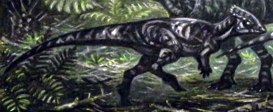
Goyocephale Restoration
''Goyocephale'' is an extinct genus of pachycephalosaurian ornithischian that lived in Mongolia during the Late Cretaceous about 76 million years ago. It was first described in 1982 by Altangerel Perle, Teresa Maryańska and Halszka Osmólska for a disarticulated skeleton with most of a skull, part of the forelimb and hindlimb, some of the pelvic girdle, and some vertebrae. Perle ''et al.'' named the remains ''Goyocephale lattimorei'', from the Mongolian гоё (''goyo''), meaning "decorated", and the Ancient Greek κεφαλή (''kephale''), for head. The species name honours Owen Lattimore. Description ''Goyocephale'' is known from a partial skull, including both mandibles, the skull roof, part of the occiput, part of the braincase region, the posterior skull, the premaxilla, and the maxilla. The posterior edge of the skull roof, at the edge of the squamosal bones, has many small bony bumps, which would have been the base of small horns in life. A feature shared with pachyc ... [...More Info...] [...Related Items...] OR: [Wikipedia] [Google] [Baidu] |
Late Cretaceous
The Late Cretaceous (100.5–66 Ma) is the younger of two epochs into which the Cretaceous Period is divided in the geologic time scale. Rock strata from this epoch form the Upper Cretaceous Series. The Cretaceous is named after ''creta'', the Latin word for the white limestone known as chalk. The chalk of northern France and the white cliffs of south-eastern England date from the Cretaceous Period. Climate During the Late Cretaceous, the climate was warmer than present, although throughout the period a cooling trend is evident. The tropics became restricted to equatorial regions and northern latitudes experienced markedly more seasonal climatic conditions. Geography Due to plate tectonics, the Americas were gradually moving westward, causing the Atlantic Ocean to expand. The Western Interior Seaway divided North America into eastern and western halves; Appalachia and Laramidia. India maintained a northward course towards Asia. In the Southern Hemisphere, Australia and Ant ... [...More Info...] [...Related Items...] OR: [Wikipedia] [Google] [Baidu] |
Owen Lattimore
Owen Lattimore (July 29, 1900 – May 31, 1989) was an American Orientalist and writer. He was an influential scholar of China and Central Asia, especially Mongolia. Although he never earned a college degree, in the 1930s he was editor of ''Pacific Affairs'', a journal published by the Institute of Pacific Relations, and then taught at Johns Hopkins University in Baltimore, Maryland, from 1938 to 1963. He was director of the Walter Hines Page School of International Relations there from 1939 to 1953. During World War II, he was an advisor to Chiang Kai-shek and the American government and contributed extensively to the public debate on American policy in Asia. From 1963 to 1970, Lattimore was the first Professor of Chinese Studies at the University of Leeds in England. In the early post-war period of McCarthyism and the Red Scare, American wartime " China Hands" were accused of being agents of the Soviet Union or under the influence of Marxism. In 1950, Senator Joseph McCarthy a ... [...More Info...] [...Related Items...] OR: [Wikipedia] [Google] [Baidu] |
Caniniform
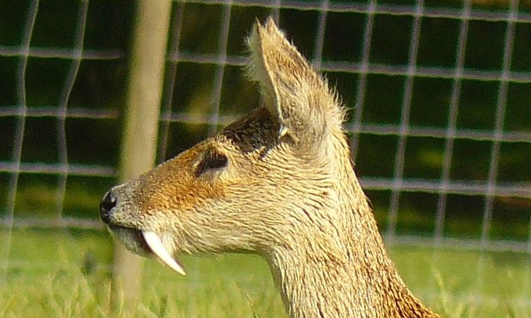
In mammalian oral anatomy, the canine teeth, also called cuspids, dog teeth, or (in the context of the upper jaw) fangs, eye teeth, vampire teeth, or vampire fangs, are the relatively long, pointed teeth. They can appear more flattened however, causing them to resemble incisors and leading them to be called ''incisiform''. They developed and are used primarily for firmly holding food in order to tear it apart, and occasionally as weapons. They are often the largest teeth in a mammal's mouth. Individuals of most species that develop them normally have four, two in the upper jaw and two in the lower, separated within each jaw by incisors; humans and dogs are examples. In most species, canines are the anterior-most teeth in the maxillary bone. The four canines in humans are the two maxillary canines and the two mandibular canines. Details There are generally four canine teeth: two in the upper (maxillary) and two in the lower (mandibular) arch. A canine is placed laterally t ... [...More Info...] [...Related Items...] OR: [Wikipedia] [Google] [Baidu] |
Heterodont
In anatomy, a heterodont (from Greek, meaning 'different teeth') is an animal which possesses more than a single tooth morphology. In vertebrates, heterodont pertains to animals where teeth are differentiated into different forms. For example, members of the Synapsida generally possess incisors, canines ("eyeteeth"), premolars, and molars. The presence of heterodont dentition is evidence of some degree of feeding and or hunting specialization in a species. In contrast, homodont or isodont dentition refers to a set of teeth that possess the same tooth morphology. In invertebrates, the term heterodont refers to a condition where teeth of differing sizes occur in the hinge plate, a part of the Bivalvia Bivalvia (), in previous centuries referred to as the Lamellibranchiata and Pelecypoda, is a class of marine and freshwater molluscs Mollusca is the second-largest phylum of invertebrate animals after the Arthropoda, the members of w .... References See also * D ... [...More Info...] [...Related Items...] OR: [Wikipedia] [Google] [Baidu] |
Squamosal Bone
The squamosal is a skull bone found in most reptiles, amphibians, and birds. In fishes, it is also called the pterotic bone. In most tetrapods, the squamosal and quadratojugal bones form the cheek series of the skull. The bone forms an ancestral component of the dermal roof and is typically thin compared to other skull bones. The squamosal bone lies ventral to the temporal series and otic notch, and is bordered anteriorly by the postorbital. Posteriorly, the squamosal articulates with the quadrate and pterygoid bones. The squamosal is bordered anteroventrally by the jugal and ventrally by the quadratojugal. Function in reptiles In reptiles, the quadrate and articular bones of the skull articulate to form the jaw joint. The squamosal bone lies anterior to the quadrate bone. Anatomy in synapsids Non-mammalian synapsids In non-mammalian synapsids, the jaw is composed of four bony elements and referred to as a quadro-articular jaw because the joint is between the articular an ... [...More Info...] [...Related Items...] OR: [Wikipedia] [Google] [Baidu] |
Maxilla
The maxilla (plural: ''maxillae'' ) in vertebrates is the upper fixed (not fixed in Neopterygii) bone of the jaw formed from the fusion of two maxillary bones. In humans, the upper jaw includes the hard palate in the front of the mouth. The two maxillary bones are fused at the intermaxillary suture, forming the anterior nasal spine. This is similar to the mandible (lower jaw), which is also a fusion of two mandibular bones at the mandibular symphysis. The mandible is the movable part of the jaw. Structure In humans, the maxilla consists of: * The body of the maxilla * Four processes ** the zygomatic process ** the frontal process of maxilla ** the alveolar process ** the palatine process * three surfaces – anterior, posterior, medial * the Infraorbital foramen * the maxillary sinus * the incisive foramen Articulations Each maxilla articulates with nine bones: * two of the cranium: the frontal and ethmoid * seven of the face: the nasal, zygomatic, lacrimal, inferior n ... [...More Info...] [...Related Items...] OR: [Wikipedia] [Google] [Baidu] |
Premaxilla
The premaxilla (or praemaxilla) is one of a pair of small cranial bones at the very tip of the upper jaw of many animals, usually, but not always, bearing teeth. In humans, they are fused with the maxilla. The "premaxilla" of therian mammal has been usually termed as the incisive bone. Other terms used for this structure include premaxillary bone or ''os premaxillare'', intermaxillary bone or ''os intermaxillare'', and Goethe's bone. Human anatomy In human anatomy, the premaxilla is referred to as the incisive bone (') and is the part of the maxilla which bears the incisor teeth, and encompasses the anterior nasal spine and alar region. In the nasal cavity, the premaxillary element projects higher than the maxillary element behind. The palatal portion of the premaxilla is a bony plate with a generally transverse orientation. The incisive foramen is bound anteriorly and laterally by the premaxilla and posteriorly by the palatine process of the maxilla. It is formed from the ... [...More Info...] [...Related Items...] OR: [Wikipedia] [Google] [Baidu] |
Anatomical Terms Of Location
Standard anatomical terms of location are used to unambiguously describe the anatomy of animals, including humans. The terms, typically derived from Latin or Greek roots, describe something in its standard anatomical position. This position provides a definition of what is at the front ("anterior"), behind ("posterior") and so on. As part of defining and describing terms, the body is described through the use of anatomical planes and anatomical axes. The meaning of terms that are used can change depending on whether an organism is bipedal or quadrupedal. Additionally, for some animals such as invertebrates, some terms may not have any meaning at all; for example, an animal that is radially symmetrical will have no anterior surface, but can still have a description that a part is close to the middle ("proximal") or further from the middle ("distal"). International organisations have determined vocabularies that are often used as standard vocabularies for subdisciplines of anatom ... [...More Info...] [...Related Items...] OR: [Wikipedia] [Google] [Baidu] |
Braincase
In human anatomy, the neurocranium, also known as the braincase, brainpan, or brain-pan is the upper and back part of the skull, which forms a protective case around the brain. In the human skull, the neurocranium includes the calvaria or skullcap. The remainder of the skull is the facial skeleton. In comparative anatomy, neurocranium is sometimes used synonymously with endocranium or chondrocranium. Structure The neurocranium is divided into two portions: * the membranous part, consisting of flat bones, which surround the brain; and * the cartilaginous part, or chondrocranium, which forms bones of the base of the skull. In humans, the neurocranium is usually considered to include the following eight bones: * 1 ethmoid bone * 1 frontal bone * 1 occipital bone * 2 parietal bones * 1 sphenoid bone * 2 temporal bones The ossicles (three on each side) are usually not included as bones of the neurocranium. There may variably also be extra sutural bones present. Below the ne ... [...More Info...] [...Related Items...] OR: [Wikipedia] [Google] [Baidu] |
Occiput
The occipital bone () is a cranial dermal bone and the main bone of the occiput (back and lower part of the skull). It is trapezoidal in shape and curved on itself like a shallow dish. The occipital bone overlies the occipital lobes of the cerebrum. At the base of skull in the occipital bone, there is a large oval opening called the foramen magnum, which allows the passage of the spinal cord. Like the other cranial bones, it is classed as a flat bone. Due to its many attachments and features, the occipital bone is described in terms of separate parts. From its front to the back is the basilar part, also called the basioccipital, at the sides of the foramen magnum are the lateral parts, also called the exoccipitals, and the back is named as the squamous part. The basilar part is a thick, somewhat quadrilateral piece in front of the foramen magnum and directed towards the pharynx. The squamous part is the curved, expanded plate behind the foramen magnum and is the largest part o ... [...More Info...] [...Related Items...] OR: [Wikipedia] [Google] [Baidu] |
Skull Roof
The skull roof, or the roofing bones of the skull, are a set of bones covering the brain, eyes and nostrils in bony fishes and all land-living vertebrates. The bones are derived from dermal bone and are part of the dermatocranium. In comparative anatomy the term is used on the full dermatocranium. Romer, A.S. & T.S. Parsons. 1977. ''The Vertebrate Body.'' 5th ed. Saunders, Philadelphia. (6th ed. 1985) In general anatomy, the roofing bones may refer specifically to the bones that form above and alongside the brain and neurocranium (i.e., excluding the marginal upper jaw bones such as the maxilla and premaxilla), and in human anatomy, the skull roof often refers specifically to the skullcap. Origin Early armoured fish did not have a skull in the common understanding of the word, but had an endocranium that was partially open above, topped by dermal bones forming armour. The dermal bones gradually evolved into a fixed unit overlaying the endocranium like a heavy "lid", protec ... [...More Info...] [...Related Items...] OR: [Wikipedia] [Google] [Baidu] |


