|
GnRH
Gonadotropin-releasing hormone (GnRH) is a releasing hormone responsible for the release of follicle-stimulating hormone (FSH) and luteinizing hormone (LH) from the anterior pituitary. GnRH is a tropic peptide hormone synthesized and released from GnRH neurons within the hypothalamus. The peptide belongs to gonadotropin-releasing hormone family. It constitutes the initial step in the hypothalamic–pituitary–gonadal axis. Structure The identity of GnRH was clarified by the 1977 Nobel Laureates Roger Guillemin and Andrew V. Schally: pyroGlu-His-Trp-Ser-Tyr-Gly-Leu-Arg-Pro-Gly-NH2 As is standard for peptide representation, the sequence is given from amino terminus to carboxyl terminus; also standard is omission of the designation of chirality, with assumption that all amino acids are in their L- form. The abbreviations are the standard abbreviations for the corresponding proteinogenic amino acids, except for ''pyroGlu'', which refers to pyroglutamic acid, a derivative of glut ... [...More Info...] [...Related Items...] OR: [Wikipedia] [Google] [Baidu] |
Gonadotropin-releasing Hormone Receptor
The gonadotropin-releasing hormone receptor (GnRHR), also known as the luteinizing hormone releasing hormone receptor (LHRHR), is a member of the seven-transmembrane, G-protein coupled receptor (GPCR) family. It is the receptor of gonadotropin-releasing hormone (GnRH). The GnRHR is expressed on the surface of pituitary gonadotrope cells as well as lymphocytes, breast, ovary, and prostate. This receptor is a 60 kDa G protein-coupled receptor and resides primarily in the pituitary and is responsible for eliciting the actions of GnRH after its release from the hypothalamus. Upon activation, the LHRHr stimulates tyrosine phosphatase and elicits the release of LH from the pituitary. Evidence exists showing the presence of GnRH and its receptor in extrapituitary tissues as well as a role in progression of some cancers. Function Following binding of GnRH, the GnRHR associates with G-proteins that activate a phosphatidylinositol (PtdIns)-calcium second messenger system. Activation ... [...More Info...] [...Related Items...] OR: [Wikipedia] [Google] [Baidu] |
Hypothalamic–pituitary–gonadal Axis
The hypothalamic–pituitary–gonadal axis (HPG axis, also known as the hypothalamic–pituitary–ovarian/testicular axis) refers to the hypothalamus, pituitary gland, and gonadal glands as if these individual endocrine glands were a single entity. Because these glands often act in concert, physiologists and endocrinologists find it convenient and descriptive to speak of them as a single system. The HPG axis plays a critical part in the development and regulation of a number of the body's systems, such as the reproductive and immune systems. Fluctuations in this axis cause changes in the hormones produced by each gland and have various local and systemic effects on the body. The axis controls development, reproduction, and aging in animals. Gonadotropin-releasing hormone (GnRH) is secreted from the hypothalamus by GnRH-expressing neurons. The anterior portion of the pituitary gland produces luteinizing hormone (LH) and follicle-stimulating hormone (FSH), and the gonads produ ... [...More Info...] [...Related Items...] OR: [Wikipedia] [Google] [Baidu] |
Luteinizing Hormone
Luteinizing hormone (LH, also known as luteinising hormone, lutropin and sometimes lutrophin) is a hormone produced by gonadotropic cells in the anterior pituitary gland. The production of LH is regulated by gonadotropin-releasing hormone (GnRH) from the hypothalamus. In females, an acute rise of LH known as an LH surge, triggers ovulation and development of the corpus luteum. In males, where LH had also been called interstitial cell–stimulating hormone (ICSH), it stimulates Leydig cell production of testosterone. It acts synergistically with follicle-stimulating hormone (Follicle-stimulating hormone, FSH). Structure LH is a heteroprotein dimer, dimeric glycoprotein. Each monomeric unit is a glycoprotein molecule; one alpha and one beta subunit make the full, functional protein. Its structure is similar to that of the other glycoprotein hormones, follicle-stimulating hormone (FSH), thyroid-stimulating hormone (TSH), and human chorionic gonadotropin (hCG). The protein dimer ... [...More Info...] [...Related Items...] OR: [Wikipedia] [Google] [Baidu] |
Follicle-stimulating Hormone
Follicle-stimulating hormone (FSH) is a gonadotropin, a glycoprotein polypeptide hormone. FSH is synthesized and secreted by the gonadotropic cells of the anterior pituitary gland and regulates the development, growth, pubertal maturation, and reproductive processes of the body. FSH and luteinizing hormone (LH) work together in the reproductive system. Structure FSH is a 35.5 kDa glycoprotein heterodimer, consisting of two polypeptide units, alpha and beta. Its structure is similar to those of luteinizing hormone (LH), thyroid-stimulating hormone (TSH), and human chorionic gonadotropin (hCG). The alpha subunits of the glycoproteins LH, FSH, TSH, and hCG are identical and consist of 96 amino acids, while the beta subunits vary. Both subunits are required for biological activity. FSH has a beta subunit of 111 amino acids (FSH β), which confers its specific biologic action, and is responsible for interaction with the follicle-stimulating hormone receptor. The sugar port ... [...More Info...] [...Related Items...] OR: [Wikipedia] [Google] [Baidu] |
Gonadotropin-releasing Hormone Family
The gonadotropin-releasing hormones (GnRH) (gonadoliberin) are a family of peptides that play a pivotal role in reproduction. The main function of GnRH is to act on the pituitary to stimulate the synthesis and secretion of luteinizing and follicle-stimulating hormones, but GnRH also acts on the brain, retina, sympathetic nervous system, gonads, and placenta in certain species. There seems to be at least three forms of GnRH. The second form is expressed in midbrain and seems to be widespread. The third form has been found so far only in fish. GnRH is a C-terminal amidated decapeptide processed from a larger precursor protein. Four of the ten residues are perfectly conserved in all species where GnRH has been sequenced. Subfamilies *Gonadoliberin I Human proteins containing this domain GNRH1, GNRH2 Progonadoliberin-2 is a protein that in humans is encoded by the ''GNRH2'' gene. The protein encoded by this gene is a preproprotein that is cleaved to form a secreted 10 aa pepti ... [...More Info...] [...Related Items...] OR: [Wikipedia] [Google] [Baidu] |
Releasing And Inhibiting Hormones
Releasing hormones and inhibiting hormones are hormones whose main purpose is to control the release of other hormones, either by stimulating or inhibiting their release. They are also called liberins () and statins () (respectively), or releasing factors and inhibiting factors. The principal examples are hypothalamic-pituitary hormones that can be classified from several viewpoints: they are hypothalamic hormones (originating in the hypothalamus), they are hypophysiotropic hormones (affecting the hypophysis, that is, the pituitary gland), and they are tropic hormones (having other endocrine glands as their target). For example, thyrotropin-releasing hormone (TRH) is released from the hypothalamus in response to low levels of secretion of thyroid-stimulating hormone (TSH) from the pituitary gland. The TSH in turn is under feedback control by the thyroid hormones T4 and T3. When the level of TSH is too high, they feed back on the brain to shut down the secretion of TRH. Synthetic ... [...More Info...] [...Related Items...] OR: [Wikipedia] [Google] [Baidu] |
Hypothalamus
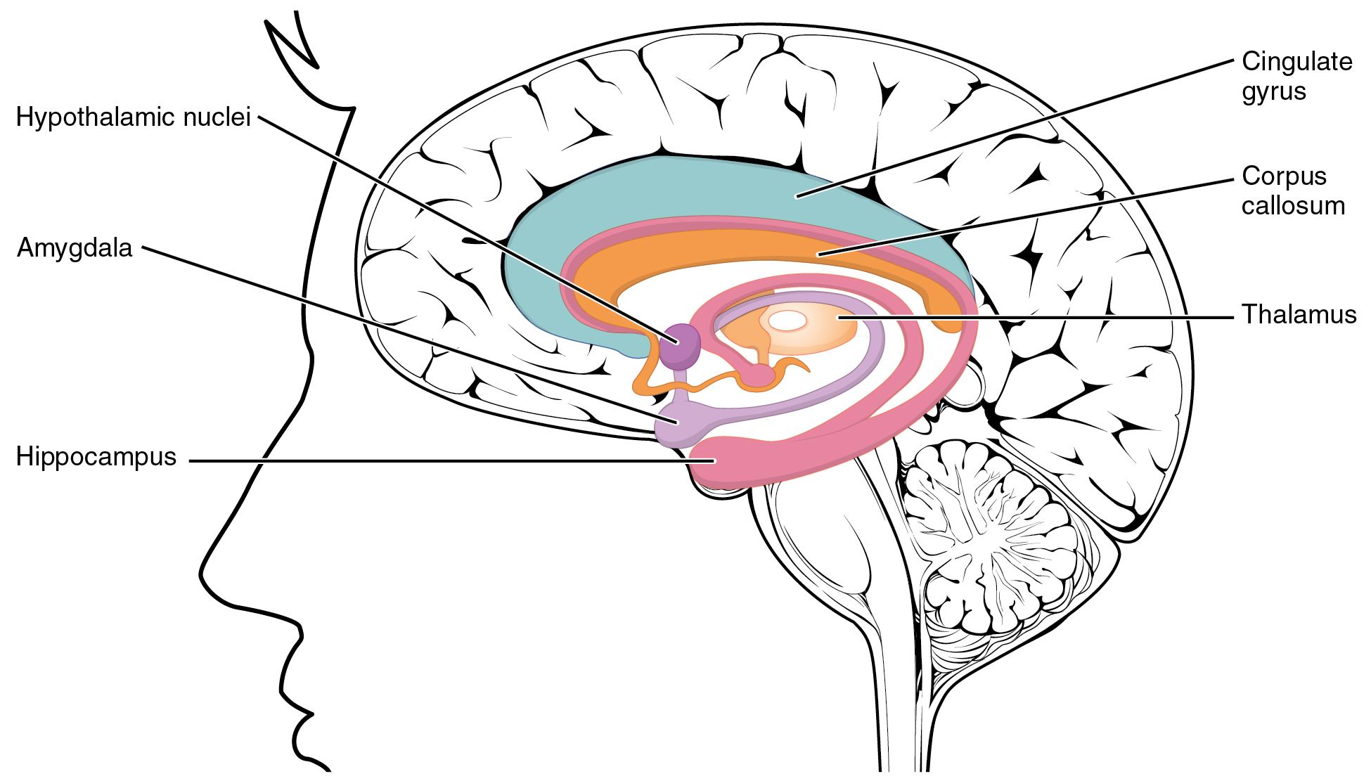
The hypothalamus () is a part of the brain that contains a number of small nuclei with a variety of functions. One of the most important functions is to link the nervous system to the endocrine system via the pituitary gland. The hypothalamus is located below the thalamus and is part of the limbic system. In the terminology of neuroanatomy, it forms the ventral part of the diencephalon. All vertebrate brains contain a hypothalamus. In humans, it is the size of an almond. The hypothalamus is responsible for regulating certain metabolic processes and other activities of the autonomic nervous system. It synthesizes and secretes certain neurohormones, called releasing hormones or hypothalamic hormones, and these in turn stimulate or inhibit the secretion of hormones from the pituitary gland. The hypothalamus controls body temperature, hunger, important aspects of parenting and maternal attachment behaviours, thirst, fatigue, sleep, and circadian rhythms. Structure T ... [...More Info...] [...Related Items...] OR: [Wikipedia] [Google] [Baidu] |
Roger Guillemin
Roger Charles Louis Guillemin (born January 11, 1924) is a French-American neuroscientist. He received the National Medal of Science in 1976, and the Nobel prize for medicine in 1977 for his work on neurohormones, sharing the prize that year with Andrew Schally and Rosalyn Sussman Yalow. Biography Completing his undergraduate work at the University of Burgundy, Guillemin received his M.D. degree from the Medical Faculty at Lyon in 1949, and went to Montreal, Quebec, Canada, to work with Hans Selye at the Institute of Experimental Medicine and Surgery at the Université de Montréal where he received a Ph.D. in 1953. The same year he moved to the United States to join the faculty at Baylor College of Medicine at Houston. In 1965, he became a naturalized citizen of the United States. In 1970 he helped to set up the Salk Institute in La Jolla, California where he worked until retirement in 1989. Guillemin and Andrew V. Schally discovered the structures of TRH and GnRH in separate ... [...More Info...] [...Related Items...] OR: [Wikipedia] [Google] [Baidu] |
Gonadotrope
Gonadotropic cells (called also Gonadotropes or Gonadotrophs or Delta Cells or Delta basophils) are endocrine cells in the anterior pituitary that produce the gonadotropins, such as the follicle-stimulating hormone (FSH) and luteinizing hormone (LH). Release of FSH and LH by gonadotropes is regulated by gonadotropin-releasing hormone (GnRH) from the hypothalamus. Gonadotropes appear basophilic Basophilic is a technical term used by pathologists. It describes the appearance of cells, tissues and cellular structures as seen through the microscope after a histological section has been stained with a basic dye. The most common such dye i ... in histological preparations. Gonadotropes have insulin receptors, which can be overstimulated by too high insulin levels. This may lead to infertility as hormone release levels are disrupted. Gonadotropes are feedback inhibited by specific hormones, including estradiol. See also * List of human cell types derived from the germ layer ... [...More Info...] [...Related Items...] OR: [Wikipedia] [Google] [Baidu] |
Pituitary Gland
In vertebrate anatomy, the pituitary gland, or hypophysis, is an endocrine gland, about the size of a chickpea and weighing, on average, in humans. It is a protrusion off the bottom of the hypothalamus at the base of the brain. The hypophysis rests upon the hypophyseal fossa of the sphenoid bone in the center of the middle cranial fossa and is surrounded by a small bony cavity (sella turcica) covered by a dural fold (diaphragma sellae). The anterior pituitary (or adenohypophysis) is a lobe of the gland that regulates several physiological processes including stress, growth, reproduction, and lactation. The intermediate lobe synthesizes and secretes melanocyte-stimulating hormone. The posterior pituitary (or neurohypophysis) is a lobe of the gland that is functionally connected to the hypothalamus by the median eminence via a small tube called the pituitary stalk (also called the infundibular stalk or the infundibulum). Hormones secreted from the pituitary gland ... [...More Info...] [...Related Items...] OR: [Wikipedia] [Google] [Baidu] |

_during_menstrual_cycle.png)

