|
Gingival
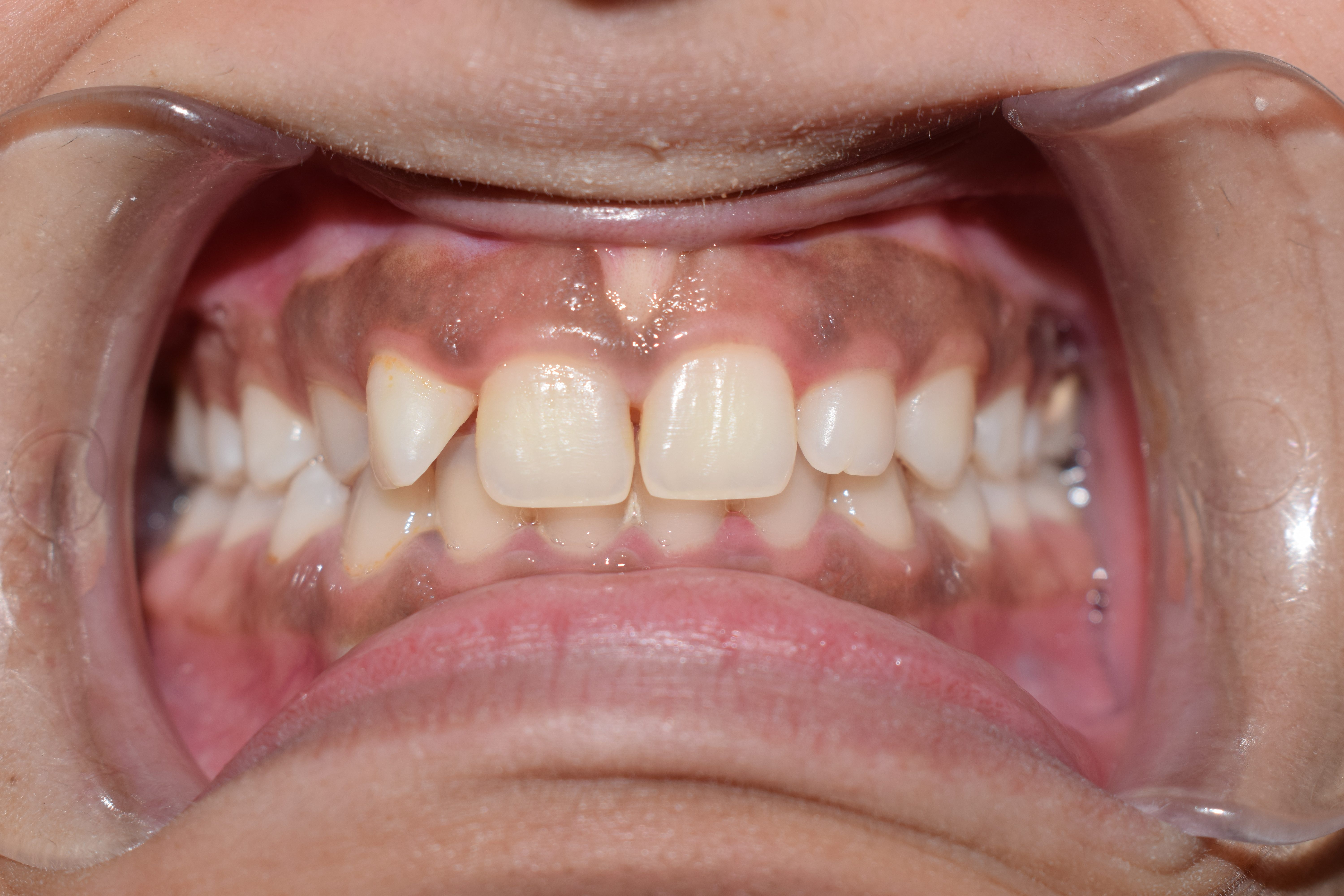
The gums or gingiva (plural: ''gingivae'') consist of the mucosal tissue that lies over the mandible and maxilla inside the mouth. Gum health and disease can have an effect on general health. Structure The gums are part of the soft tissue lining of the mouth. They surround the teeth and provide a seal around them. Unlike the soft tissue linings of the lips and cheeks, most of the gums are tightly bound to the underlying bone which helps resist the friction of food passing over them. Thus when healthy, it presents an effective barrier to the barrage of periodontal insults to deeper tissue. Healthy gums are usually coral pink in light skinned people, and may be naturally darker with melanin pigmentation. Changes in color, particularly increased redness, together with swelling and an increased tendency to bleed, suggest an inflammation that is possibly due to the accumulation of bacterial plaque. Overall, the clinical appearance of the tissue reflects the underlying histology, bo ... [...More Info...] [...Related Items...] OR: [Wikipedia] [Google] [Baidu] |
Gingival Hyperplasia
Gingival enlargement is an increase in the size of the gingiva (gums). It is a common feature of gingival disease. Gingival enlargement can be caused by a number of factors, including inflammatory conditions and the side effects of certain medications. The treatment is based on the cause. A closely related term is epulis, denoting a localized tumor (i.e. lump) on the gingiva. Classification The terms gingival hyperplasia and gingival hypertrophy have been used to describe this topic in the past. These are not precise descriptions of gingival enlargement because these terms are strictly histologic diagnoses, and such diagnoses require microscopic analysis of a tissue sample. Hyperplasia refers to an increased number of cells, and hypertrophy refers to an increase in the size of individual cells. As these identifications cannot be performed with a clinical examination and evaluation of the tissue, the term ''gingival enlargement'' is more properly applied. Gingival enlargement has ... [...More Info...] [...Related Items...] OR: [Wikipedia] [Google] [Baidu] |
Gingivitis
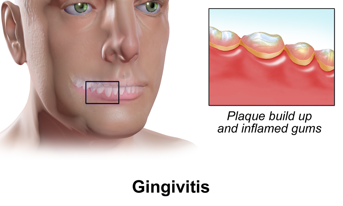
Gingivitis is a non-destructive disease that causes inflammation of the gums. The most common form of gingivitis, and the most common form of periodontal disease overall, is in response to bacterial biofilms (also called plaque) that is attached to tooth surfaces, termed ''plaque-induced gingivitis''. Most forms of gingivitis are plaque-induced. While some cases of gingivitis never progress to periodontitis, periodontitis is always preceded by gingivitis. Gingivitis is reversible with good oral hygiene; however, without treatment, gingivitis can progress to periodontitis, in which the inflammation of the gums results in tissue destruction and bone resorption around the teeth. Periodontitis can ultimately lead to tooth loss. Signs and symptoms The symptoms of gingivitis are somewhat non-specific and manifest in the gum tissue as the classic signs of inflammation: *Swollen gums *Bright red gums *Gums that are tender or painful to the touch *Bleeding gums or bleeding after bru ... [...More Info...] [...Related Items...] OR: [Wikipedia] [Google] [Baidu] |
Free Gingival Margin
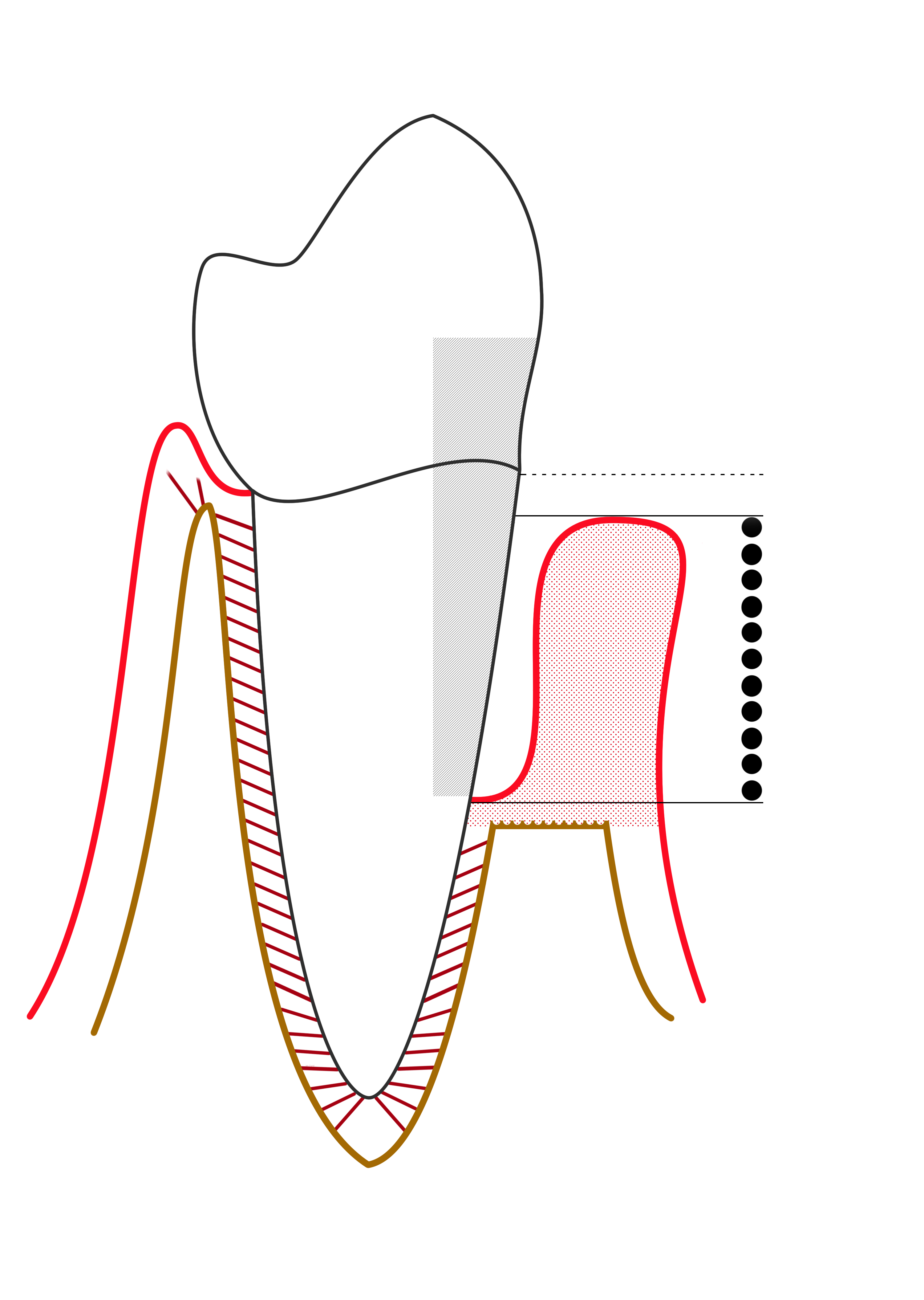
The free gingival margin is the interface between the sulcular epithelium and the epithelium of the oral cavity. This interface exists at the most coronal point of the gingiva, otherwise known as the crest of the marginal gingiva. Because the short part of gingiva existing above the height of the underlying Alveolar process of maxilla, known as the free gingiva, is not bound down to the periosteum that envelops the bone, it is moveable. However, due to the presence of gingival fibers such as the dentogingival and circular fibers, the free gingiva remains pulled up against the surface of the tooth unless being pushed away by, for example, a periodontal probe or the bristles of a toothbrush. Gingival retraction or recession ''Gingival retraction'' or ''gingival recession'' is when there is lateral movement of the gingival margin away from the tooth surface. It is usually termed ''gingival retraction'' as an intentional procedure, and in such cases it is performed by mechanical, che ... [...More Info...] [...Related Items...] OR: [Wikipedia] [Google] [Baidu] |
Gingival Fibers
The gingival fibers are the connective tissue fibers that inhabit the gingival tissue adjacent to teeth and help hold the tissue firmly against the teeth. They are primarily composed of type I collagen, although type III fibers are also involved. These fibers, unlike the fibers of the periodontal ligament, in general, attach the tooth to the gingival tissue, rather than the tooth to the alveolar bone. Functions of the gingival fibers The gingival fibers accomplish the following tasks: *They hold the marginal gingiva against the tooth *They provide the marginal gingiva with enough rigidity to withstand the forces of mastication without distorting *They serve to stabilize the marginal gingiva by uniting it with both the tissue of the more rigid attached gingiva as well as the cementum layer of the tooth. Gingival fibers and periodontitis In theory, gingival fibers are the protectors against periodontitis, as once they are breached, they cannot be regenerated. When destroyed, the ... [...More Info...] [...Related Items...] OR: [Wikipedia] [Google] [Baidu] |
Mucogingival Junction
A mucogingival junction is an anatomical feature found on the intraoral mucosa. The mucosa of the cheeks and floor of the mouth are freely moveable and fragile, whereas the mucosa around the teeth and on the palate are firm and keratinized. Where the two tissue types meet is known as a mucogingival junction. There are three mucogingival junctions: on the facial of the maxilla and on both the facial and lingual of the mandible. The palatal gingiva of the maxilla is continuous with the tissue of the palate, which is bound down to the palatal bones. Because the palate is devoid of freely moveable alveolar mucosa, there is no mucogingival junction.Carranza's Clinical Periodontology, W.B. Saunders 2002, page 17. Clinical importance The clinical importance of the mucogingival junction is in measuring the width of attached gingiva. Attached gingiva is important because it is bound very tightly to the underlying alveolar bone and provides protection to the mucosa during functional u ... [...More Info...] [...Related Items...] OR: [Wikipedia] [Google] [Baidu] |
Dental Plaque
Dental plaque is a biofilm of microorganisms (mostly bacteria, but also fungi) that grows on surfaces within the mouth. It is a sticky colorless deposit at first, but when it forms tartar, it is often brown or pale yellow. It is commonly found between the teeth, on the front of teeth, behind teeth, on chewing surfaces, along the gumline (supragingival), or below the gumline cervical margins (subgingival). Dental plaque is also known as microbial plaque, oral biofilm, dental biofilm, dental plaque biofilm or bacterial plaque biofilm. Bacterial plaque is one of the major causes for dental decay and gum disease. Progression and build-up of dental plaque can give rise to tooth decay – the localised destruction of the tissues of the tooth by acid produced from the bacterial degradation of fermentable sugar – and periodontal problems such as gingivitis and periodontitis; hence it is important to disrupt the mass of bacteria and remove it. Plaque control and removal can be achieved w ... [...More Info...] [...Related Items...] OR: [Wikipedia] [Google] [Baidu] |
Bleeding On Probing
Bleeding on probing (BoP) which is also known as bleeding gums or gingival bleeding is a term used by dentists and dental hygienists when referring to bleeding that is induced by gentle manipulation of the tissue at the depth of the gingival sulcus, or interface between the gingiva and a tooth. BoP is a sign of periodontal inflammation and indicates some sort of destruction and erosion to the lining of the sulcus or the ulceration of sulcular epithelium. The blood comes from lamina propria after the ulceration of the lining.Gingival bleeding' URL assessed on November 21, 2009 BoP seems to be correlated with Periodontal Inflamed Surface Area (PISA). Causes There are many possible causes of gingival bleeding. The main cause of gingival bleeding is the formation and accumulation of plaque at the gum line due to improper brushing and flossing of teeth. The hardened form of plaque is calculus. An advanced form of gingivitis as a result of formation of plaque is periodontitis. Other ... [...More Info...] [...Related Items...] OR: [Wikipedia] [Google] [Baidu] |
Oral Hygiene
Oral hygiene is the practice of keeping one's mouth clean and free of disease and other problems (e.g. bad breath) by regular brushing of the teeth (dental hygiene) and cleaning between the teeth. It is important that oral hygiene be carried out on a regular basis to enable prevention of dental disease and bad breath. The most common types of dental disease are tooth decay (''cavities'', ''dental caries'') and gum diseases, including gingivitis, and periodontitis. General guidelines for adults suggest brushing at least twice a day with a fluoridated toothpaste: brushing last thing at night and at least on one other occasion. Cleaning between the teeth is called interdental cleaning and is as important as tooth brushing. This is because a toothbrush cannot reach between the teeth and therefore only removes about 50% of plaque from the surface of the teeth. There are many tools to clean between the teeth, including floss, tape and interdental brushes; it is up to each individual to ... [...More Info...] [...Related Items...] OR: [Wikipedia] [Google] [Baidu] |
Periodontal Probe
A periodontal probe is an instrument in dentistry commonly used in the dental armamentarium. It is usually long, thin, and blunted at the end. The primary purpose of a periodontal probe is to measure pocket depths around a tooth in order to establish the state of health of the periodontium. There are markings inscribed onto the head of the instrument for accuracy and readability. Use Proper use of the periodontal probe is necessary to maintain accuracy. The tip of the instrument is placed with light pressure of 10-20 gramsWilkins, 1999 into the gingival sulcus, which is an area of potential space between a tooth and the surrounding tissue. It is important to keep the periodontal probe parallel to the contours of the root of the tooth and to insert the probe down to the base of the pocket. This results in obscuring a section of the periodontal probe's tip. The first marking visible above the pocket indicates the measurement of the pocket depth. It has been found that the ave ... [...More Info...] [...Related Items...] OR: [Wikipedia] [Google] [Baidu] |
Gum Depigmentation
Gum depigmentation, also known as gum bleaching, is a procedure used in cosmetic dentistry to lighten or remove black spots or patches on the gums consisting of melanin. Melanin in skin is very common in inhabitants in many parts of the world due to genetic factors. Melanin pigmentation in skin, oral mucosa, inner ear and other organs is a detoxification mechanism. Some toxic agents bind to melanin and will move out of the tissue with the ageing cells and are expelled to the tissue surfaces. Also in the gums and oral mucosa a visible pigmentation is most often caused by genetic factors, but also by tobacco smoking or in a few cases by long-term use of certain medications. If stopping smoking or change of medication do not solve the problem with a disfigurating melanin pigmentation, a surgical operation may be performed. The procedure itself can involve laser ablation techniques. Laser gum depigmentation Melanocytes are cells which reside in the basal layer of the gingival epithe ... [...More Info...] [...Related Items...] OR: [Wikipedia] [Google] [Baidu] |
Interdental Papilla
The interdental papilla, also known as the interdental gingiva, is the part of the gums (gingiva) that exists coronal to the free gingival margin on the buccal and lingual surfaces of the teeth A tooth ( : teeth) is a hard, calcified structure found in the jaws (or mouths) of many vertebrates and used to break down food. Some animals, particularly carnivores and omnivores, also use teeth to help with capturing or wounding prey, tear .... The interdental papillae fill in the area between the teeth apical to their contact areas to prevent food impaction; they assume a conical shape for the anterior teeth and a blunted shape buccolingually for the posterior teeth. A missing papilla is often visible as a small triangular gap between adjacent teeth. The relationship of interdental bone to the interproximal contact point between adjacent teeth is a determining factor in whether the interdental papilla will be present. If greater than 8mm exist between the interdental bone and t ... [...More Info...] [...Related Items...] OR: [Wikipedia] [Google] [Baidu] |
Cementoenamel Junction
The cementoenamel junction, frequently abbreviated as the CEJ, is a slightly visible anatomical border identified on a tooth. It is the location where the enamel, which covers the anatomical crown of a tooth, and the cementum, which covers the anatomical root of a tooth, meet. Informally it is known as the neck of the tooth. The border created by these two dental tissues has much significance as it is usually the location where the gingiva attaches to a healthy tooth by fibers called the gingival fibers The gingival fibers are the connective tissue fibers that inhabit the gingival tissue adjacent to teeth and help hold the tissue firmly against the teeth. They are primarily composed of type I collagen, although type III fibers are also involved .... Active recession of the gingiva reveals the cementoenamel junction in the mouth and is usually a sign of an unhealthy condition. There exists a normal variation in the relationship of the cementum and the enamel at the cementoe ... [...More Info...] [...Related Items...] OR: [Wikipedia] [Google] [Baidu] |


