|
Flexor Carpi Radialis
In anatomy, flexor carpi radialis is a muscle of the human forearm that acts to flex and (radially) abduct the hand. The Latin ''carpus'' means wrist; hence flexor carpi is a flexor of the wrist. Origin and insertion The flexor carpi radialis is one of four muscles in the superficial layer of the anterior compartment of the forearm. This muscle originates from the medial epicondyle of the humerus as part of the common flexor tendon. It runs just laterally of flexor digitorum superficialis and inserts on the anterior aspect of the base of the second metacarpal, and has small slips to both the third metacarpal and trapezium tuberosity. The tendon of the flexor carpi radialis is visible on the anterior surface of the forearm, just proximal to the wrist, when the wrist is flexed. It is the tendon seen most lateral, closest to the thumb. Nerve and artery Like most flexors of the anterior compartment of the forearm, FCR is innervated by the median nerve, specifically by axons from c ... [...More Info...] [...Related Items...] OR: [Wikipedia] [Google] [Baidu] |
Medial Epicondyle Of Humerus
The medial epicondyle of the humerus is an epicondyle of the humerus bone of the upper arm in humans. It is larger and more prominent than the lateral epicondyle and is directed slightly more posteriorly in the anatomical position. In birds, where the arm is somewhat rotated compared to other tetrapods, it is called the ventral epicondyle of the humerus. In comparative anatomy, the more neutral term entepicondyle is used. The medial epicondyle gives attachment to the ulnar collateral ligament of elbow joint, to the pronator teres, and to a common tendon of origin (the common flexor tendon) of some of the flexor muscles of the forearm: the flexor carpi radialis, the flexor carpi ulnaris, the flexor digitorum superficialis, and the palmaris longus. The medial epicondyle is located on the distal end of the humerus. Additionally, the medial epicondyle is inferior to the medial supracondylar ridge. It is also proximal to the olecranon fossa. The medial epicondyle protects the u ... [...More Info...] [...Related Items...] OR: [Wikipedia] [Google] [Baidu] |
Humerus
The humerus (; ) is a long bone in the arm that runs from the shoulder to the elbow. It connects the scapula and the two bones of the lower arm, the radius and ulna, and consists of three sections. The humeral upper extremity consists of a rounded head, a narrow neck, and two short processes (tubercles, sometimes called tuberosities). The body is cylindrical in its upper portion, and more prismatic below. The lower extremity consists of 2 epicondyles, 2 processes (trochlea & capitulum), and 3 fossae (radial fossa, coronoid fossa, and olecranon fossa). As well as its true anatomical neck, the constriction below the greater and lesser tubercles of the humerus is referred to as its surgical neck due to its tendency to fracture, thus often becoming the focus of surgeons. Etymology The word "humerus" is derived from la, humerus, umerus meaning upper arm, shoulder, and is linguistically related to Gothic ''ams'' shoulder and Greek ''ōmos''. Structure Upper extremity The upper or pr ... [...More Info...] [...Related Items...] OR: [Wikipedia] [Google] [Baidu] |
Flexor Carpi Ulnaris Muscle
The flexor carpi ulnaris (FCU) is a muscle of the forearm that flexes and adducts at the wrist joint. Structure Origin The flexor carpi ulnaris has two heads; a humeral head and ulnar head. The humeral head originates from the medial epicondyle of the humerus via the common flexor tendon. The ulnar head originates from the medial margin of the olecranon of the ulnar and the upper two-thirds of the dorsal border of the ulnar by an aponeurosis. Between the two heads passes the ulnar nerve and ulnar artery. Insertion The flexor carpi ulnaris inserts onto the pisiform, hook of the hamate (via the pisohamate ligament) and the anterior surface of the base of the fifth metacarpal (via the pisometacarpal ligament). Action The flexor carpi ulnaris flexes and adducts at the wrist joint. Innervation The flexor carpi ulnaris is innervated by the ulnar nerve. The corresponding spinal nerves are C8 and T1. Tendon The tendon of flexor carpi ulnaris can be seen on the anterior surface of t ... [...More Info...] [...Related Items...] OR: [Wikipedia] [Google] [Baidu] |
Flexor Retinaculum Of The Hand
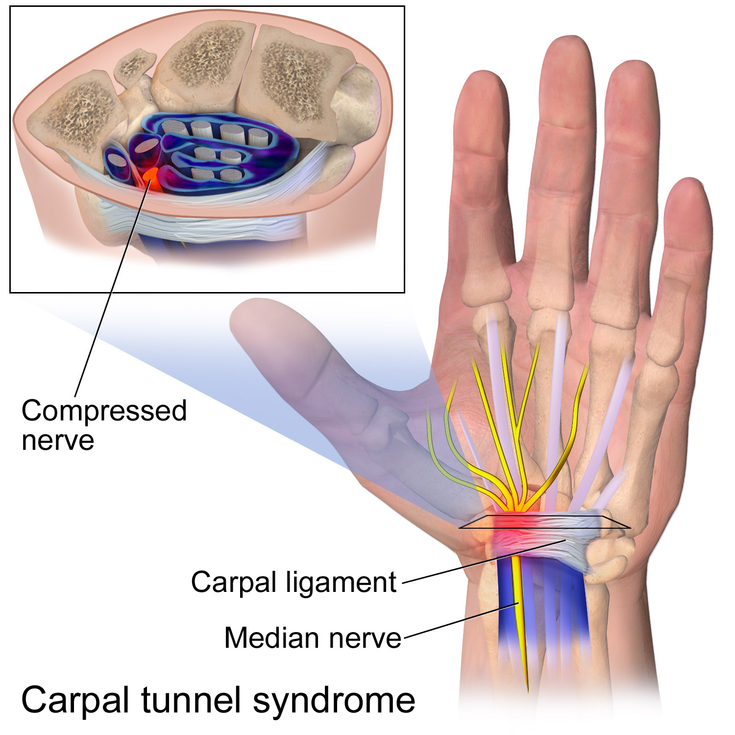
The flexor retinaculum (transverse carpal ligament, or anterior annular ligament) is a fibrous band on the palmar side of the hand near the wrist. It arches over the carpal bones of the hands, covering them and forming the carpal tunnel. Structure The flexor retinaculum is a strong, fibrous band that covers the carpal bones on the palmar side of the hand near the wrist. It attaches to the bones near the radius and ulna. On the ulnar side, the flexor retinaculum attaches to the pisiform bone and the hook of the hamate bone. On the radial side, it attaches to the tubercle of the scaphoid bone, and to the medial part of the palmar surface and the ridge of the trapezium bone. The flexor retinaculum is continuous with the palmar carpal ligament, and deeper with the palmar aponeurosis. The ulnar artery and ulnar nerve, and the cutaneous branches of the median and ulnar nerves, pass on top of the flexor retinaculum. On the radial side of the retinaculum is the tendon of the flexor c ... [...More Info...] [...Related Items...] OR: [Wikipedia] [Google] [Baidu] |
Dumbbell
The dumbbell, a type of free weight, is a piece of equipment used in weight training. It can be used individually or in pairs, with one in each hand. History The forerunner of the dumbbell, halteres, were used in ancient Greece as lifting weights and also as weights in the ancient Greek version of the long jump. A kind of dumbbell was also used in India for more than a millennium, shaped like a club – so it was named Indian club. The design of the "Nal", as the equipment was referred to, can be seen as a halfway point between a barbell and a dumbbell. It was generally used in pairs, in workouts by wrestlers, bodybuilders, sports players, and others wishing to increase strength and muscle size. Etymology The term "dumbbell" or "dumb bell" originated in late Stuart England. In 1711 the poet Joseph Addison mentioned exercising with a "dumb bell" in an essay published in ''The Spectator''. Although Addison elsewhere in the same publication describes having used equipment sim ... [...More Info...] [...Related Items...] OR: [Wikipedia] [Google] [Baidu] |
Wrist Curls
The wrist curl is a weight training exercise for developing just the wrist flexor muscles of the forearm. It is therefore an isolation exercise. Ideally, it should be done in combination with the "reverse wrist curl" (also called wrist extension) to ensure equal development of the wrist flexor and wrist extensor muscles. Wrist curls can be performed with a dumbbell or with both hands holding a barbell. To perform a seated wrist curl, the lifter should be seated on a bench with knees bent and the forearm(s) resting on the thigh, or with forearms on a bench and hands hanging off the edge. The palm should be facing up and the hand should be free to move completely up and down. At the starting point, the wrist should be bent back so that the finger A finger is a limb of the body and a type of digit, an organ of manipulation and sensation found in the hands of most of the Tetrapods, so also with humans and other primates. Most land vertebrates have five fingers ( Pentadactyly). C ... [...More Info...] [...Related Items...] OR: [Wikipedia] [Google] [Baidu] |
Wrist Roller
The wrist roller is a device designed for strengthening the forearm muscles together in a rolling-pulling motion. It consists of a bar of varying lengths, with a cord or rope attached, which the user rolls and unrolls. This is resisted by the weight of a mass at the bottom of the cord. Rolled and unrolled, it has a concentric and eccentric proportion. Different forearm muscles, specifically the flexors and extensors can be targeted by rolling the bar in opposite directions; that is by having the rope either on your side or on the opposite one respectively. It can even be performed (with lighter weights) manipulated by the fingertips to develop dexterity. The length of the rope determines the length of time one will take to vary between the concentric and eccentric portions (though it will always be the same length, if rolled and unrolled at the same pace). Strength is built by using a larger weight. The focus on gripping muscles versus wrist extensor muscles can be varied based upo ... [...More Info...] [...Related Items...] OR: [Wikipedia] [Google] [Baidu] |
Strength Training
Strength training or resistance training involves the performance of physical exercises that are designed to improve strength and endurance. It is often associated with the lifting of weights. It can also incorporate a variety of training techniques such as bodyweight exercises, isometrics, and plyometrics. Training works by progressively increasing the force output of the muscles and uses a variety of exercises and types of equipment. Strength training is primarily an anaerobic activity, although circuit training also is a form of aerobic exercise. Strength training can increase muscle, tendon, and ligament strength as well as bone density, metabolism, and the lactate threshold; improve joint and cardiac function; and reduce the risk of injury in athletes and the elderly. For many sports and physical activities, strength training is central or is used as part of their training regimen. Principles and training methods The basic principles of strength training involve ... [...More Info...] [...Related Items...] OR: [Wikipedia] [Google] [Baidu] |
Ulnar Artery
The ulnar artery is the main blood vessel, with oxygenated blood, of the medial aspects of the forearm. It arises from the brachial artery and terminates in the superficial palmar arch, which joins with the superficial branch of the radial artery. It is palpable on the anterior and medial aspect of the wrist. Along its course, it is accompanied by a similarly named vein or veins, the ulnar vein or ulnar veins. The ulnar artery, the larger of the two terminal branches of the brachial, begins a little below the bend of the elbow in the cubital fossa, and, passing obliquely downward, reaches the ulnar side of the forearm at a point about midway between the elbow and the wrist. It then runs along the ulnar border to the wrist, crosses the transverse carpal ligament on the radial side of the pisiform bone, and immediately beyond this bone divides into two branches, which enter into the formation of the superficial and deep volar arches. Branches Forearm: Anterior ulnar recurrent ... [...More Info...] [...Related Items...] OR: [Wikipedia] [Google] [Baidu] |
Median Nerve
The median nerve is a nerve in humans and other animals in the upper limb. It is one of the five main nerves originating from the brachial plexus. The median nerve originates from the lateral and medial cords of the brachial plexus, and has contributions from ventral roots of C5-C7 (lateral cord) and C8 and T1 (medial cord). The median nerve is the only nerve that passes through the carpal tunnel. Carpal tunnel syndrome is the disability that results from the median nerve being pressed in the carpal tunnel. Structure The median nerve arises from the branches from lateral and medial cords of the brachial plexus, courses through the anterior part of arm, forearm, and hand, and terminates by supplying the muscles of the hand. Arm After receiving inputs from both the lateral and medial cords of the brachial plexus, the median nerve enters the arm from the axilla at the inferior margin of the teres major muscle. It then passes vertically down and courses lateral to the brachial ar ... [...More Info...] [...Related Items...] OR: [Wikipedia] [Google] [Baidu] |
Metacarpus
In human anatomy, the metacarpal bones or metacarpus form the intermediate part of the skeletal hand located between the phalanges of the fingers and the carpal bones of the wrist, which forms the connection to the forearm. The metacarpal bones are analogous to the metatarsal bones in the foot. Structure The metacarpals form a transverse arch to which the rigid row of distal carpal bones are fixed. The peripheral metacarpals (those of the thumb and little finger) form the sides of the cup of the palmar gutter and as they are brought together they deepen this concavity. The index metacarpal is the most firmly fixed, while the thumb metacarpal articulates with the trapezium and acts independently from the others. The middle metacarpals are tightly united to the carpus by intrinsic interlocking bone elements at their bases. The ring metacarpal is somewhat more mobile while the fifth metacarpal is semi-independent.Tubiana ''et al'' 1998, p 11 Each metacarpal bone consists of a bod ... [...More Info...] [...Related Items...] OR: [Wikipedia] [Google] [Baidu] |
Third Metacarpal Bone
The third metacarpal bone (metacarpal bone of the middle finger) is a little smaller than the second. The dorsal aspect of its base presents on its radial side a pyramidal eminence, the styloid process, which extends upward behind the capitate; immediately distal to this is a rough surface for the attachment of the extensor carpi radialis brevis muscle. The carpal articular facet is concave behind, flat in front, and articulates with the capitate. On the radial side is a smooth, concave facet for articulation with the second metacarpal, and on the ulnar side two small oval facets for the fourth metacarpal. Ossification The ossification process begins in the shaft during prenatal life, and in the head between 11th and 27th months. Additional images File:Third metacarpal bone (left hand) - animation01.gif, Third metacarpal bone of the left hand (shown in red). Animation. File:Third metacarpal bone (left hand) - animation02.gif, Third metacarpal bone of the left hand. Close ... [...More Info...] [...Related Items...] OR: [Wikipedia] [Google] [Baidu] |


_dorsal_view.png)