|
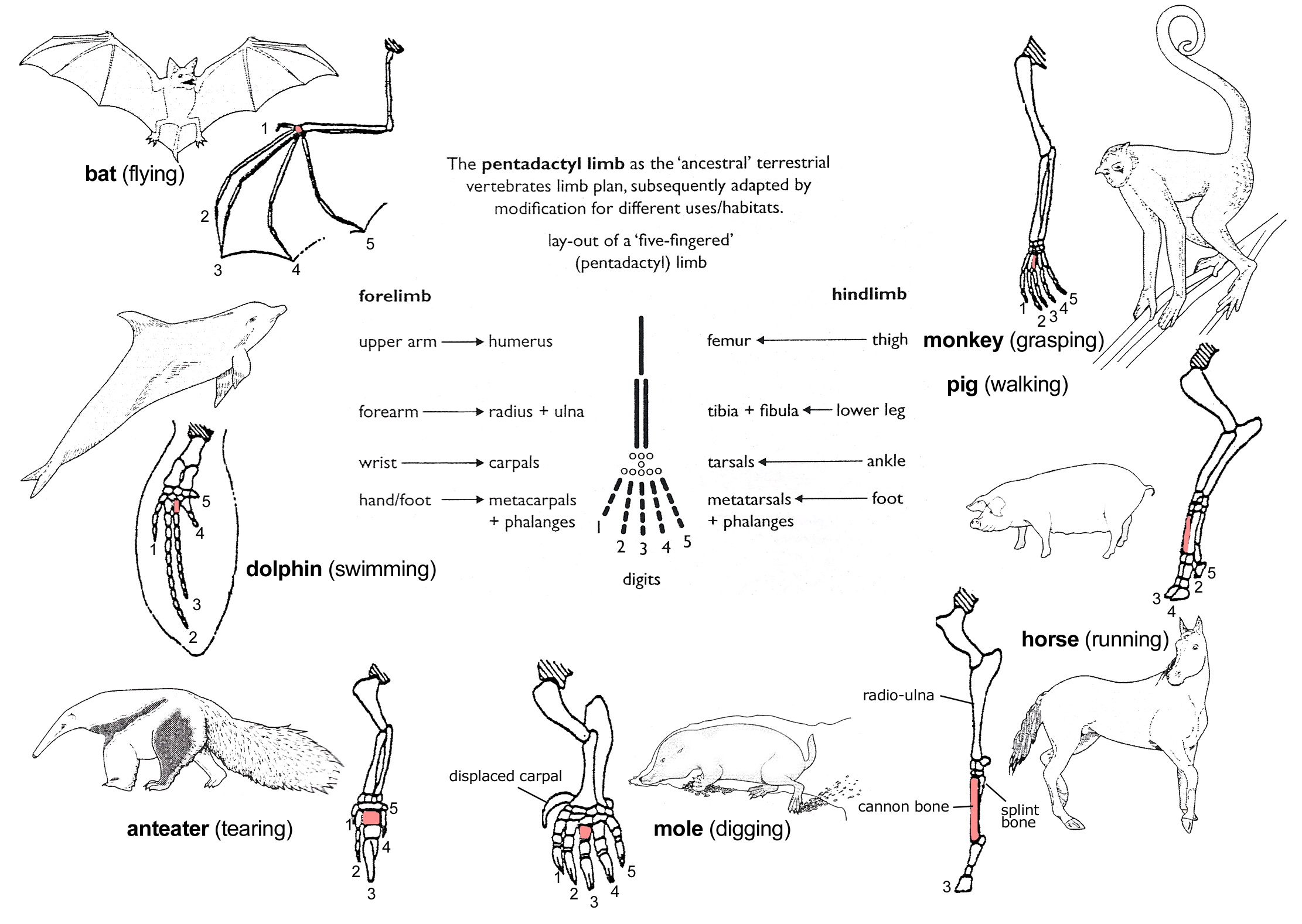
Third Metacarpal Bone
The third metacarpal bone (metacarpal bone of the middle finger) is a little smaller than the second. The dorsal aspect of its base presents on its radial side a pyramidal eminence, the styloid process, which extends upward behind the capitate; immediately distal to this is a rough surface for the attachment of the extensor carpi radialis brevis muscle. The carpal articular facet is concave behind, flat in front, and articulates with the capitate. On the radial side is a smooth, concave facet for articulation with the second metacarpal, and on the ulnar side two small oval facets for the fourth metacarpal. Ossification The ossification process begins in the shaft during prenatal life, and in the head between the 11th and 27th months. Additional images File:Third metacarpal bone (left hand) - animation01.gif, Third metacarpal bone of the left hand (shown in red). Animation. File:Third metacarpal bone (left hand) - animation02.gif, Third metacarpal bone of the left hand. ... [...More Info...] [...Related Items...] OR: [Wikipedia] [Google] [Baidu] |
Middle Finger
The middle finger, long finger, second finger, third finger, toll finger or tall man is the third digit of the human hand, typically located between the index finger and the ring finger. It is typically the longest digit. In anatomy, it is also called ''the third finger'', ''digitus medius'', ''digitus tertius'' or ''digitus III''. Overview In Western countries, The finger, extending the middle finger (either by itself, or along with the index finger in the United Kingdom: see V sign) is an offensive and obscene gesture, widely recognized as a form of insult, due to its resemblance of an Erection, erect penis. It is known, colloquially, as "flipping the bird", "flipping (someone) off", or "giving (someone) the finger". The middle finger is often used for finger snapping together with the thumb. See also * Finger numbering * Galileo's middle finger References External links * Fingers Hand gestures {{Anatomy-stub ... [...More Info...] [...Related Items...] OR: [Wikipedia] [Google] [Baidu] |
Third Metacarpal Styloid Process
The third metacarpal styloid process enables the hand bone to lock into the wrist bones, allowing for greater amounts of pressure to be applied to the wrist and hand from a grasping thumb and fingers. It allows humans Humans (''Homo sapiens'') or modern humans are the most common and widespread species of primate, and the last surviving species of the genus ''Homo''. They are Hominidae, great apes characterized by their Prehistory of nakedness and clothing ... the dexterity and strength to make and use complex tools. This unique anatomical feature separates humans from apes and other nonhuman primates, and is not seen in human fossils older than 1.8 million years. References Bones of the hand {{musculoskeletal-stub ... [...More Info...] [...Related Items...] OR: [Wikipedia] [Google] [Baidu] |
Capitate
The capitate bone is a bone in the human wrist found in the center of the carpal bone region, located at the distal end of the radius and ulna bones. It articulates with the third metacarpal bone (the middle finger) and forms the third carpometacarpal joint. The capitate bone is the largest of the carpal bones in the human hand. It presents, above, a rounded portion or head, which is received into the concavity formed by the scaphoid and lunate bones; a constricted portion or neck; and below this, the body.''Gray's Anatomy'' (1918). See infobox. The bone is also found in many other mammals, and is homologous with the "third distal carpal" of reptiles and amphibians. Structure The capitate is the largest carpal bone found within the hand. The capitate is found within the distal row of carpal bones. The capitate lies directly adjacent to the metacarpal of the ring finger on its distal surface, has the hamate on its ulnar surface and trapezoid on its radial surface, and abuts the ... [...More Info...] [...Related Items...] OR: [Wikipedia] [Google] [Baidu] |
Extensor Carpi Radialis Brevis Muscle
In human anatomy, extensor carpi radialis brevis is a muscle in the forearm that acts to extend and abduct the wrist. It is shorter and thicker than its namesake extensor carpi radialis longus which can be found above the proximal end of the extensor carpi radialis brevis. Origin and insertion It arises from the lateral epicondyle of the humerus, by the common extensor tendon; from the radial collateral ligament of the elbow-joint; from a strong aponeurosis which covers its surface; and from the intermuscular septa between it and the adjacent muscles.''Gray's Anatomy'' 1918, see infobox The fibres end approximately at the middle of the forearm in the form of a flat tendon, which is closely connected with that of the extensor carpi radialis longus, and accompanies it to the wrist; it passes beneath the abductor pollicis longus and extensor pollicis brevis, beneath the extensor retinaculum, and inserts into the lateral dorsal surface of the base of the third metacarpal bone, ... [...More Info...] [...Related Items...] OR: [Wikipedia] [Google] [Baidu] |
Second Metacarpal
The second metacarpal bone (metacarpal bone of the index finger) is the longest, and its base the largest, of all the metacarpal bones.''Gray's Anatomy'' (1918). See infobox. Human anatomy Its base is prolonged upward and medialward, forming a prominent ridge. It presents four articular facets, three on the upper surface and one on the ulnar side: * Of the facets on the upper surface: ** the ''intermediate'' is the largest and is concave from side to side, convex from before backward for articulation with the lesser multangular; ** the ''lateral'' is small, flat and oval for articulation with the greater multangular; ** the ''medial'', on the summit of the ridge, is long and narrow for articulation with the capitate. * The facet on the ulnar side articulates with the third metacarpal. The extensor carpi radialis longus muscle is inserted on the dorsal surface and the flexor carpi radialis muscle on the volar surface of the base. The shaft gives origin to the first palma ... [...More Info...] [...Related Items...] OR: [Wikipedia] [Google] [Baidu] |
Fourth Metacarpal
The fourth metacarpal bone (metacarpal bone of the ring finger) is shorter and smaller than the third. The base is small and quadrilateral; its superior surface presents two facets, a large one medially for articulation with the hamate, and a small one laterally for the capitate. On the radial side are two oval facets, for articulation with the third metacarpal; and on the ulnar side a single concave facet, for the fifth metacarpal. Clinical relevance A shortened fourth metacarpal bone can be a symptom of Kallmann syndrome, a genetic condition which results in the failure to commence or the non-completion of puberty. A short fourth metacarpal bone can also be found in Turner syndrome, a disorder involving sex chromosomes. A fracture of the fourth and/or fifth metacarpal bones transverse neck secondary due to axial loading is known as a boxer's fracture.Shultz, S. J., Houglum, P. A., Perrin, D. H. (2010). Examination of Musculoskeletal Injuries. Chicago: Human Kinetics Ossific ... [...More Info...] [...Related Items...] OR: [Wikipedia] [Google] [Baidu] |
Metacarpus
In human anatomy, the metacarpal bones or metacarpus, also known as the "palm bones", are the appendicular skeleton, appendicular bones that form the intermediate part of the hand between the phalanges (fingers) and the carpal bones (wrist, wrist bones), which joint, articulate with the forearm. The metacarpal bones are homologous to the metatarsal bones in the foot. Structure The metacarpals form a transverse arch to which the rigid row of distal carpal bones are fixed. The peripheral metacarpals (those of the thumb and little finger) form the sides of the cup of the palmar gutter and as they are brought together they deepen this concavity. The index metacarpal is the most firmly fixed, while the thumb metacarpal articulates with the trapezium and acts independently from the others. The middle metacarpals are tightly united to the carpus by intrinsic interlocking bone elements at their bases. The ring metacarpal is somewhat more mobile while the fifth metacarpal is semi-indepen ... [...More Info...] [...Related Items...] OR: [Wikipedia] [Google] [Baidu] |
First Metacarpal Bone
The first metacarpal bone or the metacarpal bone of the thumb is the first bone proximal to the thumb. It is connected to the trapezium of the carpus at the first carpometacarpal joint and to the proximal thumb phalanx at the first metacarpophalangeal joint. Characteristics The first metacarpal bone is short and thick with a shaft thicker and broader than those of the other metacarpal bones. Its narrow shaft connects its widened base and rounded head; the former consisting of a thick cortical bone surrounding the open medullary canal; the latter two consisting of cancellous bone surrounded by a thin cortical shell. Head The head is less rounded and less spherical than those of the other metacarpals, making it better suited for a hinge-like articulation. The distal articular surface is quadrilateral, wide, and flat; thicker and broader transversely and extends much further palmarly than dorsally. On the palmar aspect of the articular surface there is a pair of emin ... [...More Info...] [...Related Items...] OR: [Wikipedia] [Google] [Baidu] |
Second Metacarpal Bone
The second metacarpal bone (metacarpal bone of the index finger) is the longest, and its base the largest, of all the Metacarpus, metacarpal bones.''Gray's Anatomy'' (1918). See infobox. Human anatomy Its base is prolonged upward and medialward, forming a prominent ridge. It presents four articular facets, three on the upper surface and one on the ulnar side: * Of the facets on the upper surface: ** the ''intermediate'' is the largest and is concave from side to side, convex from before backward for articulation with the lesser multangular; ** the ''lateral'' is small, flat and oval for articulation with the greater multangular; ** the ''medial'', on the summit of the ridge, is long and narrow for articulation with the capitate. * The facet on the ulnar side articulates with the third metacarpal. The extensor carpi radialis longus muscle is inserted on the dorsal surface and the flexor carpi radialis muscle on the Anatomical terms of location#Hands and feet, volar surface of t ... [...More Info...] [...Related Items...] OR: [Wikipedia] [Google] [Baidu] |
Fourth Metacarpal Bone
The fourth metacarpal bone (metacarpal bone of the ring finger) is shorter and smaller than the third. The base is small and quadrilateral; its superior surface presents two facets, a large one medially for articulation with the hamate, and a small one laterally for the capitate. On the radial side are two oval facets, for articulation with the third metacarpal; and on the ulnar side a single concave facet, for the fifth metacarpal. Clinical relevance A shortened fourth metacarpal bone can be a symptom of Kallmann syndrome, a genetic condition which results in the failure to commence or the non-completion of puberty. A short fourth metacarpal bone can also be found in Turner syndrome, a disorder involving sex chromosomes. A fracture of the fourth and/or fifth metacarpal bones transverse neck secondary due to axial loading is known as a boxer's fracture.Shultz, S. J., Houglum, P. A., Perrin, D. H. (2010). Examination of Musculoskeletal Injuries. Chicago: Human Kinetics Ossi ... [...More Info...] [...Related Items...] OR: [Wikipedia] [Google] [Baidu] |
Fifth Metacarpal Bone
The fifth metacarpal bone (metacarpal bone of the little finger or pinky finger) is the most medial and second-shortest of the metacarpal bones. Surfaces It presents on its base one facet on its superior surface, which is concavo-convex and articulates with the hamate, and one on its radial side, which articulates with the fourth metacarpal. On its ulnar side is a prominent tubercle for the insertion of the tendon of the extensor carpi ulnaris muscle. The dorsal surface of the body is divided by an oblique ridge, which extends from near the ulnar side of the base to the radial side of the head. The lateral part of this surface serves for the attachment of the fourth interosseus dorsalis; the medial part is smooth, triangular, and covered by the extensor tendons of the little finger. The palmar surface is similarly divided: Its lateral side (facing the fourth metacarpal) provides the origin for the third palmar interosseus, its medial side contains the insertion of opponens ... [...More Info...] [...Related Items...] OR: [Wikipedia] [Google] [Baidu] |
Skeletal System
A skeleton is the structural frame that supports the body of most animals. There are several types of skeletons, including the exoskeleton, which is a rigid outer shell that holds up an organism's shape; the endoskeleton, a rigid internal frame to which the organs and soft tissues attach; and the hydroskeleton, a flexible internal structure supported by the hydrostatic pressure of body fluids. Vertebrates are animals with an endoskeleton centered around an axial vertebral column, and their skeletons are typically composed of bones and cartilages. Invertebrates are other animals that lack a vertebral column, and their skeletons vary, including hard-shelled exoskeleton (arthropods and most molluscs), plated internal shells (e.g. cuttlebones in some cephalopods) or rods (e.g. ossicles in echinoderms), hydrostatically supported body cavities (most), and spicules (sponges). Cartilage is a rigid connective tissue that is found in the skeletal systems of vertebrates and invert ... [...More Info...] [...Related Items...] OR: [Wikipedia] [Google] [Baidu] |


