|
Fibrous Papule Of The Nose
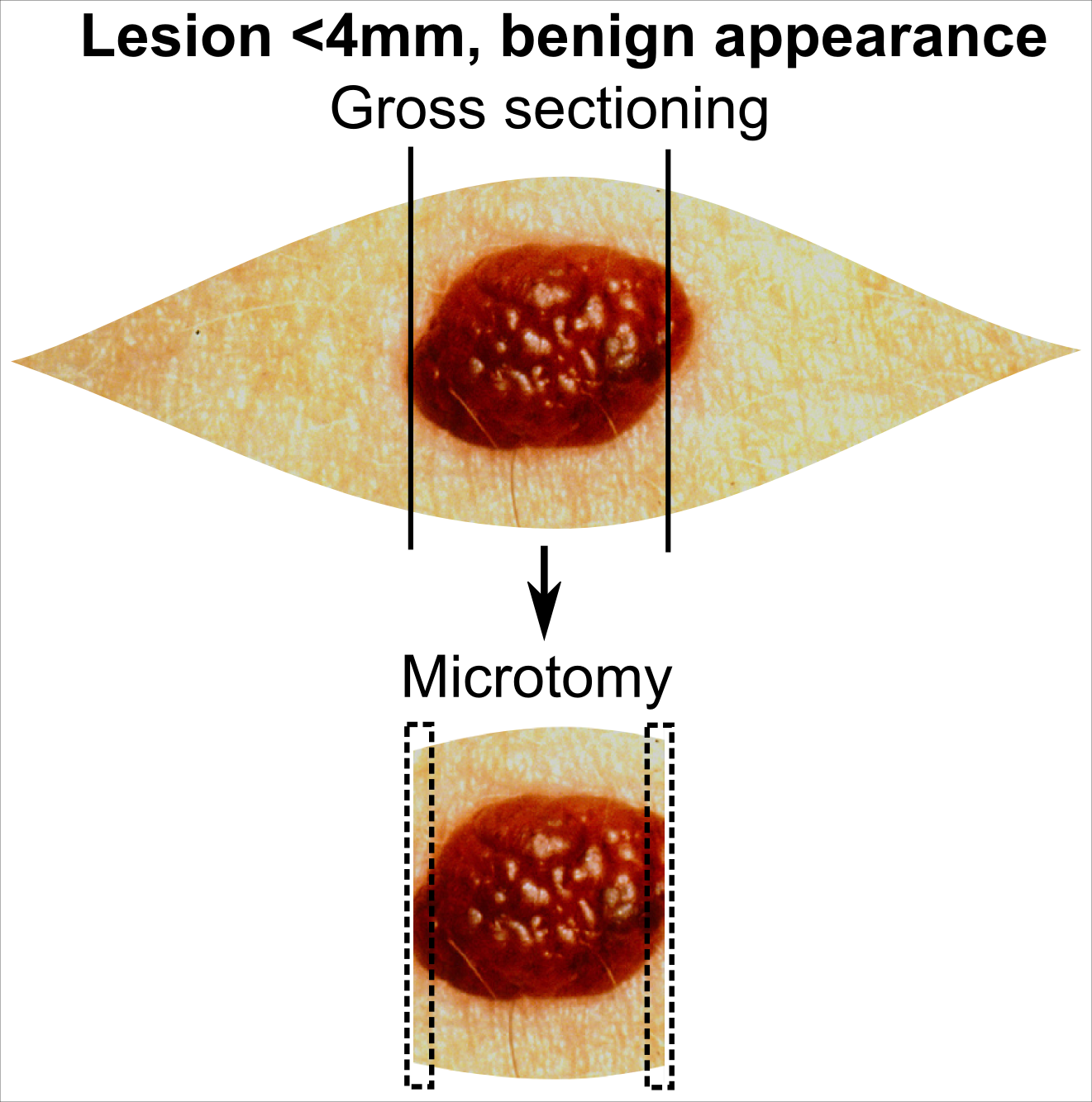
Fibrous papule of the nose is a harmless small bump on or near the nose. It is typically dome-shaped, skin-colored, white or reddish, smooth and firm. Less frequently it can occur elsewhere on the face. Sometimes there are a few. It may be shiny and remains unchanged for life. There may be a central hair. The precise cause is unknown. It is a type of angiofibroma which originates in a dendrocyte in skin. Diagnosis is by visualisation, skin biopsy or histopathology of one that has been surgically cut out. Histopathology shows fibroblasts, fibrotic stroma and large blood vessels. It can appear similar to a benign melanocytic naevus or an early BCC. It may be mistaken for a nevocytic nevus, neurofibroma and pyogenic granuloma. The presence of several should alert to seeking for other signs of tuberous sclerosis. Usually no treatment is necessary. Treatment for cosmetic reasons include shave excision, and electrosurgery. Following treatment, recurrence is rare. It is common ... [...More Info...] [...Related Items...] OR: [Wikipedia] [Google] [Baidu] |
Dermatology
Dermatology is the branch of medicine dealing with the skin.''Random House Webster's Unabridged Dictionary.'' Random House, Inc. 2001. Page 537. . It is a speciality with both medical and surgical aspects. A dermatologist is a specialist medical doctor who manages diseases related to skin, hair, nails, and some cosmetic problems. Etymology Attested in English in 1819, the word "dermatology" derives from the Greek δέρματος (''dermatos''), genitive of δέρμα (''derma''), "skin" (itself from δέρω ''dero'', "to flay") and -λογία '' -logia''. Neo-Latin ''dermatologia'' was coined in 1630, an anatomical term with various French and German uses attested from the 1730s. History In 1708, the first great school of dermatology became a reality at the famous Hôpital Saint-Louis in Paris, and the first textbooks (Willan's, 1798–1808) and atlases ( Alibert's, 1806–1816) appeared in print around the same time.Freedberg, et al. (2003). ''Fitzpatrick's Dermatology in ... [...More Info...] [...Related Items...] OR: [Wikipedia] [Google] [Baidu] |
Papule
A papule is a small, well-defined bump in the skin. It may have a rounded, pointed or flat top, and may have a dip. It can appear with a stalk, be thread-like or look warty. It can be soft or firm and its surface may be rough or smooth. Some have crusts or scales. A papule can be flesh colored, yellow, white, brown, red, blue or purplish. There may be just one or many, and they may occur irregularly in different parts of the body or appear in clusters. It does not contain fluid but may progress to a pustule or vesicle. A papule is smaller than a nodule; it can be as tiny as a pinhead and is typically less than 1 cm in width, according to some sources, and 0.5 cm according to others. When merged together, it appears as a plaque. Its color might indicate its cause, such as white in milia, red in eczema, yellowish in xanthoma and black in melanoma. They may open when scratched and become infected and crusty. Definition A papule is a small, well-defined bump in the s ... [...More Info...] [...Related Items...] OR: [Wikipedia] [Google] [Baidu] |
Dendritic Cell
Dendritic cells (DCs) are antigen-presenting cells (also known as ''accessory cells'') of the mammalian immune system. Their main function is to process antigen material and present it on the cell surface to the T cells of the immune system. They act as messengers between the innate and the adaptive immune systems. Dendritic cells are present in those tissues that are in contact with the external environment, such as the skin (where there is a specialized dendritic cell type called the Langerhans cell) and the inner lining of the nose, lungs, stomach and intestines. They can also be found in an immature state in the blood. Once activated, they migrate to the lymph nodes where they interact with T cells and B cells to initiate and shape the adaptive immune response. At certain development stages they grow branched projections, the ''dendrites'' that give the cell its name (δένδρον or déndron being Greek for 'tree'). While similar in appearance, these are structures ... [...More Info...] [...Related Items...] OR: [Wikipedia] [Google] [Baidu] |
Skin Biopsy
Skin biopsy is a biopsy technique in which a skin lesion is removed to be sent to a pathologist to render a microscopic diagnosis. It is usually done under local anesthetic in a physician's office, and results are often available in 4 to 10 days. It is commonly performed by dermatologists. Skin biopsies are also done by family physicians, internists, surgeons, and other specialties. However, performed incorrectly, and without appropriate clinical information, a pathologist's interpretation of a skin biopsy can be severely limited, and therefore doctors and patients may forgo traditional biopsy techniques and instead choose Mohs surgery. There are four main types of skin biopsies: shave biopsy, punch biopsy, excisional biopsy, and incisional biopsy. The choice of the different skin biopsies is dependent on the suspected diagnosis of the skin lesion. Like most biopsies, patient consent and anesthesia (usually lidocaine injected into the skin) are prerequisites. Types Shave biop ... [...More Info...] [...Related Items...] OR: [Wikipedia] [Google] [Baidu] |
Histopathology
Histopathology (compound of three Greek words: ''histos'' "tissue", πάθος ''pathos'' "suffering", and -λογία '' -logia'' "study of") refers to the microscopic examination of tissue in order to study the manifestations of disease. Specifically, in clinical medicine, histopathology refers to the examination of a biopsy or surgical specimen by a pathologist, after the specimen has been processed and histological sections have been placed onto glass slides. In contrast, cytopathology examines free cells or tissue micro-fragments (as "cell blocks"). Collection of tissues Histopathological examination of tissues starts with surgery, biopsy, or autopsy. The tissue is removed from the body or plant, and then, often following expert dissection in the fresh state, placed in a fixative which stabilizes the tissues to prevent decay. The most common fixative is 10% neutral buffered formalin (corresponding to 3.7% w/v formaldehyde in neutral buffered water, such as phosphate buf ... [...More Info...] [...Related Items...] OR: [Wikipedia] [Google] [Baidu] |
Basal Cell Carcinoma
Basal-cell carcinoma (BCC), also known as basal-cell cancer, is the most common type of skin cancer. It often appears as a painless raised area of skin, which may be shiny with small blood vessels running over it. It may also present as a raised area with ulceration. Basal-cell cancer grows slowly and can damage the tissue around it, but it is unlikely to spread to distant areas or result in death. Risk factors include exposure to ultraviolet light, having lighter skin, radiation therapy, long-term exposure to arsenic and poor immune-system function. Exposure to UV light during childhood is particularly harmful. Tanning beds have become another common source of ultraviolet radiation. Diagnosis often depends on skin examination, confirmed by tissue biopsy. It remains unclear whether sunscreen affects the risk of basal-cell cancer. Treatment is typically by surgical removal. This can be by simple excision if the cancer is small; otherwise, Mohs surgery is generally recomme ... [...More Info...] [...Related Items...] OR: [Wikipedia] [Google] [Baidu] |
Electrosurgery
Electrosurgery is the application of a high-frequency (radio frequency) alternating polarity, electrical current to biological tissue as a means to cut, coagulate, desiccate, or fulgurate tissue.Hainer BL, "Fundamentals of electrosurgery", ''Journal of the American Board of Family Practice'', 4(6):419–26, 1991 Nov.–Dec. 400 V peak-to-peak) the vapor sheath is ionized, forming conductive plasma. Electric current continues to flow from the metal electrode through the ionized gas into the tissue. Rapid overheating of tissue results in its vaporization, fragmentation and ejection of fragments, allowing for tissue cutting. In applications of a continuous wave the heat diffusion typically leads to formation of a significant thermal damage zone at the edges of the lesion. Open circuit voltage in electrosurgical waveforms is typically in the range of 300–10,000 V peak-to-peak. Higher precision can be achieved with pulsed waveforms. Using bursts of several tens of microseconds in d ... [...More Info...] [...Related Items...] OR: [Wikipedia] [Google] [Baidu] |
Benign
Malignancy () is the tendency of a medical condition to become progressively worse. Malignancy is most familiar as a characterization of cancer. A ''malignant'' tumor contrasts with a non-cancerous benign tumor, ''benign'' tumor in that a malignancy is not self-limited in its growth, is capable of invading into adjacent tissues, and may be capable of spreading to distant tissues. A benign tumor has none of those properties. Malignancy in cancers is characterized by anaplasia, invasiveness, and metastasis. Malignant tumors are also characterized by genome instability, so that cancers, as assessed by whole genome sequencing, frequently have between 10,000 and 100,000 mutations in their entire genomes. Cancers usually show tumour heterogeneity, containing multiple subclones. They also frequently have reduced expression of DNA repair enzymes due to Epigenetics#DNA repair epigenetics in cancer, epigenetic methylation of DNA repair genes or altered MicroRNA#DNA repair and cancer, micr ... [...More Info...] [...Related Items...] OR: [Wikipedia] [Google] [Baidu] |
Angiofibroma
Angiofibroma (AGF) is a descriptive term for a wide range of benign skin or mucous membrane (i.e. the outer membrane lining body cavities such as the mouth and nose) lesions in which individuals have: 1) benign papules, i.e. pinhead-sized elevations that lack visible evidence of containing fluid; 2) nodules, i.e. small firm lumps usually >0.1 cm in diameter; and/or 3) tumors, i.e. masses often regarded as ~0.8 cm or larger. AGF lesions share common macroscopic (i.e. gross) and microscopic appearances. Grossly, AGF lesions consist of multiple papules, one or more skin-colored to erythematous, dome-shaped nodules, or usually just a single tumor. Microscopically, they consist of spindle-shaped and stellate-shaped cells centered around dilated and thin-walled blood vessels in a background of coarse bundles of collagen (i.e. the main fibrous component of connective tissue). Angiofibromas have been divided into different types but commonly a specific type was given multiple a ... [...More Info...] [...Related Items...] OR: [Wikipedia] [Google] [Baidu] |
Fibroblasts
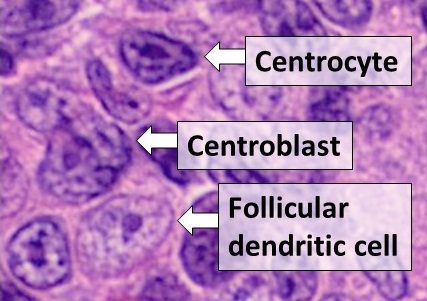
A fibroblast is a type of biological cell that synthesizes the extracellular matrix and collagen, produces the structural framework ( stroma) for animal tissues, and plays a critical role in wound healing. Fibroblasts are the most common cells of connective tissue in animals. Structure Fibroblasts have a branched cytoplasm surrounding an elliptical, speckled nucleus having two or more nucleoli. Active fibroblasts can be recognized by their abundant rough endoplasmic reticulum. Inactive fibroblasts (called fibrocytes) are smaller, spindle-shaped, and have a reduced amount of rough endoplasmic reticulum. Although disjointed and scattered when they have to cover a large space, fibroblasts, when crowded, often locally align in parallel clusters. Unlike the epithelial cells lining the body structures, fibroblasts do not form flat monolayers and are not restricted by a polarizing attachment to a basal lamina on one side, although they may contribute to basal lamina components in s ... [...More Info...] [...Related Items...] OR: [Wikipedia] [Google] [Baidu] |
Neurofibroma
A neurofibroma is a benign nerve-sheath tumor in the peripheral nervous system. In 90% of cases, they are found as stand-alone tumors (solitary neurofibroma, solitary nerve sheath tumor or sporadic neurofibroma), while the remainder are found in persons with neurofibromatosis type I (NF1), an autosomal-dominant genetically inherited disease. They can result in a range of symptoms from physical disfiguration and pain to cognitive disability. Neurofibromas arise from nonmyelinating-type Schwann cells that exhibit biallelic inactivation of the ''NF1'' gene that codes for the protein neurofibromin. This protein is responsible for regulating the RAS-mediated cell growth signaling pathway. In contrast to schwannomas, another type of tumor arising from Schwann cells, neurofibromas incorporate many additional types of cells and structural elements in addition to Schwann cells, making it difficult to identify and understand all the mechanisms through which they originate and develop. ... [...More Info...] [...Related Items...] OR: [Wikipedia] [Google] [Baidu] |


.jpg)
