|
Fascial Manipulation
Fascial Manipulation is a manual therapy technique developed by Italian physiotherapist Luigi Stecco in the 1980s, aimed at evaluating and treating global fascial dysfunction by restoring normal motion/gliding to the system. The method is based on a biomechanical model which lays an emphasis on the significant role of fascia, particularly deep muscular fascia in treating musculoskeletal disorders, and internal organ disfunction. The fascial system consists of a three-dimensional continuum of soft, collagen-containing, loose and dense fibrous connective tissues that permeate the body. History In the 1980s, Stecco focused his fascial research on the treatment of recurring pain, pain which could not be alleviated by other treatments, and the recovery time of the injury. He developed a soft tissue manual technique aimed at treating myofascial dysfunction, and consequently, musculoskeletal disease. He named the technique Fascial Manipulation. He continued to focus his research on the m ... [...More Info...] [...Related Items...] OR: [Wikipedia] [Google] [Baidu] |
Fascia
A fascia (; plural fasciae or fascias; adjective fascial; from Latin: "band") is a band or sheet of connective tissue, primarily collagen, beneath the skin that attaches to, stabilizes, encloses, and separates muscles and other internal organs. Fascia is classified by layer, as superficial fascia, deep fascia, and ''visceral'' or ''parietal'' fascia, or by its function and anatomical location. Like ligaments, aponeuroses, and tendons, fascia is made up of fibrous connective tissue containing closely packed bundles of collagen fibers oriented in a wavy pattern parallel to the direction of pull. Fascia is consequently flexible and able to resist great unidirectional tension forces until the wavy pattern of fibers has been straightened out by the pulling force. These collagen fibers are produced by fibroblasts located within the fascia. Fasciae are similar to ligaments and tendons as they have collagen as their major component. They differ in their location and function: ligament ... [...More Info...] [...Related Items...] OR: [Wikipedia] [Google] [Baidu] |
Human Musculoskeletal System
The human musculoskeletal system (also known as the human locomotor system, and previously the activity system) is an organ system that gives humans the ability to move using their muscular and skeletal systems. The musculoskeletal system provides form, support, stability, and movement to the body. It is made up of the bones of the skeleton, muscles, cartilage, tendons, ligaments, joints, and other connective tissue that supports and binds tissues and organs together. The musculoskeletal system's primary functions include supporting the body, allowing motion, and protecting vital organs. The skeletal portion of the system serves as the main storage system for calcium and phosphorus and contains critical components of the hematopoietic system. This system describes how bones are connected to other bones and muscle fibers via connective tissue such as tendons and ligaments. The bones provide stability to the body. Muscles keep bones in place and also play a role in the movement ... [...More Info...] [...Related Items...] OR: [Wikipedia] [Google] [Baidu] |
New York University Grossman School Of Medicine
NYU Grossman School of Medicine is a medical school of New York University, a private research university in New York City. It was founded in 1841 and is one of two medical schools of the university, with the other being the Long Island School of Medicine. NYU Grossman School of Medicine is part of NYU Langone Health, named after Kenneth Langone, the investment banker and financial backer of The Home Depot. In 2022, U.S''. News & World Report'' ranked NYU Grossman School of Medicine as No. 2 “Best Graduate Schools". History New York University College of Medicine was established in 1841. The medical school merged with Bellevue Hospital Medical College in 1898 to form the University and Bellevue Hospital Medical College. The name NYU Grossman School of Medicine was adopted in 2019. NYU Grossman School of Medicine is home to many key advancements in medical education. In 1854, human dissection in New York was legalized due to efforts of the faculty. In 1866, NYU professors pro ... [...More Info...] [...Related Items...] OR: [Wikipedia] [Google] [Baidu] |
Mechanoreceptor
A mechanoreceptor, also called mechanoceptor, is a sensory receptor that responds to mechanical pressure or distortion. Mechanoreceptors are innervated by sensory neurons that convert mechanical pressure into electrical signals that, in animals, are sent to the central nervous system. Vertebrate mechanoreceptors Cutaneous mechanoreceptors Cutaneous mechanoreceptors respond to mechanical stimuli that result from physical interaction, including pressure and vibration. They are located in the skin, like other cutaneous receptors. They are all innervated by Aβ fibers, except the mechanorecepting free nerve endings, which are innervated by Aδ fibers. Cutaneous mechanoreceptors can be categorized by what kind of sensation they perceive, by the rate of adaptation, and by morphology. Furthermore, each has a different receptive field. By sensation *The Slowly Adapting type 1 (SA1) mechanoreceptor, with the Merkel corpuscle end-organ (also known as Merkel discs) detect sustained p ... [...More Info...] [...Related Items...] OR: [Wikipedia] [Google] [Baidu] |
Fibular Retinacula
The fibular retinacula (also known as peroneal retinacula) are fibrous retaining bands that bind down the tendons of the fibularis longus and fibularis brevis muscles as they run across the side of the ankle. (''Retinaculum'' is Latin for "retainer.") These bands consist of the superior fibular retinaculum and the inferior fibular retinaculum. The superior fibers are attached above to the lateral malleolus and below to the lateral surface of the calcaneus. The inferior fibers are continuous in front with those of the inferior extensor retinaculum of the foot; behind they are attached to the lateral surface of the calcaneus; some of the fibers are fixed to the calcaneal tubercle, forming a septum between the tendons of the fibularis longus and fibularis brevis. See also * Fibularis longus * Fibularis brevis In human anatomy, the fibularis brevis (or peroneus brevis) is a muscle that lies underneath the fibularis longus within the lateral compartment of the leg. It acts to tilt th ... [...More Info...] [...Related Items...] OR: [Wikipedia] [Google] [Baidu] |
Retinaculum
A retinaculum (plural ''retinacula'') is a band of thickened deep fascia around tendons that holds them in place. It is not part of any muscle. Its function is mostly to stabilize a tendon. The term retinaculum is New Latin, derived from the Latin verb ''retinere'' (to retain). Specific retinacula include: * In the wrist: ** Flexor retinaculum of the hand ** Extensor retinaculum of the hand * In the ankle: ** Flexor retinaculum of foot ** Superior extensor retinaculum of foot ** Inferior extensor retinaculum of foot ** Superior fibular retinaculum ** Inferior fibular retinaculum * In the knee: ** Lateral retinaculum The lateral retinaculum is the fibrous tissue on the lateral (outer) side of the kneecap (patella). The kneecap has both a medial (on the inner aspect) and a lateral (on the outer side) retinaculum, and these help to support the kneecap in its posi ... ** Medial patellar retinaculum References {{reflist Tendons ... [...More Info...] [...Related Items...] OR: [Wikipedia] [Google] [Baidu] |
Joint Mobilization
Joint mobilization is a manual therapy intervention, a type of straight-lined, passive movement of a skeletal joint that addresses arthrokinematic joint motion (joint gliding) rather than osteokinematic joint motion. It is usually aimed at a 'target' synovial joint with the aim of achieving a therapeutic effect. These techniques are used by a variety of health care professionals with specific training in manual therapy assessment and treatment techniques. IFOMPT defines joint mobilization as "a manual therapy technique comprising a continuum of skilled passive movements that are applied at varying speeds and amplitudes to joints, muscles or nerves with the intent to restore optimal motion, function, and/or to reduce pain." The APTA Guide to Physical Therapist Practice defines mobilization/manipulation as “a manual therapy technique a continuum of skilled passive movements that are applied at varying speeds and amplitudes, including a small amplitude/ high velocity therapeuti ... [...More Info...] [...Related Items...] OR: [Wikipedia] [Google] [Baidu] |
Connective Tissue
Connective tissue is one of the four primary types of animal tissue, along with epithelial tissue, muscle tissue, and nervous tissue. It develops from the mesenchyme derived from the mesoderm the middle embryonic germ layer. Connective tissue is found in between other tissues everywhere in the body, including the nervous system. The three meninges, membranes that envelop the brain and spinal cord are composed of connective tissue. Most types of connective tissue consists of three main components: elastic and collagen fibers, ground substance, and cells. Blood, and lymph are classed as specialized fluid connective tissues that do not contain fiber. All are immersed in the body water. The cells of connective tissue include fibroblasts, adipocytes, macrophages, mast cells and leucocytes. The term "connective tissue" (in German, ''Bindegewebe'') was introduced in 1830 by Johannes Peter Müller. The tissue was already recognized as a distinct class in the 18th century. ... [...More Info...] [...Related Items...] OR: [Wikipedia] [Google] [Baidu] |
Rotator Cuff Tear
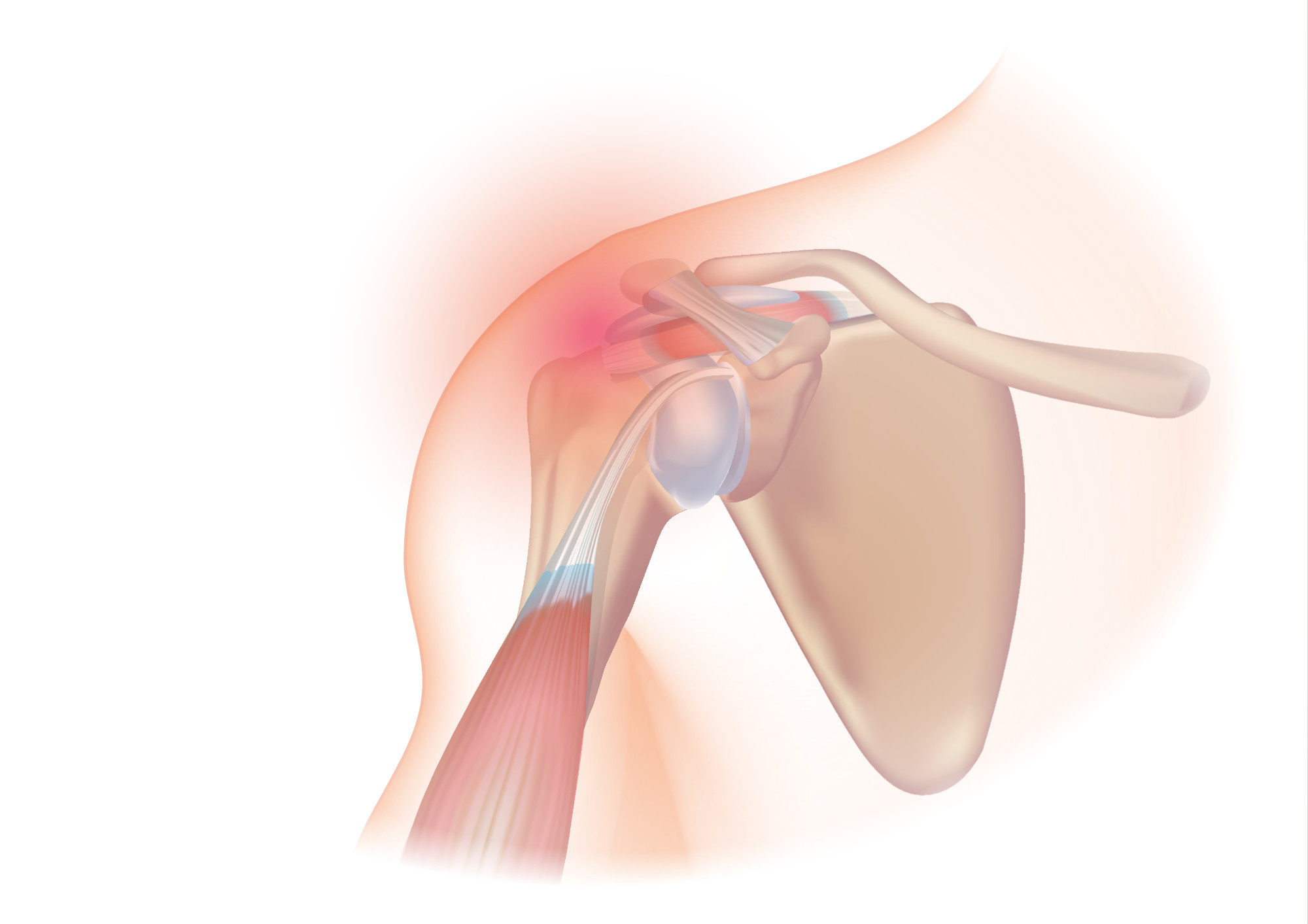
A rotator cuff tear is an injury where one or more of the tendons or muscles of the rotator cuff of the shoulder get torn. Symptoms may include shoulder pain, which is often worse with movement, limited range of motion, or weakness. This may limit people's ability to brush their hair or put on clothing. Clicking may also occur with movement of the arm. Tears may occur as the result of a sudden force or gradually over time. Risk factors include certain repetitive activities, smoking, and a family history of the condition. Diagnosis is based on symptoms, examination, and medical imaging. The rotator cuff is made up of the supraspinatus, infraspinatus, teres minor, and subscapularis. The supraspinatus is the most commonly affected. Treatment may include pain medication such as NSAIDs and specific exercises. It is recommended that people who are unable to raise their arm above 90 degrees after 2 weeks should be further assessed. In severe cases surgery may be tried, however ben ... [...More Info...] [...Related Items...] OR: [Wikipedia] [Google] [Baidu] |
Arthroplasty
Arthroplasty (literally " e-orming of joint") is an orthopedic surgical procedure where the articular surface of a musculoskeletal joint is replaced, remodeled, or realigned by osteotomy or some other procedure. It is an elective procedure that is done to relieve pain and restore function to the joint after damage by arthritis or some other type of trauma. Types For the last 45 years, the most successful and common form of arthroplasty is the surgical replacement of arthritic or destructive or necrotic joint or joint surface with a prosthesis. For example, a hip joint that is affected by osteoarthritis may be replaced entirely (total hip arthroplasty) with a prosthetic hip. This would involve replacing both the acetabulum (hip socket) and the head and neck of the femur. The purpose of this procedure is to relieve pain, to restore range of motion and to improve walking ability, thus leading to the improvement of muscle strength. Other types of arthroplasty * Interposition ... [...More Info...] [...Related Items...] OR: [Wikipedia] [Google] [Baidu] |


