|
Fabella With Arrow
The fabella is a small sesamoid bone found in some mammals embedded in the tendon of the lateral head of the gastrocnemius muscle behind the lateral condyle of the femur. It is an accessory bone, an anatomical variation present in 39% of humans. Rarely, there are two or three of these bones (fabella bi- or tripartita). It can be mistaken for a loose body or osteophyte. The word ''fabella'' is a Latin diminutive of ''faba'' 'bean'. In humans, it is more common in men than women, older individuals compared to younger, and there is high regional variation, with fabellae being most common in people living in Asia and Oceania and least common in people living in North America and Africa. Bilateral cases (one per knee) are more common than unilateral ones (one per individual), and within individual cases, fabellae are equally likely to be present in right or left knees. Taken together, these data suggest the ability to form a fabella may be genetically controlled, but fabella ossifica ... [...More Info...] [...Related Items...] OR: [Wikipedia] [Google] [Baidu] |
Sesamoid Bone
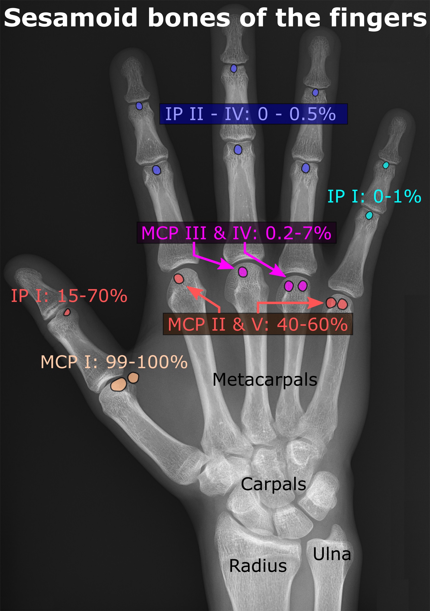
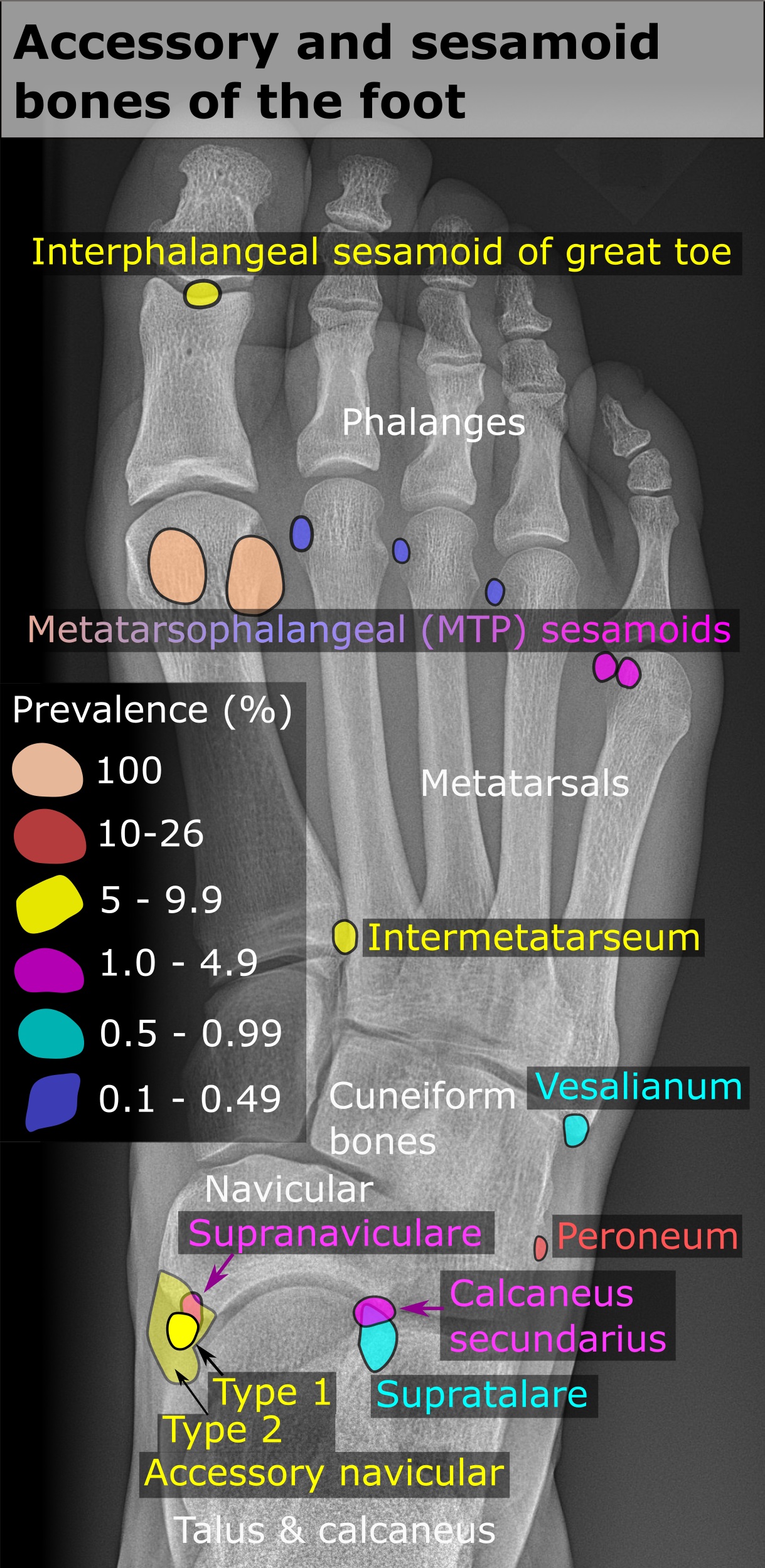
In anatomy, a sesamoid bone () is a bone embedded within a tendon or a muscle. Its name is derived from the Arabic word for ' sesame seed', indicating the small size of most sesamoids. Often, these bones form in response to strain, or can be present as a normal variant. The patella is the largest sesamoid bone in the body. Sesamoids act like pulleys, providing a smooth surface for tendons to slide over, increasing the tendon's ability to transmit muscular forces. Structure Sesamoid bones can be found on joints throughout the body, including: * In the knee—the patella (within the quadriceps tendon). This is the largest sesamoid bone. * In the hand—two sesamoid bones are commonly found in the distal portions of the first metacarpal bone (within the tendons of adductor pollicis and flexor pollicis brevis). There is also commonly a sesamoid bone in distal portions of the second metacarpal bone. * In the wrist—The pisiform of the wrist is a sesamoid bone (within the tend ... [...More Info...] [...Related Items...] OR: [Wikipedia] [Google] [Baidu] |
Ossification
Ossification (also called osteogenesis or bone mineralization) in bone remodeling is the process of laying down new bone material by cells named osteoblasts. It is synonymous with bone tissue formation. There are two processes resulting in the formation of normal, healthy bone tissue: Intramembranous ossification is the direct laying down of bone into the primitive connective tissue ( mesenchyme), while endochondral ossification involves cartilage as a precursor. In fracture healing, endochondral osteogenesis is the most commonly occurring process, for example in fractures of long bones treated by plaster of Paris, whereas fractures treated by open reduction and internal fixation with metal plates, screws, pins, rods and nails may heal by intramembranous osteogenesis. Heterotopic ossification is a process resulting in the formation of bone tissue that is often atypical, at an extraskeletal location. Calcification is often confused with ossification. Calcification is sy ... [...More Info...] [...Related Items...] OR: [Wikipedia] [Google] [Baidu] |
Sesamoid Bones
In anatomy, a sesamoid bone () is a bone embedded within a tendon or a muscle. Its name is derived from the Arabic word for 'sesame seed', indicating the small size of most sesamoids. Often, these bones form in response to strain, or can be present as a normal variant. The patella is the largest sesamoid bone in the body. Sesamoids act like pulleys, providing a smooth surface for tendons to slide over, increasing the tendon's ability to transmit muscular forces. Structure Sesamoid bones can be found on joints throughout the body, including: * In the knee—the patella (within the quadriceps tendon). This is the largest sesamoid bone. * In the hand—two sesamoid bones are commonly found in the distal portions of the first metacarpal bone (within the tendons of adductor pollicis and flexor pollicis brevis). There is also commonly a sesamoid bone in distal portions of the second metacarpal bone. * In the wrist—The pisiform of the wrist is a sesamoid bone (within the tendon o ... [...More Info...] [...Related Items...] OR: [Wikipedia] [Google] [Baidu] |
Thieme Medical Publishers
Thieme Medical Publishers is a German medical and science publisher in the Thieme Publishing Group. It produces professional journals, textbooks, atlases, monographs and reference books in both German and English covering a variety of medical specialties, including neurosurgery, orthopaedics, endocrinology, urology, radiology, anatomy, chemistry, otolaryngology, ophthalmology, audiology and speech-language pathology, complementary and alternative medicine. Thieme has more than 1,000 employees and maintains offices in seven cities worldwide, including New York City, Beijing, Delhi, Stuttgart, and three other cities in Germany. History Georg Thieme Verlag was founded in 1886 in Leipzig, Germany, by Georg Thieme when he was 26 years old. Thieme remains privately held and family-owned. The company received some early success in 1896 by publishing Wilhelm Röntgen's famous picture of his wife's hand in what is still one of Thieme's and Germany's oldest journals, the ''Deutsche ... [...More Info...] [...Related Items...] OR: [Wikipedia] [Google] [Baidu] |
Fabella Sign
The fabella sign is displacement of the fabella that is seen in cases of synovial effusion and popliteal fossa masses. The fabella is an accessory ossicle located inside the gastrocnemius lateral head tendon on the posterior side of the knee, in about 25% of people. It can thus serve as a surrogate radio-opaque marker of the posterior border of the knee's synovium. On a lateral radiograph of the knee, an increase in the distance from the fabella to the femur or to the tibia The tibia (; ), also known as the shinbone or shankbone, is the larger, stronger, and anterior (frontal) of the two bones in the leg below the knee in vertebrates (the other being the fibula, behind and to the outside of the tibia); it connects ... can be suggestive of fluid or of a mass within the synovial fossa. This is of particular use in radiographic detection of knee effusions, as the cause for the effusion may obscure the subcutaneous planes on x-ray that can also be used to determine presence of e ... [...More Info...] [...Related Items...] OR: [Wikipedia] [Google] [Baidu] |
The Anatomical Record
''The Anatomical Record'' is a peer-reviewed scientific journal covering anatomy. It was established by the American Association of Anatomists in 1906 and is published by John Wiley & Sons. According to the ''Journal Citation Reports'', the journal has a 2020 impact factor of 2.064. See also * List of biology journals This is a list of articles about scientific journals in biology and its various subfields. General Agriculture *''Journal of Animal Science'' *''Journal of Dairy Science'' *''Journal of Food Science'' *'' Poultry Science'' *'' Animal Feed S ... References External links * Anatomy journals Wiley (publisher) academic journals Publications established in 1906 Monthly journals English-language journals 1906 establishments in the United States {{biology-journal-stub ... [...More Info...] [...Related Items...] OR: [Wikipedia] [Google] [Baidu] |
Chimpanzee–human Last Common Ancestor
The chimpanzee–human last common ancestor (CHLCA) is the last common ancestor shared by the extant ''Homo'' (human) and '' Pan'' (chimpanzee and bonobo) genera of Hominini. Due to complex hybrid speciation, it is not currently possible to give a precise estimate on the age of this ancestral population. While "original divergence" between populations may have occurred as early as 13 million years ago (Miocene), hybridization may have been ongoing until as recently as 4 million years ago (Pliocene). In human genetic studies, the CHLCA is useful as an anchor point for calculating single-nucleotide polymorphism (SNP) rates in human populations where chimpanzees are used as an outgroup, that is, as the extant species most genetically similar to ''Homo sapiens''. Taxonomy The taxon tribe ''Hominini'' was proposed to separate humans (genus ''Homo'') from chimpanzees ('' Pan'') and gorillas (genus ''Gorilla'') on the notion that the least similar species should be separat ... [...More Info...] [...Related Items...] OR: [Wikipedia] [Google] [Baidu] |
Human Anatomy
The human body is the structure of a human being. It is composed of many different types of cells that together create tissues and subsequently organ systems. They ensure homeostasis and the viability of the human body. It comprises a head, hair, neck, trunk (which includes the thorax and abdomen), arms and hands, legs and feet. The study of the human body involves anatomy, physiology, histology and embryology. The body varies anatomically in known ways. Physiology focuses on the systems and organs of the human body and their functions. Many systems and mechanisms interact in order to maintain homeostasis, with safe levels of substances such as sugar and oxygen in the blood. The body is studied by health professionals, physiologists, anatomists, and by artists to assist them in their work. Composition The human body is composed of elements including hydrogen, oxygen, carbon, calcium and phosphorus. These elements reside in trillions of cells and non-cellula ... [...More Info...] [...Related Items...] OR: [Wikipedia] [Google] [Baidu] |
Hominidae
The Hominidae (), whose members are known as the great apes or hominids (), are a taxonomic family of primates that includes eight extant species in four genera: '' Pongo'' (the Bornean, Sumatran and Tapanuli orangutan); ''Gorilla'' (the eastern and western gorilla); '' Pan'' (the chimpanzee and the bonobo); and ''Homo'', of which only modern humans remain. Several revisions in classifying the great apes have caused the use of the term ''"hominid"'' to vary over time. The original meaning of "hominid" referred only to humans (''Homo'') and their closest extinct relatives. However, by the 1990s humans, apes, and their ancestors were considered to be "hominids". The earlier restrictive meaning has now been largely assumed by the term ''"hominin"'', which comprises all members of the human clade after the split from the chimpanzees (''Pan''). The current meaning of "hominid" includes all the great apes including humans. Usage still varies, however, and some scientists and la ... [...More Info...] [...Related Items...] OR: [Wikipedia] [Google] [Baidu] |
Human Evolution
Human evolution is the evolutionary process within the history of primates that led to the emergence of ''Homo sapiens'' as a distinct species of the hominid family, which includes the great apes. This process involved the gradual development of traits such as human bipedalism and language, as well as interbreeding with other hominins, which indicate that human evolution was not linear but a web.Human Hybrids (PDF). Michael F. Hammer. ''Scientific American'', May 2013. The study of human evolution involves scientific disciplines, including [...More Info...] [...Related Items...] OR: [Wikipedia] [Google] [Baidu] |
Osteophyte
Osteophytes are exostoses (bony projections) that form along joint margins. They should not be confused with enthesophytes, which are bony projections that form at the attachment of a tendon or ligament. Osteophytes are not always distinguished from exostoses in any definite way, although in many cases there are a number of differences. Osteophytes are typically intra-articular (within the joint capsule). Cause A range of bone-formation processes are associated with aging, degeneration, mechanical instability, and disease (such as diffuse idiopathic skeletal hyperostosis). Osteophyte formation has classically been related to sequential and consequential changes in such processes. Often osteophytes form in osteoarthritic joints as a result of damage and wear from inflammation. Calcification and new bone formation can also occur in response to mechanical damage in joints. Pathophysiology Osteophytes form because of the increase in a damaged joint's surface area. This is most co ... [...More Info...] [...Related Items...] OR: [Wikipedia] [Google] [Baidu] |
Tendon
A tendon or sinew is a tough, high-tensile-strength band of dense fibrous connective tissue that connects muscle to bone. It is able to transmit the mechanical forces of muscle contraction to the skeletal system without sacrificing its ability to withstand significant amounts of tension. Tendons are similar to ligaments; both are made of collagen. Ligaments connect one bone to another, while tendons connect muscle to bone. Structure Histologically, tendons consist of dense regular connective tissue. The main cellular component of tendons are specialized fibroblasts called tendon cells (tenocytes). Tenocytes synthesize the extracellular matrix of tendons, abundant in densely packed collagen fibers. The collagen fibers are parallel to each other and organized into tendon fascicles. Individual fascicles are bound by the endotendineum, which is a delicate loose connective tissue containing thin collagen fibrils and elastic fibres. Groups of fascicles are bounded by the epitenon, ... [...More Info...] [...Related Items...] OR: [Wikipedia] [Google] [Baidu] |

.jpg)


