|
Exophthalmia
Exophthalmos (also called exophthalmus, exophthalmia, proptosis, or exorbitism) is a bulging of the eye anteriorly out of the orbit. Exophthalmos can be either bilateral (as is often seen in Graves' disease) or unilateral (as is often seen in an orbital tumor). Complete or partial dislocation from the orbit is also possible from trauma or swelling of surrounding tissue resulting from trauma. In the case of Graves' disease, the displacement of the eye results from abnormal connective tissue deposition in the orbit and extraocular muscles, which can be visualized by CT or MRI. If left untreated, exophthalmos can cause the eyelids to fail to close during sleep, leading to corneal dryness and damage. Another possible complication is a form of redness or irritation called superior limbic keratoconjunctivitis, in which the area above the cornea becomes inflamed as a result of increased friction when blinking. The process that is causing the displacement of the eye may also compres ... [...More Info...] [...Related Items...] OR: [Wikipedia] [Google] [Baidu] |
Exophthalmus
''Exophthalmus'' is a genus of broad-nosed weevils in the family Curculionidae. It contains 85 described species. Taxonomy ''Exophthalmus'' was named for the first time by Carl Johan Schönherr in 1823 (column 1140). It belongs in the tribe Eustylini. In revising the Jamaican species, Vaurie offers an overview to the genus and its taxonomic conflicts. A preliminary phylogeny for ''Exophthalmus'' and its allies was presented by Franz. It is part of the so-called ''"Exophthalmus'' genus complex" which involves members of the genera ''Diaprepes'', ''Compsus'', ''Lachnopus'', among others. Based on morphological and molecuelar evidence, it has been proposed that the genus needs to be reclassified to better reflect the actual distribution of natural clades. Description In general, ''Exophthalmus'' species are characterized by the smooth and broad longitudinal bridge running longitudinally across the rostrum. There is a lot of variation in sizes, density, coloration, and patt ... [...More Info...] [...Related Items...] OR: [Wikipedia] [Google] [Baidu] |
Idiopathic Orbital Inflammatory Disease
Idiopathic orbital inflammatory (IOI) disease refers to a marginated mass-like enhancing soft tissue involving any area of the orbit. It is the most common painful orbital mass in the adult population, and is associated with proptosis, cranial nerve palsy (Tolosa–Hunt syndrome), uveitis, and retinal detachment. Idiopathic orbital inflammatory syndrome, also known as orbital pseudotumor, was first described by GleasonGleason JE. Idipathic myositis involving the extraocular muscles. Ophthalmol Rec.12:471–478, 1903 in 1903 and by Busse and Hochheim.Busse O, Hochheim W. cited by Dunnington JH, Berke RN. Exophthalmos due to chronic orbital myositic. Arch Ophthal . 30:446–466, 1943 It was then characterized as a distinct entity in 1905 by Birch-Hirschfeld.Birch-Hirschfeld A. Zur diagnostic and pathologic der orbital tumoren. Ber Dtsch Ophthalmol Ges. 32: 127–135, 1905Birch-Hirschfeld A. Handbuch der gesamten augenheilkunde, vol. 9. Berlin: Julius Springer. p. 251–253, 1930 It ... [...More Info...] [...Related Items...] OR: [Wikipedia] [Google] [Baidu] |
Cushing's Syndrome
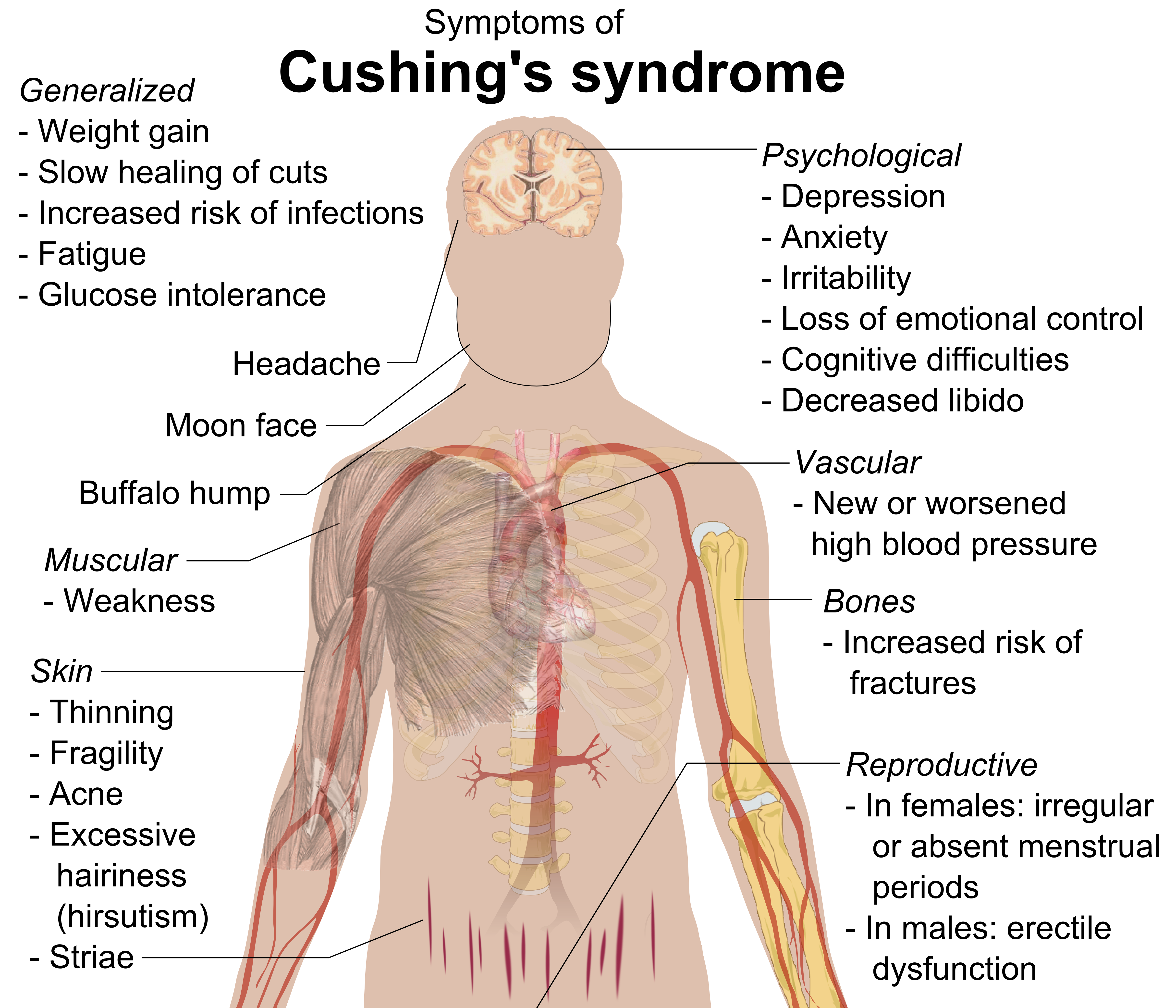
Cushing's syndrome is a collection of signs and symptoms due to prolonged exposure to glucocorticoids such as cortisol. Signs and symptoms may include high blood pressure, abdominal obesity but with thin arms and legs, reddish stretch marks, a round red face, a fat lump between the shoulders, weak muscles, weak bones, acne, and fragile skin that heals poorly. Women may have more hair and irregular menstruation. Occasionally there may be changes in mood, headaches, and a chronic feeling of tiredness. Cushing's syndrome is caused by either excessive cortisol-like medication, such as prednisone, or a tumor that either produces or results in the production of excessive cortisol by the adrenal glands. Cases due to a pituitary adenoma are known as Cushing's disease, which is the second most common cause of Cushing's syndrome after medication. A number of other tumors, often referred to as ectopic due to their placement outside the pituitary, may also cause Cushing's. Some of ... [...More Info...] [...Related Items...] OR: [Wikipedia] [Google] [Baidu] |
Retrobulbar Hemorrhage
Retrobulbar bleeding, also known as retrobulbar hemorrhage, is when bleeding occurs behind the eye. Symptoms may include pain, bruising around the eye, the eye bulging outwards, vomiting, and vision loss. Retrobulbar bleeding can occur as a result of trauma to the eye, surgery to the eye, blood thinners, or an arteriovenous malformation Arteriovenous malformation is an abnormal connection between arteries and veins, bypassing the capillary system. This vascular anomaly is widely known because of its occurrence in the central nervous system (usually cerebral AVM), but can appea .... In those with significant symptoms lateral canthotomy with cantholysis is indicated. This is recommended to be carried out within two hours. The condition is rare. References {{reflist Eye injury Gross pathology ... [...More Info...] [...Related Items...] OR: [Wikipedia] [Google] [Baidu] |
Orbital Fracture
Facial trauma, also called maxillofacial trauma, is any physical trauma to the face. Facial trauma can involve soft tissue injuries such as burns, lacerations and bruises, or fractures of the facial bones such as nasal fractures and fractures of the jaw, as well as trauma such as eye injuries. Symptoms are specific to the type of injury; for example, fractures may involve pain, swelling, loss of function, or changes in the shape of facial structures. Facial injuries have the potential to cause disfigurement and loss of function; for example, blindness or difficulty moving the jaw can result. Although it is seldom life-threatening, facial trauma can also be deadly, because it can cause severe bleeding or interference with the airway; thus a primary concern in treatment is ensuring that the airway is open and not threatened so that the patient can breathe. Depending on the type of facial injury, treatment may include bandaging and suturing of open wounds, administration of i ... [...More Info...] [...Related Items...] OR: [Wikipedia] [Google] [Baidu] |
Aortic Insufficiency
Aortic regurgitation (AR), also known as aortic insufficiency (AI), is the leaking of the aortic valve of the heart that causes blood to flow in the reverse direction during ventricular diastole, from the aorta into the left ventricle. As a consequence, the cardiac muscle is forced to work harder than normal. Signs and symptoms Symptoms of aortic regurgitation are similar to those of heart failure and include the following: * Dyspnea on exertion * Orthopnea * Paroxysmal nocturnal dyspnea * Palpitations * Angina pectoris * Cyanosis (in acute cases) Causes In terms of the cause of aortic regurgitation, is often due to the aortic root dilation ('' annuloaortic ectasia''), which is idiopathic in over 80% of cases, but otherwise may result from aging, syphilitic aortitis, osteogenesis imperfecta, aortic dissection, Behçet's disease, reactive arthritis and systemic hypertension.Chapter 1: Diseases of the Cardiovascular system > Section: Valvular Heart Disease in: Aortic root dilation ... [...More Info...] [...Related Items...] OR: [Wikipedia] [Google] [Baidu] |
Carotid-cavernous Fistula
A carotid-cavernous fistula results from an abnormal communication between the arterial and venous systems within the cavernous sinus in the skull. It is a type of arteriovenous fistula. As arterial blood under high pressure enters the cavernous sinus, the normal venous return to the cavernous sinus is impeded and this causes engorgement of the draining veins, manifesting most dramatically as a sudden engorgement and redness of the eye of the same side. Presentation CCF symptoms include bruit (a humming sound within the skull due to high blood flow through the arteriovenous fistula), progressive visual loss, and pulsatile proptosis Exophthalmos (also called exophthalmus, exophthalmia, proptosis, or exorbitism) is a bulging of the eye anteriorly out of the orbit. Exophthalmos can be either bilateral (as is often seen in Graves' disease) or unilateral (as is often seen i ... or progressive bulging of the eye due to dilatation of the veins draining the eye. Pain is the sympto ... [...More Info...] [...Related Items...] OR: [Wikipedia] [Google] [Baidu] |
Dermoid Cyst
A dermoid cyst is a teratoma of a cystic nature that contains an array of developmentally mature, solid tissues. It frequently consists of skin, hair follicles, and sweat glands, while other commonly found components include clumps of long hair, pockets of sebum, blood, fat, bone, nail, teeth, eyes, cartilage, and thyroid tissue. As dermoid cysts grow slowly and contain mature tissue, this type of cystic teratoma is nearly always benign. In those rare cases wherein the dermoid cyst is malignant, a squamous cell carcinoma usually develops in adults, while infants and children usually present with an endodermal sinus tumor.Freedberg, et al. (2003). ''Fitzpatrick's Dermatology in General Medicine''. (6th ed.). McGraw-Hill. . Location Due to its classification, a dermoid cyst can occur wherever a teratoma can occur. Vaginal and ovarian dermoid cysts Ovaries normally grow cyst-like structures called follicles each month. Once an egg is released from its follicle during ovulation, ... [...More Info...] [...Related Items...] OR: [Wikipedia] [Google] [Baidu] |
Hemangioma
A hemangioma or haemangioma is a usually benign vascular tumor derived from blood vessel cell types. The most common form, seen in infants, is an infantile hemangioma, known colloquially as a "strawberry mark", most commonly presenting on the skin at birth or in the first weeks of life. A hemangioma can occur anywhere on the body, but most commonly appears on the face, scalp, chest or back. They tend to grow for up to a year before gradually shrinking as the child gets older. A hemangioma may need to be treated if it interferes with vision or breathing or is likely to cause long-term disfigurement. In rare cases internal hemangiomas can cause or contribute to other medical problems. Most of the time they tend to disappear in 10 years. The first line treatment option is beta blockers, which are highly effective in the majority of cases. Ones that form at birth are called congenital hemangiomas while ones that form later in life are called infantile hemangiomas. Types Hema ... [...More Info...] [...Related Items...] OR: [Wikipedia] [Google] [Baidu] |
Hand–Schüller–Christian Disease
Chronic multifocal Langerhans cell histiocytosis, previously known as Hand–Schüller–Christian disease, is a type of Langerhans cell histiocytosis (LCH), which can affect multiple organs. The condition is traditionally associated with a combination of three features; bulging eyes, breakdown of bone (lytic bone lesions often in the skull), and diabetes insipidus (excessive thirst and passing urine), although around 75% of cases do not have all three features. Other features may include a fever and weight loss, and depending on the organs involved there maybe rashes, asymmetry of the face, ear infections, signs in the mouth and the appearance of advanced gum disease. Features relating to lung and liver disease may occur. It is due to a genetic mutation in the MAPKinase pathway that occurs during early development. The diagnosis may be suspected based on symptoms and MRI and confirmed by tissue biopsy. Blood tests may show anaemia, and less commonly a low white blood cell count ... [...More Info...] [...Related Items...] OR: [Wikipedia] [Google] [Baidu] |
Nasopharyngeal Angiofibroma
Nasopharyngeal angiofibroma is an angiofibroma also known as juvenile nasal angiofibroma, fibromatous hamartoma, and angiofibromatous hamartoma of the nasal cavity. It is a histologically benign but locally aggressive vascular tumor of the nasopharynx that arises from the superior margin of the sphenopalatine foramen and grows in the back of the nasal cavity. It most commonly affects adolescent males (because it is a hormone-sensitive tumor). Though it is a benign tumor, it is locally invasive and can invade the nose, cheek, orbit (frog face deformity), or brain. Patients with nasopharyngeal angiofibroma usually present with one-sided nasal obstruction with profuse epistaxis. Signs and symptoms * Frequent chronic epistaxis or blood-tinged nasal discharge * Nasal obstruction and rhinorrhea * Facial dysmorphism (when locally invasive) * Conductive hearing loss from eustachian-tube obstruction * Diplopia, which occurs secondary to erosion into superior orbital fissure and due to third ... [...More Info...] [...Related Items...] OR: [Wikipedia] [Google] [Baidu] |
Meningioma
Meningioma, also known as meningeal tumor, is typically a slow-growing tumor that forms from the meninges, the membranous layers surrounding the brain and spinal cord. Symptoms depend on the location and occur as a result of the tumor pressing on nearby tissue. Many cases never produce symptoms. Occasionally seizures, dementia, trouble talking, vision problems, one sided weakness, or loss of bladder control may occur. Risk factors include exposure to ionizing radiation such as during radiation therapy, a family history of the condition, and neurofibromatosis type 2. As of 2014 they do not appear to be related to cell phone use. They appear to be able to form from a number of different types of cells including arachnoid cells. Diagnosis is typically by medical imaging. If there are no symptoms, periodic observation may be all that is required. Most cases that result in symptoms can be cured by surgery. Following complete removal fewer than 20% recur. If surgery is not possibl ... [...More Info...] [...Related Items...] OR: [Wikipedia] [Google] [Baidu] |


