|
Entoloma Austroprunicolor
''Entoloma austroprunicolor'' is a species of agaric fungus in the family Entolomataceae. Described as new to science in 2007, it is found in Tasmania, where it fruits on the ground of wet sclerophyll forests in late spring to early winter (usually between January and March). The fruit bodies (mushrooms) have reddish-purple caps measuring up to in diameter supported by whitish stipes measuring long by thick. On the cap underside, the crowded gills are initially white before turning pink as the spores mature. Taxonomy The species was first formally described in 2007 by Australian mycologist Genevieve Gates and Dutch mycologist Machiel Noordeloos, from collections made in Tasmania, Australia. The specific epithet ''austroprunicolor '' is derived from the Latin prefix ''austro-'', meaning "southern", and the Latin word ''prunicolor'', meaning "plum-coloured". The type collection was made in January 2002 at Kermandie Falls, near Geeveston in southern Tasmania. The species was ... [...More Info...] [...Related Items...] OR: [Wikipedia] [Google] [Baidu] |
Wielangta Forest
The Wielangta forest is in south-east Tasmania, Australia. It is notable for its role in a 2006 court case that called into question the effectiveness of Australia's cooperative Commonwealth-State forest management regime known as Regional Forest Agreements. Name Wielangta is an Aboriginal name of high trees. Environment The Wielangta forest is part of remnant glacial refugia forest and contains blue gum eucalypt forest and pockets of cool temperate rainforest. The forest is a key habitat of rare and threatened species, including the Tasmanian wedge-tailed eagle, swift parrot, Wielangta stag beetle, spotted-tail quoll and eastern barred bandicoot. A rare orchid (''Genoplesium nudum'') has also been discovered in the forest. The forest forms part of the South-east Tasmania Important Bird Area, identified as such by BirdLife International because of its importance in the conservation of a range of woodland birds. Logging controversy The forest is under the control of For ... [...More Info...] [...Related Items...] OR: [Wikipedia] [Google] [Baidu] |
Entoloma
''Entoloma'' is a large genus of terrestrial pink-gilled mushrooms, with about 1,000 species. Most have a drab appearance, pink gills which are attached to the stem, a smooth thick cap, and angular spores. Many entolomas are saprobic but some are mycorrhizal. The best-known member of the genus is the livid agaric (''Entoloma sinuatum''), responsible for a number of poisonings over the years in Europe and North America, and '' Entoloma rhodopolium'' in Japan. Some southern hemisphere species such as ''Entoloma rodwayi'' and '' Entoloma viridomarginatum'' from Australia, and ''Entoloma hochstetteri'' from New Zealand, are very colourful, with caps of unusual shades of green and blue-green. Most entolomas are dull shades of olive, brown, or grey. Etymology The part '' ἐντός'' means "within, inside". The part "loma" is a noun-forming element derived from Greek '' λῶμ(α)'', "fringe, hem" and used in the botanical taxonomy for naming plants distinguished by having a fri ... [...More Info...] [...Related Items...] OR: [Wikipedia] [Google] [Baidu] |
Spore Print
300px, Making a spore print of the mushroom ''Volvariella volvacea'' shown in composite: (photo lower half) mushroom cap laid on white and dark paper; (photo upper half) cap removed after 24 hours showing pinkish-tan spore print. A 3.5-centimeter glass slide placed in middle allows for examination of spore characteristics under a microscope. image:spore Print ID.gif, 300px, A printable chart to make a spore print and start identification The spore print is the powdery deposit obtained by allowing spores of a fungal sporocarp (fungi), fruit body to fall onto a surface underneath. It is an important diagnostic character in most handbooks for identifying mushrooms. It shows the colour of the mushroom spores if viewed en masse. Method A spore print is made by placing the spore-producing surface flat on a sheet of dark and white paper or on a sheet of clear, stiff plastic, which facilitates moving the spore print to a darker or lighter surface for improved contrast; for example, it ... [...More Info...] [...Related Items...] OR: [Wikipedia] [Google] [Baidu] |
Edible Mushroom
Edible mushrooms are the fleshy and edible fruit bodies of several species of macrofungi (fungi which bear fruiting structures that are large enough to be seen with the naked eye). They can appear either below ground (hypogeous) or above ground (epigeous) where they may be picked by hand. Edibility may be defined by criteria that include absence of poisonous effects on humans and desirable taste and aroma. Edible mushrooms are consumed for their nutritional and culinary value. Mushrooms, especially dried shiitake, are sources of umami flavor. Edible mushrooms include many fungal species that are either harvested wild or cultivated. Easily cultivated and common wild mushrooms are often available in markets, and those that are more difficult to obtain (such as the prized truffle, matsutake, and morel) may be collected on a smaller scale by private gatherers. Some preparations may render certain poisonous mushrooms fit for consumption. Before assuming that any wild mushroom is ... [...More Info...] [...Related Items...] OR: [Wikipedia] [Google] [Baidu] |
Trama (mycology)
In mycology, the term trama is used in two ways. In the broad sense, it is the inner, fleshy portion of a mushroom's basidiocarp, or fruit body. It is distinct from the outer layer of tissue, known as the pileipellis or cuticle, and from the spore-bearing tissue layer known as the hymenium. In essence, the trama is the tissue that is commonly referred to as the "flesh" of mushrooms and similar fungi.Largent D, Johnson D, Watling R. 1977. ''How to Identify Mushrooms to Genus III: Microscopic Features''. Arcata, CA: Mad River Press. . pp. 60–70. The second use is more specific, and refers to the "hymenophoral trama" that supports the hymenium. It is similarly interior, connective tissue, but it is more specifically the central layer of hyphae running from the underside of the mushroom cap to the lamella or gill, upon which the hymenium rests. Various types have been classified by their structure, including trametoid, cantharelloid, boletoid, and agaricoid, with agaricoid the ... [...More Info...] [...Related Items...] OR: [Wikipedia] [Google] [Baidu] |
Adnation
Adnation in Angiosperms is the fusion of two or more whorls of a flower, e.g. stamens to petals". This is in contrast to connation, the fusion among a single whorl A whorl ( or ) is an individual circle, oval, volution or equivalent in a whorled pattern, which consists of a spiral or multiple concentric objects (including circles, ovals and arcs). Whorls in nature File:Photograph and axial plane floral .... References Plant anatomy {{botany-stub ... [...More Info...] [...Related Items...] OR: [Wikipedia] [Google] [Baidu] |
Lamella (mycology)
In mycology, a lamella, or gill, is a papery hymenophore rib under the cap of some mushroom species, most often agarics. The gills are used by the mushrooms as a means of spore dispersal, and are important for species identification. The attachment of the gills to the stem is classified based on the shape of the gills when viewed from the side, while color, crowding and the shape of individual gills can also be important features. Additionally, gills can have distinctive microscopic or macroscopic features. For instance, ''Lactarius'' species typically seep latex from their gills. It was originally believed that all gilled fungi were Agaricales, but as fungi were studied in more detail, some gilled species were demonstrated not to be. It is now clear that this is a case of convergent evolution (i.e. gill-like structures evolved separately) rather than being an anatomic feature that evolved only once. The apparent reason that various basidiomycetes have evolved gills is that ... [...More Info...] [...Related Items...] OR: [Wikipedia] [Google] [Baidu] |
Umbo (mycology)
'' Cantharellula umbonata'' has an umbo. The cap of '' Psilocybe makarorae'' is acutely papillate.">papillate.html" ;"title="Psilocybe makarorae'' is acutely papillate">Psilocybe makarorae'' is acutely papillate. An umbo is a raised area in the center of a mushroom cap. pileus (mycology), Caps that possess this feature are called ''umbonate''. Umbos that are sharply pointed are called ''acute'', while those that are more rounded are ''broadly umbonate''. If the umbo is elongated, it is ''cuspidate'', and if the umbo is sharply delineated but not elongated (somewhat resembling the shape of a human areola The human areola (''areola mammae'', or ) is the pigmented area on the breast around the nipple. Areola, more generally, is a small circular area on the body with a different histology from the surrounding tissue, or other small circular ar ...), it is called '' mammilate'' or ''papillate''. References {{reflist Fungal morphology and anatomy Mycology ... [...More Info...] [...Related Items...] OR: [Wikipedia] [Google] [Baidu] |
Biological Pigment
Biological pigments, also known simply as pigments or biochromes, are substances produced by living organisms that have a color resulting from selective color absorption. Biological pigments include plant pigments and flower pigments. Many biological structures, such as skin, eyes, feathers, fur and hair contain pigments such as melanin in specialized cells called chromatophores. In some species, pigments accrue over very long periods during an individual's lifespan. Pigment color differs from structural color in that it is the same for all viewing angles, whereas structural color is the result of selective reflection or iridescence, usually because of multilayer structures. For example, butterfly wings typically contain structural color, although many butterflies have cells that contain pigment as well. Biological pigments See conjugated systems for electron bond chemistry that causes these molecules to have pigment. * Heme/porphyrin-based: chlorophyll, bilirubin, hemocy ... [...More Info...] [...Related Items...] OR: [Wikipedia] [Google] [Baidu] |
Granule (cell Biology)
In cell biology, a granule is a small particle. It can be any structure barely visible by light microscopy. The term is most often used to describe a secretory vesicle. In leukocytes A group of leukocytes, called granulocytes, contain granules and play an important role in the immune system. The granules of certain cells, such as natural killer cells, contain components which can lead to the lysis of neighboring cells. The granules of leukocytes are classified as azurophilic granules or specific granules. Leukocyte granules are released in response to immunological stimuli during a process known as degranulation. In platelets The granules of platelets are classified as dense granules and alpha granules. α-Granules are unique to platelets and are the most abundant of the platelet granules, numbering 50–80 per platelet 2. These granules measure 200–500 nm in diameter and account for about 10% of platelet volume. They contain mainly proteins, both membrane-associated ... [...More Info...] [...Related Items...] OR: [Wikipedia] [Google] [Baidu] |
Hypha
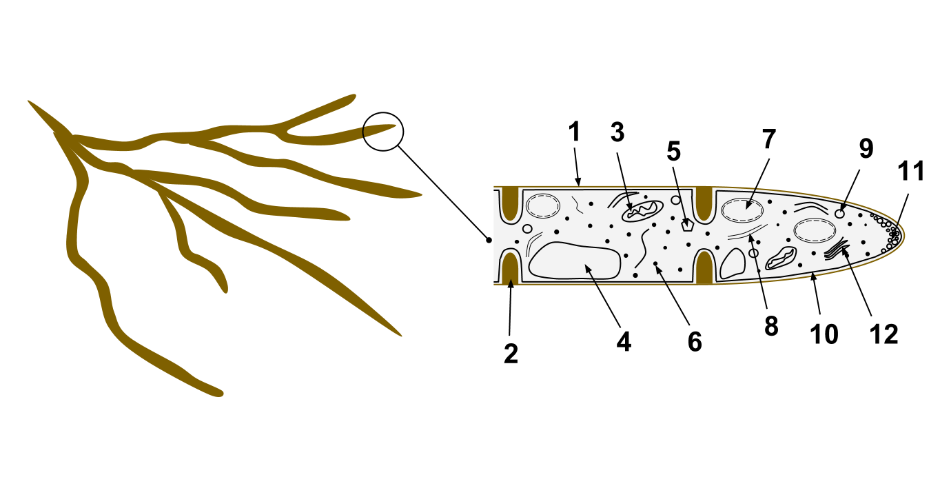
A hypha (; ) is a long, branching, filamentous structure of a fungus, oomycete, or actinobacterium. In most fungi, hyphae are the main mode of vegetative growth, and are collectively called a mycelium. Structure A hypha consists of one or more cells surrounded by a tubular cell wall. In most fungi, hyphae are divided into cells by internal cross-walls called "septa" (singular septum). Septa are usually perforated by pores large enough for ribosomes, mitochondria, and sometimes nuclei to flow between cells. The major structural polymer in fungal cell walls is typically chitin, in contrast to plants and oomycetes that have cellulosic cell walls. Some fungi have aseptate hyphae, meaning their hyphae are not partitioned by septa. Hyphae have an average diameter of 4–6 µm. Growth Hyphae grow at their tips. During tip growth, cell walls are extended by the external assembly and polymerization of cell wall components, and the internal production of new cell membrane. The S ... [...More Info...] [...Related Items...] OR: [Wikipedia] [Google] [Baidu] |
Clamp Connection
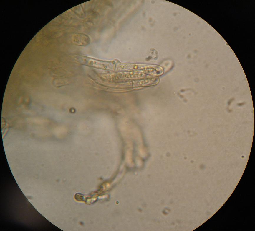
A clamp connection is a hook-like structure formed by growing hyphal cells of certain fungi. It is a characteristic feature of Basidiomycetes fungi. It is created to ensure that each cell, or segment of hypha separated by septa (cross walls), receives a set of differing nuclei, which are obtained through mating of hyphae of differing sexual types. It is used to maintain genetic variation within the hypha much like the mechanisms found in crozier (hook) during sexual reproduction. Formation Clamp connections are formed by the terminal hypha during elongation. Before the clamp connection is formed this terminal segment contains two nuclei. Once the terminal segment is long enough it begins to form the clamp connection. At the same time, each nucleus undergoes mitotic division to produce two daughter nuclei. As the clamp continues to develop it uptakes one of the daughter (green circle) nuclei and separates it from its sister nucleus. While this is occurring the remaining nuclei ... [...More Info...] [...Related Items...] OR: [Wikipedia] [Google] [Baidu] |
_Noordeloos_195666.jpg)


