|
Emotional Lateralization
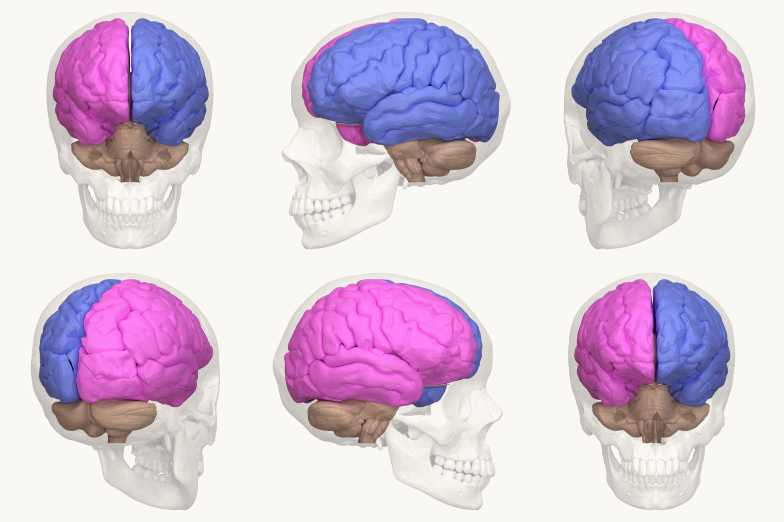
Emotional lateralization is the asymmetrical representation of emotional control and processing in the brain. There is evidence for the Lateralization of brain function, lateralization of other brain functions as well. Emotions are complex and involve a variety of physical and cognitive responses, many of which are not well understood. The general purpose of emotions is to produce a specific response to a stimulus. Feelings are the conscious perception of emotions, and when an emotion occurs frequently or continuously this is called a mood. A variety of scientific studies have found lateralization of emotions. FMRI and lesion studies have shown asymmetrical activation of brain regions while thinking of emotions, responding to extreme emotional stimuli, and viewing emotional situations. Processing and production of facial expressions also appear to be asymmetric in nature. Many theories of lateralization have been proposed and some of those specific to emotions. Please keep in mind t ... [...More Info...] [...Related Items...] OR: [Wikipedia] [Google] [Baidu] |
Lateralization Of Brain Function
The lateralization of brain function is the tendency for some neural functions or cognitive processes to be specialized to one side of the brain or the other. The median longitudinal fissure separates the human brain into two distinct cerebral hemispheres, connected by the corpus callosum. Although the macrostructure of the two hemispheres appears to be almost identical, different composition of neuronal networks allows for specialized function that is different in each hemisphere. Lateralization of brain structures is based on general trends expressed in healthy patients; however, there are numerous counterexamples to each generalization. Each human's brain develops differently, leading to unique lateralization in individuals. This is different from specialization, as lateralization refers only to the function of one structure divided between two hemispheres. Specialization is much easier to observe as a trend, since it has a stronger anthropological history. The best examp ... [...More Info...] [...Related Items...] OR: [Wikipedia] [Google] [Baidu] |
Neuroanatomical Basis For Emotional Lateralization
Neuroanatomy is the study of the structure and organization of the nervous system. In contrast to animals with radial symmetry, whose nervous system consists of a distributed network of cells, animals with bilateral symmetry have segregated, defined nervous systems. Their neuroanatomy is therefore better understood. In vertebrates, the nervous system is segregated into the internal structure of the brain and spinal cord (together called the central nervous system, or CNS) and the routes of the nerves that connect to the rest of the body (known as the peripheral nervous system, or PNS). The delineation of distinct structures and regions of the nervous system has been critical in investigating how it works. For example, much of what neuroscientists have learned comes from observing how damage or "lesions" to specific brain areas affects behavior or other neural functions. For information about the composition of non-human animal nervous systems, see nervous system. For information ab ... [...More Info...] [...Related Items...] OR: [Wikipedia] [Google] [Baidu] |
Parahippocampal Gyrus
The parahippocampal gyrus (or hippocampal gyrus') is a grey matter cortical region of the brain that surrounds the hippocampus and is part of the limbic system. The region plays an important role in memory encoding and retrieval. It has been involved in some cases of hippocampal sclerosis. Asymmetry has been observed in schizophrenia. Structure The anterior part of the gyrus includes the perirhinal and entorhinal cortices. The term parahippocampal cortex is used to refer to an area that encompasses both the posterior parahippocampal gyrus and the medial portion of the fusiform gyrus. Function Scene recognition The parahippocampal place area (PPA) is a sub-region of the parahippocampal cortex that lies medially in the inferior temporo-occipital cortex. PPA plays an important role in the encoding and recognition of environmental scenes (rather than faces). fMRI studies indicate that this region of the brain becomes highly active when human subjects view topographical scene sti ... [...More Info...] [...Related Items...] OR: [Wikipedia] [Google] [Baidu] |
Chimeric Faces
Chimera, Chimaera, or Chimaira (Greek for " she-goat") originally referred to: * Chimera (mythology), a fire-breathing monster of Ancient Lycia said to combine parts from multiple animals * Mount Chimaera, a fire-spewing region of Lycia or Cilicia typically considered the inspiration for the myth As such, chimera, Chimera, chimère, Chimaira, etc. may also refer to: Biology * Chimaera, any of various cartilaginous fishes of the order Chimaeriformes ** ''Chimaera'' (genus), the eponymous genus of the order Chimaeriformes * Chimera (EST), a single cDNA sequence originating from two transcripts * Chimera (genetics), a single animal or plant with genetically distinct cells from two different zygotes * Chimera (virus), a virus containing genetic material from other organisms * Chimera, or fusion protein, a hybrid protein made by the splicing of two genes * Chimera (paleontology), a fossil which was reconstructed with parts from different animals * Chimera Project, a Soviet biological ... [...More Info...] [...Related Items...] OR: [Wikipedia] [Google] [Baidu] |
Functional Imaging
Functional imaging (or physiological imaging) is a medical imaging technique of detecting or measuring changes in metabolism, blood flow, regional chemical composition, and absorption. As opposed to structural imaging, functional imaging centers on revealing physiological activities within a certain tissue or organ by employing medical image modalities that very often use tracers or probes to reflect spatial distribution of them within the body. These tracers are often analogous to some chemical compounds, like glucose, within the body. To achieve this, isotopes are used because they have similar chemical and biological characteristics. By appropriate proportionality, the nuclear medicine physicians can determine the real intensity of certain substance within the body to evaluate the risk or danger of developing some diseases. Modalities * Positron emission tomography (PET) ** Fludeoxyglucose for Glucose metabolism ** O-15 as a flow tracer * Single-photon emission computed t ... [...More Info...] [...Related Items...] OR: [Wikipedia] [Google] [Baidu] |
Frontal Lobes
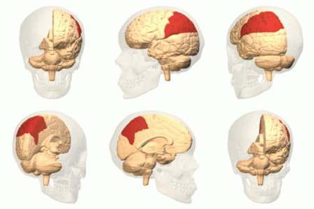
The frontal lobe is the largest of the four major lobes of the brain in mammals, and is located at the front of each cerebral hemisphere (in front of the parietal lobe and the temporal lobe). It is parted from the parietal lobe by a groove between tissues called the central sulcus and from the temporal lobe by a deeper groove called the lateral sulcus (Sylvian fissure). The most anterior rounded part of the frontal lobe (though not well-defined) is known as the frontal pole, one of the three poles of the cerebrum. The frontal lobe is covered by the frontal cortex. The frontal cortex includes the premotor cortex, and the primary motor cortex – parts of the motor cortex. The front part of the frontal cortex is covered by the prefrontal cortex. There are four principal gyri in the frontal lobe. The precentral gyrus is directly anterior to the central sulcus, running parallel to it and contains the primary motor cortex, which controls voluntary movements of specific body parts. T ... [...More Info...] [...Related Items...] OR: [Wikipedia] [Google] [Baidu] |
Parietal Lobe
The parietal lobe is one of the four major lobes of the cerebral cortex in the brain of mammals. The parietal lobe is positioned above the temporal lobe and behind the frontal lobe and central sulcus. The parietal lobe integrates sensory information among various modalities, including spatial sense and navigation (proprioception), the main sensory receptive area for the sense of touch in the somatosensory cortex which is just posterior to the central sulcus in the postcentral gyrus, and the dorsal stream of the visual system. The major sensory inputs from the skin (touch, temperature, and pain receptors), relay through the thalamus to the parietal lobe. Several areas of the parietal lobe are important in language processing. The somatosensory cortex can be illustrated as a distorted figure – the cortical homunculus (Latin: "little man") in which the body parts are rendered according to how much of the somatosensory cortex is devoted to them. The superior parietal lobule and in ... [...More Info...] [...Related Items...] OR: [Wikipedia] [Google] [Baidu] |
Face Perception
Facial perception is an individual's understanding and interpretation of the face. Here, perception implies the presence of consciousness and hence excludes automated facial recognition systems. Although facial recognition is found in other species, this article focuses on facial perception in humans. The perception of facial features is an important part of social cognition. Information gathered from the face helps people understand each other's identity, what they are thinking and feeling, anticipate their actions, recognize their emotions, build connections, and communicate through body language. Developing facial recognition is a necessary building block for complex societal constructs. Being able to perceive identity, mood, age, sex, and race lets people mold the way we interact with one another, and understand our immediate surroundings. Though facial perception is mainly considered to stem from visual intake, studies have shown that even people born blind can learn face ... [...More Info...] [...Related Items...] OR: [Wikipedia] [Google] [Baidu] |
Language Processing In The Brain
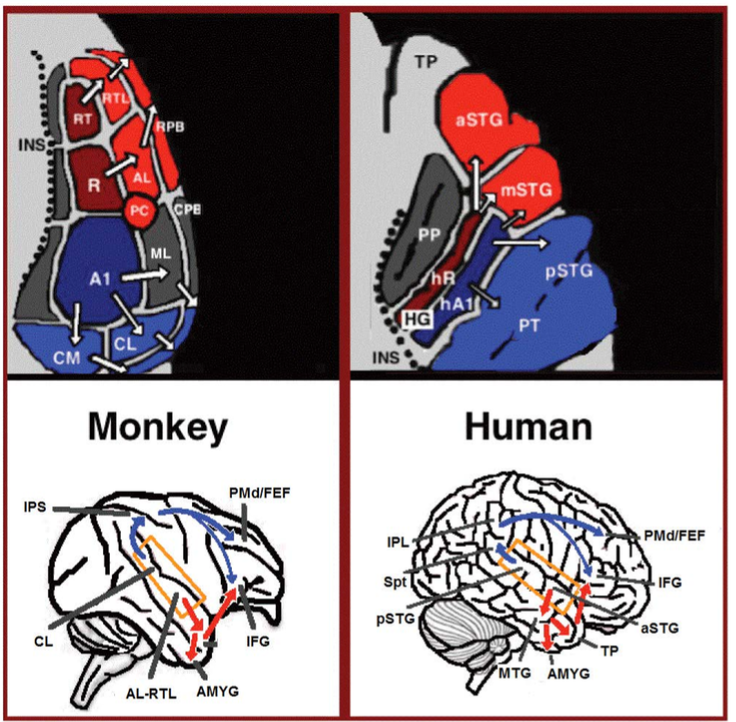
Language processing refers to the way humans use words to communicate ideas and feelings, and how such communications are processed and understood. Language processing is considered to be a uniquely human ability that is not produced with the same grammatical understanding or systematicity in even human's closest primate relatives. Throughout the 20th century the dominant model for language processing in the brain was the Geschwind-Lichteim-Wernicke model, which is based primarily on the analysis of brain damaged patients. However, due to improvements in intra-cortical electrophysiological recordings of monkey and human brains, as well non-invasive techniques such as fMRI, PET, MEG and EEG, a dual auditory pathway has been revealed and a two-streams model has been developed. In accordance with this model, there are two pathways that connect the auditory cortex to the frontal lobe, each pathway accounting for different linguistic roles. The auditory ventral stream pathway is re ... [...More Info...] [...Related Items...] OR: [Wikipedia] [Google] [Baidu] |
Temporal Cortex
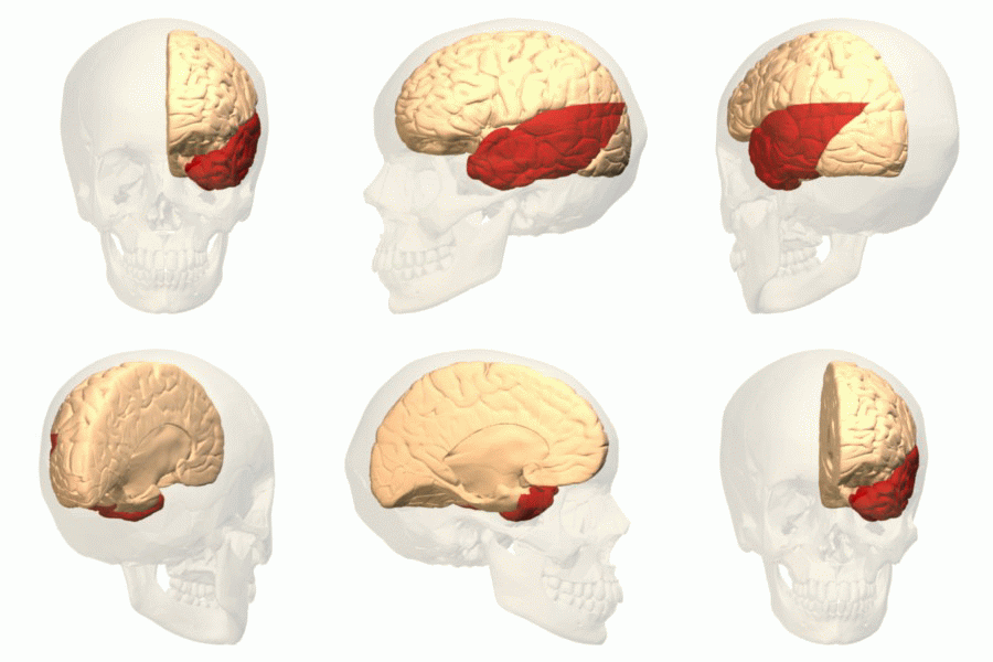
The temporal lobe is one of the four major lobes of the cerebral cortex in the brain of mammals. The temporal lobe is located beneath the lateral fissure on both cerebral hemispheres of the mammalian brain. The temporal lobe is involved in processing sensory input into derived meanings for the appropriate retention of visual memory, language comprehension, and emotion association. ''Temporal'' refers to the head's temples. Structure The temporal lobe consists of structures that are vital for declarative or long-term memory. Declarative (denotative) or explicit memory is conscious memory divided into semantic memory (facts) and episodic memory (events). Medial temporal lobe structures that are critical for long-term memory include the hippocampus, along with the surrounding hippocampal region consisting of the perirhinal, parahippocampal, and entorhinal neocortical regions. The hippocampus is critical for memory formation, and the surrounding medial temporal cortex is curre ... [...More Info...] [...Related Items...] OR: [Wikipedia] [Google] [Baidu] |
Insular Cortex
The insular cortex (also insula and insular lobe) is a portion of the cerebral cortex folded deep within the lateral sulcus (the fissure separating the temporal lobe from the parietal and frontal lobes) within each hemisphere of the mammalian brain. The insulae are believed to be involved in consciousness and play a role in diverse functions usually linked to emotion or the regulation of the body's homeostasis. These functions include compassion, empathy, taste, perception, motor control, self-awareness, cognitive functioning, interpersonal experience, and awareness of homeostatic emotions such as hunger, pain and fatigue. In relation to these, it is involved in psychopathology. The insular cortex is divided into two parts: the anterior insula and the posterior insula in which more than a dozen field areas have been identified. The cortical area overlying the insula toward the lateral surface of the brain is the operculum (meaning ''lid''). The opercula are formed from parts o ... [...More Info...] [...Related Items...] OR: [Wikipedia] [Google] [Baidu] |
Anterior Cingulate Cortex
In the human brain, the anterior cingulate cortex (ACC) is the frontal part of the cingulate cortex that resembles a "collar" surrounding the frontal part of the corpus callosum. It consists of Brodmann areas 24, 32, and 33. It is involved in certain higher-level functions, such as attention allocation, reward anticipation, decision-making, ethics and morality, impulse control (e.g. performance monitoring and error detection), and emotion. Anatomy The anterior cingulate cortex can be divided anatomically based on cognitive (dorsal), and emotional (ventral) components. The dorsal part of the ACC is connected with the prefrontal cortex and parietal cortex, as well as the motor system and the frontal eye fields, making it a central station for processing top-down and bottom-up stimuli and assigning appropriate control to other areas in the brain. By contrast, the ventral part of the ACC is connected with the amygdala, nucleus accumbens, hypothalamus, hippocampus, and ant ... [...More Info...] [...Related Items...] OR: [Wikipedia] [Google] [Baidu] |


