|
Functional Imaging
Functional imaging (or physiological imaging) is a medical imaging technique of detecting or measuring changes in metabolism, blood flow, regional chemical composition, and absorption. As opposed to structural imaging, functional imaging centers on revealing physiological activities within a certain tissue or organ by employing medical image modalities that very often use radioactive tracers, tracers or probes to reflect spatial distribution of them within the body. These tracers are often structural analog, analogous to some chemical compounds, like glucose, within the body. To achieve this, isotopes are used because they have similar chemical and biological characteristics. By appropriate proportionality, the nuclear medicine physicians can determine the real intensity of certain substance within the body to evaluate the risk or danger of developing some diseases. Modalities * Positron emission tomography (PET) ** Fludeoxyglucose for Glucose metabolism ** O-15 as a flow tracer ... [...More Info...] [...Related Items...] OR: [Wikipedia] [Google] [Baidu] |
Medical Imaging
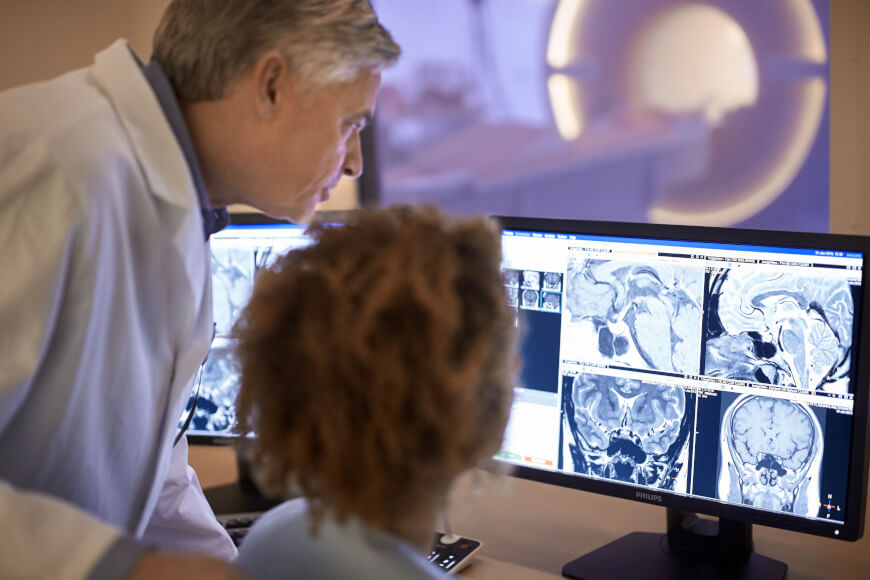
Medical imaging is the technique and process of imaging the interior of a body for clinical analysis and medical intervention, as well as visual representation of the function of some organs or tissues ( physiology). Medical imaging seeks to reveal internal structures hidden by the skin and bones, as well as to diagnose and treat disease. Medical imaging also establishes a database of normal anatomy and physiology to make it possible to identify abnormalities. Although imaging of removed organs and tissues can be performed for medical reasons, such procedures are usually considered part of pathology instead of medical imaging. Measurement and recording techniques that are not primarily designed to produce images, such as electroencephalography (EEG), magnetoencephalography (MEG), electrocardiography (ECG), and others, represent other technologies that produce data susceptible to representation as a parameter graph versus time or maps that contain data about the measurement ... [...More Info...] [...Related Items...] OR: [Wikipedia] [Google] [Baidu] |
Arterial Spin Labeling
Arterial spin labeling (ASL), also known as arterial spin tagging, is a magnetic resonance imaging technique used to quantify cerebral blood perfusion by labelling blood water as it flows throughout the brain. ASL specifically refers to magnetic labeling of arterial blood below or in the imaging slab, without the need of gadolinium contrast. A number of ASL schemes are possible, the simplest being flow alternating inversion recovery (FAIR) which requires two acquisitions of identical parameters with the exception of the out-of-slice saturation; the difference in the two images is theoretically only from inflowing spins, and may be considered a 'perfusion map'. The ASL technique was developed by Alan P. Koretsky, Donald S. Williams, John A. Detre and John S. Leigh, Jr in 1992. Physics Arterial spin labeling utilizes the water molecules circulating with the brain, and using a radiofrequency pulse, tracks the blood water as it circulates throughout the brain. After a period of time in ... [...More Info...] [...Related Items...] OR: [Wikipedia] [Google] [Baidu] |
Radiology
Radiology ( ) is the medical discipline that uses medical imaging to diagnose diseases and guide their treatment, within the bodies of humans and other animals. It began with radiography (which is why its name has a root referring to radiation), but today it includes all imaging modalities, including those that use no electromagnetic radiation (such as ultrasonography and magnetic resonance imaging), as well as others that do, such as computed tomography (CT), fluoroscopy, and nuclear medicine including positron emission tomography (PET). Interventional radiology is the performance of usually minimally invasive medical procedures with the guidance of imaging technologies such as those mentioned above. The modern practice of radiology involves several different healthcare professions working as a team. The radiologist is a medical doctor who has completed the appropriate post-graduate training and interprets medical images, communicates these findings to other physicians b ... [...More Info...] [...Related Items...] OR: [Wikipedia] [Google] [Baidu] |
PET-CT
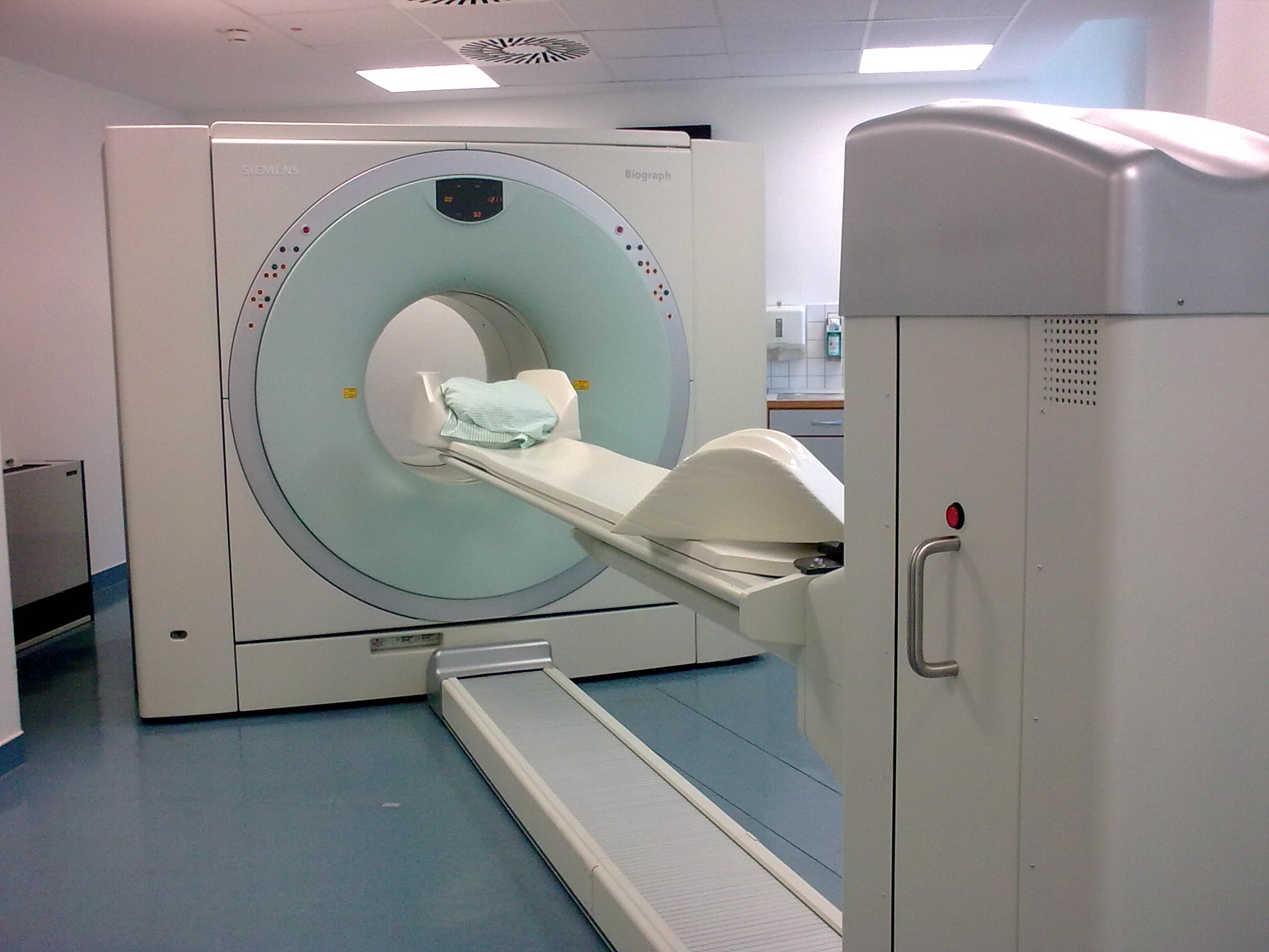
Positron emission tomography–computed tomography (better known as PET-CT or PET/CT) is a nuclear medicine technique which combines, in a single gantry, a positron emission tomography (PET) scanner and an x-ray computed tomography (CT) scanner, to acquire sequential images from both devices in the same session, which are combined into a single superposed ( co-registered) image. Thus, functional imaging obtained by PET, which depicts the spatial distribution of metabolic or biochemical activity in the body can be more precisely aligned or correlated with anatomic imaging obtained by CT scanning. Two- and three-dimensional image reconstruction may be rendered as a function of a common software and control system. PET-CT has revolutionized medical diagnosis in many fields, by adding precision of anatomic localization to functional imaging, which was previously lacking from pure PET imaging. For example, many diagnostic imaging procedures in oncology, surgical planning, radi ... [...More Info...] [...Related Items...] OR: [Wikipedia] [Google] [Baidu] |
Medical Imaging
Medical imaging is the technique and process of imaging the interior of a body for clinical analysis and medical intervention, as well as visual representation of the function of some organs or tissues ( physiology). Medical imaging seeks to reveal internal structures hidden by the skin and bones, as well as to diagnose and treat disease. Medical imaging also establishes a database of normal anatomy and physiology to make it possible to identify abnormalities. Although imaging of removed organs and tissues can be performed for medical reasons, such procedures are usually considered part of pathology instead of medical imaging. Measurement and recording techniques that are not primarily designed to produce images, such as electroencephalography (EEG), magnetoencephalography (MEG), electrocardiography (ECG), and others, represent other technologies that produce data susceptible to representation as a parameter graph versus time or maps that contain data about the measurement ... [...More Info...] [...Related Items...] OR: [Wikipedia] [Google] [Baidu] |
Biomedical Engineering
Biomedical engineering (BME) or medical engineering is the application of engineering principles and design concepts to medicine and biology for healthcare purposes (e.g., diagnostic or therapeutic). BME is also traditionally logical sciences to advance health care treatment, including diagnosis, monitoring, and therapy. Also included under the scope of a biomedical engineer is the management of current medical equipment in hospitals while adhering to relevant industry standards. This involves procurement, routine testing, preventive maintenance, and making equipment recommendations, a role also known as a Biomedical Equipment Technician (BMET) or as clinical engineering. Biomedical engineering has recently emerged as its own study, as compared to many other engineering fields. Such an evolution is common as a new field transition from being an interdisciplinary specialization among already-established fields to being considered a field in itself. Much of the work in biomedi ... [...More Info...] [...Related Items...] OR: [Wikipedia] [Google] [Baidu] |
Near-infrared Spectroscopy
Near-infrared spectroscopy (NIRS) is a spectroscopic method that uses the near-infrared region of the electromagnetic spectrum (from 780 nm to 2500 nm). Typical applications include medical and physiological diagnostics and research including blood sugar, pulse oximetry, functional neuroimaging, sports medicine, elite sports training, ergonomics, rehabilitation, neonatal research, brain computer interface, urology (bladder contraction), and neurology (neurovascular coupling). There are also applications in other areas as well such as pharmaceutical, food and agrochemical quality control, atmospheric chemistry, combustion research and astronomy. Theory Near-infrared spectroscopy is based on molecular overtone and combination vibrations. Such transitions are forbidden by the selection rules of quantum mechanics. As a result, the molar absorptivity in the near-IR region is typically quite small. (NIR absorption bands are typically 10–100 times weaker than the ... [...More Info...] [...Related Items...] OR: [Wikipedia] [Google] [Baidu] |
Optical Imaging
Medical optical imaging is the use of light as an investigational imaging technique for medical applications. Examples include optical microscopy, spectroscopy, endoscopy, scanning laser ophthalmoscopy, laser Doppler imaging, and optical coherence tomography. Because light is an electromagnetic wave, similar phenomena occur in X-rays, microwaves, and radio waves. Optical imaging systems may be divided into diffusive and ballistic imaging systems. A model for photon migration in turbid biological media has been developed by Bonner et al. Such a model can be applied for interpretation data obtained from laser Doppler blood-flow monitors and for designing protocols for therapeutic excitation of tissue chromophores. Diffusive optical imaging Diffuse optical imaging (DOI) is a method of imaging using near-infrared spectroscopy (NIRS) or fluorescence-based methods. When used to create 3D volumetric models of the imaged material DOI is referred to as diffuse optical tomography, whereas ... [...More Info...] [...Related Items...] OR: [Wikipedia] [Google] [Baidu] |
Magnetic Particle Imaging
Magnetic particle imaging (MPI) is an emerging non-invasive tomographic technique that directly detects superparamagnetic nanoparticle tracers. The technology has potential applications in diagnostic imaging and material science. Currently, it is used in medical research to measure the 3-D location and concentration of nanoparticles. Imaging does not use ionizing radiation and can produce a signal at any depth within the body. MPI was first conceived in 2001 by scientists working at the Royal Philips Research lab in Hamburg. The first system was established and reported in 2005. Since then, the technology has been advanced by academic researchers at several universities around the world. The first commercial MPI scanners have recently become available froMagnetic Insightan The hardware used for MPI is very different from MRI. MPI systems use changing magnetic fields to generate a signal from superparamagnetic iron oxide (SPIO) nanoparticles. These fields are specificall ... [...More Info...] [...Related Items...] OR: [Wikipedia] [Google] [Baidu] |
Photoacoustic Imaging In Biomedicine
Photoacoustic imaging or optoacoustic imaging is a biomedical imaging modality based on the photoacoustic effect. Non-ionizing laser pulses are delivered into biological tissues and part of the energy will be absorbed and converted into heat, leading to transient thermoelastic expansion and thus wideband (i.e. MHz) ultrasonic emission. The generated ultrasonic waves are detected by ultrasonic transducers and then analyzed to produce images. It is known that optical absorption is closely associated with physiological properties, such as hemoglobin concentration and oxygen saturation. As a result, the magnitude of the ultrasonic emission (i.e. photoacoustic signal), which is proportional to the local energy deposition, reveals physiologically specific optical absorption contrast. 2D or 3D images of the targeted areas can then be formed. Biomedical imaging The optical absorption in biological tissues can be due to endogenous molecules such as hemoglobin or melanin, or exogenously ... [...More Info...] [...Related Items...] OR: [Wikipedia] [Google] [Baidu] |
Hyperpolarized Carbon-13 MRI
Hyperpolarized carbon-13 MRI is a functional medical imaging technique for probing perfusion and metabolism using injected substrates. It is enabled by techniques for hyperpolarization of carbon-13-containing molecules using dynamic nuclear polarization and rapid dissolution to create an injectable solution. Following the injection of a hyperpolarized substrate, metabolic activity can be mapped based on enzymatic conversion of the injected molecule. In contrast with other metabolic imaging methods such as positron emission tomography, hyperpolarized carbon-13 MRI provides chemical as well as spatial information, allowing this technique to be used to probe the activity of specific metabolic pathways. This has led to new ways of imaging disease. For example, metabolic conversion of hyperpolarized pyruvate into lactate is increasingly being used to image cancerous tissues via the Warburg effect. Hyperpolarization While hyperpolarization of inorganic small molecules (like 3He and ... [...More Info...] [...Related Items...] OR: [Wikipedia] [Google] [Baidu] |
Blood Volume MRI
Blood is a body fluid in the circulatory system of humans and other vertebrates that delivers necessary substances such as nutrients and oxygen to the cells, and transports metabolic waste products away from those same cells. Blood in the circulatory system is also known as ''peripheral blood'', and the blood cells it carries, ''peripheral blood cells''. Blood is composed of blood cells suspended in blood plasma. Plasma, which constitutes 55% of blood fluid, is mostly water (92% by volume), and contains proteins, glucose, mineral ions, hormones, carbon dioxide (plasma being the main medium for excretory product transportation), and blood cells themselves. Albumin is the main protein in plasma, and it functions to regulate the colloidal osmotic pressure of blood. The blood cells are mainly red blood cells (also called RBCs or erythrocytes), white blood cells (also called WBCs or leukocytes) and platelets (also called thrombocytes). The most abundant cells in vertebrate blood ar ... [...More Info...] [...Related Items...] OR: [Wikipedia] [Google] [Baidu] |


