|
Embryonic Cell
In biology, a blastomere is a type of Cell (biology), cell produced by cleavage (embryo), cell division (cleavage) of the zygote after fertilization; blastomeres are an essential part of blastula formation, and blastocyst formation in mammals. Human blastomere characteristics In humans, blastomere formation begins immediately following fertilization and continues through the first week of embryonic development. About 90 minutes after fertilization, the zygote divides into two cells. The two-cell blastomere state, present after the zygote first divides, is considered the earliest mitotic product of the fertilized oocyte. These mitosis, mitotic divisions continue and result in a grouping of cells called blastomeres. During this process, the total size of the embryo does not increase, so each division results in smaller and smaller cells. When the zygote contains 16 to 32 blastomeres it is referred to as a morula. These are the preliminary stages in the embryo beginning to form. O ... [...More Info...] [...Related Items...] OR: [Wikipedia] [Google] [Baidu] |
Cell (biology)
The cell is the basic structural and functional unit of life forms. Every cell consists of a cytoplasm enclosed within a membrane, and contains many biomolecules such as proteins, DNA and RNA, as well as many small molecules of nutrients and metabolites.Cell Movements and the Shaping of the Vertebrate Body in Chapter 21 of Molecular Biology of the Cell '' fourth edition, edited by Bruce Alberts (2002) published by Garland Science. The Alberts text discusses how the "cellular building blocks" move to shape developing embryos. It is also common to describe small molecules such as ... [...More Info...] [...Related Items...] OR: [Wikipedia] [Google] [Baidu] |
Cell Potency
Pluripotency: These are the cells that can generate into any of the three Germ layers which imply Endodermal, Mesodermal, and Ectodermal cells except tissues like the placenta. According to Latin terms, Pluripotentia means the ability for many things. We can generate Induced Pluripotent cells by using the Induced pluripotency technique by triggering or expressing the genes or the transcription factors of the normal somatic cells. They are abbreviated as iPSC or IPS. We can forcefully express the transcription factors like Oct4, Sox2, Klf4, and c-Myc of a non-pluripotent cell and convert them into a stem cell. This procedure is first studied in a Mouse fibroblast cell in 2006 and followed the same instructions in developing a Human pluripotent cell from a Human epidermal fibroblast cell. The technique is called Regeneration. Though the iPSC has similar properties to embryonic stem cells they were never approved for clinical stage research because they are highly Tumerogenic, hav ... [...More Info...] [...Related Items...] OR: [Wikipedia] [Google] [Baidu] |
Mosaicism
Mosaicism or genetic mosaicism is a condition in multicellular organisms in which a single organism possesses more than one genetic line as the result of genetic mutation. This means that various genetic lines resulted from a single fertilized egg. Genetic mosaics may often be confused with chimerism, in which two or more genotypes arise in one individual similarly to mosaicism. In chimerism, though, the two genotypes arise from the fusion of more than one fertilized zygote in the early stages of embryonic development, rather than from a mutation or chromosome loss. Genetic mosaicism can result from many different mechanisms including chromosome nondisjunction, anaphase lag, and endoreplication. Anaphase lagging is the most common way by which mosaicism arises in the preimplantation embryo. Mosaicism can also result from a mutation in one cell during development, in which case the mutation will be passed on only to its daughter cells (and will be present only in certain adult cel ... [...More Info...] [...Related Items...] OR: [Wikipedia] [Google] [Baidu] |
Blastocyst Biopsy
Preimplantation genetic diagnosis (PGD or PIGD) is the genetic profiling of embryos prior to implantation (as a form of embryo profiling), and sometimes even of oocytes prior to fertilization. PGD is considered in a similar fashion to prenatal diagnosis. When used to screen for a specific genetic disease, its main advantage is that it avoids selective abortion, as the method makes it highly likely that the baby will be free of the disease under consideration. PGD thus is an adjunct to assisted reproductive technology, and requires in vitro fertilization (IVF) to obtain oocytes or embryos for evaluation. Embryos are generally obtained through blastomere or blastocyst biopsy. The latter technique has proved to be less deleterious for the embryo, therefore it is advisable to perform the biopsy around day 5 or 6 of development. The world's first PGD was performed by Handyside, Kontogianni and Winston at the Hammersmith Hospital in London. Female embryos were selectively transferred ... [...More Info...] [...Related Items...] OR: [Wikipedia] [Google] [Baidu] |
Polyploidy
Polyploidy is a condition in which the cells of an organism have more than one pair of ( homologous) chromosomes. Most species whose cells have nuclei ( eukaryotes) are diploid, meaning they have two sets of chromosomes, where each set contains one or more chromosomes and comes from each of two parents, resulting in pairs of homologous chromosomes between sets. However, some organisms are polyploid. Polyploidy is especially common in plants. Most eukaryotes have diploid somatic cells, but produce haploid gametes (eggs and sperm) by meiosis. A monoploid has only one set of chromosomes, and the term is usually only applied to cells or organisms that are normally diploid. Males of bees and other Hymenoptera, for example, are monoploid. Unlike animals, plants and multicellular algae have life cycles with two alternating multicellular generations. The gametophyte generation is haploid, and produces gametes by mitosis, the sporophyte generation is diploid and produces spores by mei ... [...More Info...] [...Related Items...] OR: [Wikipedia] [Google] [Baidu] |
Diploidy
Ploidy () is the number of complete sets of chromosomes in a cell, and hence the number of possible alleles for autosomal and pseudoautosomal genes. Sets of chromosomes refer to the number of maternal and paternal chromosome copies, respectively, in each homologous chromosome pair, which chromosomes naturally exist as. Somatic cells, tissues, and individual organisms can be described according to the number of sets of chromosomes present (the "ploidy level"): monoploid (1 set), diploid (2 sets), triploid (3 sets), tetraploid (4 sets), pentaploid (5 sets), hexaploid (6 sets), heptaploid or septaploid (7 sets), etc. The generic term polyploid is often used to describe cells with three or more chromosome sets. Virtually all sexually reproducing organisms are made up of somatic cells that are diploid or greater, but ploidy level may vary widely between different organisms, between different tissues within the same organism, and at different stages in an organism's life cycle. Half ... [...More Info...] [...Related Items...] OR: [Wikipedia] [Google] [Baidu] |
Mosaic (genetics)
Mosaicism or genetic mosaicism is a condition in multicellular organisms in which a single organism possesses more than one genetic line as the result of genetic mutation. This means that various genetic lines resulted from a single fertilized egg. Genetic mosaics may often be confused with chimerism, in which two or more genotypes arise in one individual similarly to mosaicism. In chimerism, though, the two genotypes arise from the fusion of more than one fertilized zygote in the early stages of embryonic development, rather than from a mutation or chromosome loss. Genetic mosaicism can result from many different mechanisms including chromosome nondisjunction, anaphase lag, and endoreplication. Anaphase lagging is the most common way by which mosaicism arises in the preimplantation embryo. Mosaicism can also result from a mutation in one cell during development, in which case the mutation will be passed on only to its daughter cells (and will be present only in certain adult ce ... [...More Info...] [...Related Items...] OR: [Wikipedia] [Google] [Baidu] |
Nondisjunction
Nondisjunction is the failure of homologous chromosomes or sister chromatids to separate properly during cell division (mitosis/meiosis). There are three forms of nondisjunction: failure of a pair of homologous chromosomes to separate in meiosis I, failure of sister chromatids to separate during meiosis II, and failure of sister chromatids to separate during mitosis. Nondisjunction results in daughter cells with abnormal chromosome numbers (aneuploidy). Calvin Bridges and Thomas Hunt Morgan are credited with discovering nondisjunction in ''Drosophila melanogaster'' sex chromosomes in the spring of 1910, while working in the Zoological Laboratory of Columbia University. Types In general, nondisjunction can occur in any form of cell division that involves ordered distribution of chromosomal material. Higher animals have three distinct forms of such cell divisions: Meiosis I and meiosis II are specialized forms of cell division occurring during generation of gametes (eggs and sperm) ... [...More Info...] [...Related Items...] OR: [Wikipedia] [Google] [Baidu] |
Adherens Junction
Adherens junctions (or zonula adherens, intermediate junction, or "belt desmosome") are protein complexes that occur at cell–cell junctions, cell–matrix junctions in epithelial and endothelial tissues, usually more basal than tight junctions. An adherens junction is defined as a cell junction whose cytoplasmic face is linked to the actin cytoskeleton. They can appear as bands encircling the cell (zonula adherens) or as spots of attachment to the extracellular matrix (focal adhesion). Adherens junctions uniquely disassemble in uterine epithelial cells to allow the blastocyst to penetrate between epithelial cells. A similar cell junction in non-epithelial, non-endothelial cells is the fascia adherens. It is structurally the same, but appears in ribbonlike patterns that do not completely encircle the cells. One example is in cardiomyocytes. Proteins Adherens junctions are composed of the following proteins: * cadherins. The cadherins are a family of transmembrane proteins tha ... [...More Info...] [...Related Items...] OR: [Wikipedia] [Google] [Baidu] |
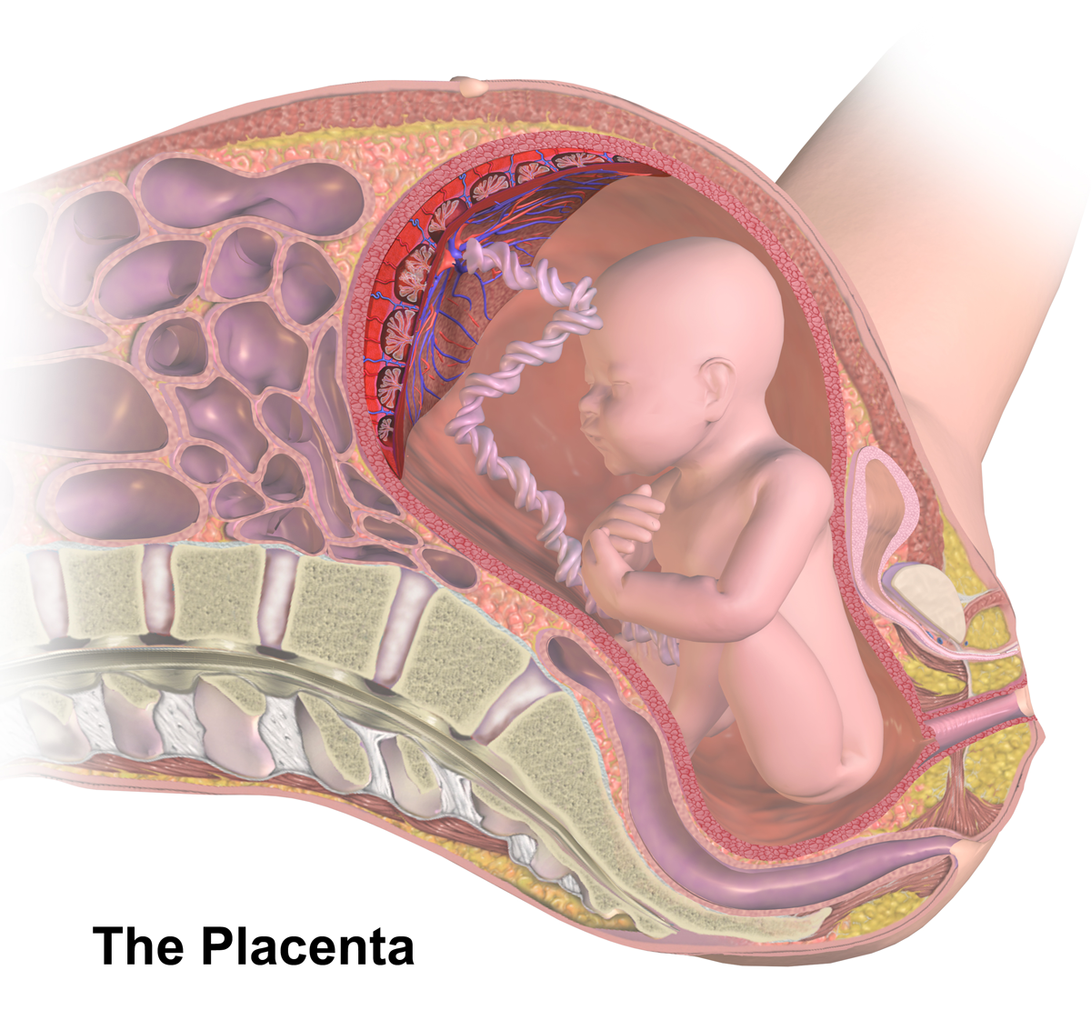
Placenta
The placenta is a temporary embryonic and later fetal organ that begins developing from the blastocyst shortly after implantation. It plays critical roles in facilitating nutrient, gas and waste exchange between the physically separate maternal and fetal circulations, and is an important endocrine organ, producing hormones that regulate both maternal and fetal physiology during pregnancy. The placenta connects to the fetus via the umbilical cord, and on the opposite aspect to the maternal uterus in a species-dependent manner. In humans, a thin layer of maternal decidual (endometrial) tissue comes away with the placenta when it is expelled from the uterus following birth (sometimes incorrectly referred to as the 'maternal part' of the placenta). Placentas are a defining characteristic of placental mammals, but are also found in marsupials and some non-mammals with varying levels of development. Mammalian placentas probably first evolved about 150 million to 200 million years ... [...More Info...] [...Related Items...] OR: [Wikipedia] [Google] [Baidu] |
Trophoblast
The trophoblast (from Greek : to feed; and : germinator) is the outer layer of cells of the blastocyst. Trophoblasts are present four days after fertilization in humans. They provide nutrients to the embryo and develop into a large part of the placenta. They form during the first stage of pregnancy and are the first cells to differentiate from the fertilized egg to become extraembryonic structures that do not directly contribute to the embryo. After gastrulation, the trophoblast is contiguous with the ectoderm of the embryo and is referred to as the trophectoderm. After the first differentiation, the cells in the human embryo lose their totipotency and are no longer totipotent stem cells because they cannot form a trophoblast. They are now pluripotent stem cells. Structure The trophoblast proliferates and differentiates into two cell layers at approximately six days after fertilization for humans. Function Trophoblasts are specialized cells of the placenta that play an i ... [...More Info...] [...Related Items...] OR: [Wikipedia] [Google] [Baidu] |
Inner Cell Mass
The inner cell mass (ICM) or embryoblast (known as the pluriblast in marsupials) is a structure in the early development of an embryo. It is the mass of cells inside the blastocyst that will eventually give rise to the definitive structures of the fetus. The inner cell mass forms in the earliest stages of embryonic development, before implantation into the endometrium of the uterus. The ICM is entirely surrounded by the single layer of trophoblast cells of the trophectoderm. Further development The physical and functional separation of the inner cell mass from the trophectoderm (TE) is a special feature of mammalian development and is the first cell lineage specification in these embryos. Following fertilization in the oviduct, the mammalian embryo undergoes a relatively slow round of cleavages to produce an eight-cell morula. Each cell of the morula, called a blastomere, increases surface contact with its neighbors in a process called compaction. This results in a pola ... [...More Info...] [...Related Items...] OR: [Wikipedia] [Google] [Baidu] |


