|
Ectopic Salivary Gland Tissue
Ectopic salivary gland tissue which is located in sites other than the normal location is variously described as aberrant, accessory, ectopic, heterotopic or salivary gland choristoma. Accessory salivary glands An ''accessory salivary gland'' is ectopic salivary gland tissue with a salivary gland duct system. The most common location of accessory salivary gland tissue is an extra major salivary gland in front of the parotid gland. It is typically about 3 cm or less in size, and drains into the parotid duct via a single tributary. Accessory parotid tissue is found in 21-56% of adults. Any disease process which affects the salivary glands, including cancer, may also occur within an accessory salivary gland tissue. Heterotopic salivary gland tissue ''Salivary gland heterotopia'' is where salivary gland acini cells are present in an abnormal location without any duct system. The most common location is the cervical lymph nodes. Other reported sites of heterotopic salivary gland ... [...More Info...] [...Related Items...] OR: [Wikipedia] [Google] [Baidu] |
Ectopia (medicine)
An ectopia () is a displacement or malposition of an organ or other body part, which is then referred to as ectopic ({{IPAc-en, ɛ, k, ˈ, t, ɒ, p, ɪ, k). Most ectopias are congenital, but some may happen later in life. Examples *Ectopic ACTH syndrome, also known as small-cell carcinoma. *Ectopic calcification, a pathologic deposition of calcium salts in tissues or bone growth in soft tissues * Cerebellar tonsillar ectopia, aka Chiari malformation, a herniation of the brain through the foramen magnum, which may be congenital or caused by trauma. * Ectopic cilia, a hair growing where it isn't supposed to be, commonly an eyelash on an abnormal spot on the eyelid, distichia *Ectopia cordis, the displacement of the heart outside the body during fetal development * Ectopic enamel, a tooth abnormality, where enamel is found in an unusual location, such as at the root of a tooth *Ectopic expression, the expression of a gene in an abnormal place in an organism * Ectopic hormone, a horm ... [...More Info...] [...Related Items...] OR: [Wikipedia] [Google] [Baidu] |
Pituitary Gland
In vertebrate anatomy, the pituitary gland, or hypophysis, is an endocrine gland, about the size of a chickpea and weighing, on average, in humans. It is a protrusion off the bottom of the hypothalamus at the base of the brain. The hypophysis rests upon the hypophyseal fossa of the sphenoid bone in the center of the middle cranial fossa and is surrounded by a small bony cavity (sella turcica) covered by a dural fold (diaphragma sellae). The anterior pituitary (or adenohypophysis) is a lobe of the gland that regulates several physiological processes including stress, growth, reproduction, and lactation. The intermediate lobe synthesizes and secretes melanocyte-stimulating hormone. The posterior pituitary (or neurohypophysis) is a lobe of the gland that is functionally connected to the hypothalamus by the median eminence via a small tube called the pituitary stalk (also called the infundibular stalk or the infundibulum). Hormones secreted from the pituitary gland ... [...More Info...] [...Related Items...] OR: [Wikipedia] [Google] [Baidu] |
Salivary Gland Neoplasm
Salivary gland tumours, also known as mucous gland adenomas or neoplasms, are tumours that form in the tissues of salivary glands. The salivary glands are classified as major or minor. The major salivary glands consist of the parotid, submandibular, and sublingual glands. The minor salivary glands consist of 800 to 1000 small mucus-secreting glands located throughout the lining of the oral cavity. Patients with these types of tumours may be asymptomatic. Presentation Salivary gland tumours usually present as a lump or swelling in the affected gland which may or may not have been present for a long time. The lump may be accompanied by symptoms of duct blockage (e.g. xerostomia). Usually, in their early stages it is not possible to distinguish a benign tumour from a malignant one. One of the key differentiating symptoms of a malignant growth is nerve involvement; for example, signs of facial nerve damage (e.g. facial palsy) are associated with malignant parotid tumours. Facial pain ... [...More Info...] [...Related Items...] OR: [Wikipedia] [Google] [Baidu] |
Vulva
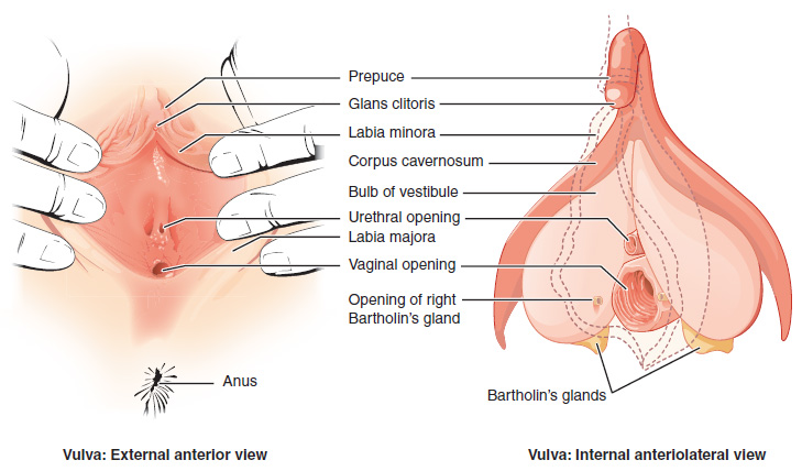
The vulva (plural: vulvas or vulvae; derived from Latin for wrapper or covering) consists of the external sex organ, female sex organs. The vulva includes the mons pubis (or mons veneris), labia majora, labia minora, clitoris, bulb of vestibule, vestibular bulbs, vulval vestibule, urinary meatus, the Vagina#Vaginal opening and hymen, vaginal opening, hymen, and Bartholin's gland, Bartholin's and Skene's gland, Skene's vestibular glands. The urinary meatus is also included as it opens into the vulval vestibule. Other features of the vulva include the pudendal cleft, sebaceous glands, the urogenital triangle (anterior part of the perineum), and pubic hair. The vulva includes the entrance to the vagina, which leads to the uterus, and provides a double layer of protection for this by the folds of the outer and inner labia. Pelvic floor muscles support the structures of the vulva. Other muscles of the urogenital triangle also give support. Blood supply to the vulva comes from the t ... [...More Info...] [...Related Items...] OR: [Wikipedia] [Google] [Baidu] |
Rectum
The rectum is the final straight portion of the large intestine in humans and some other mammals, and the Gastrointestinal tract, gut in others. The adult human rectum is about long, and begins at the rectosigmoid junction (the end of the sigmoid colon) at the level of the third sacral vertebra or the sacral promontory depending upon what definition is used. Its diameter is similar to that of the sigmoid colon at its commencement, but it is dilated near its termination, forming the rectal ampulla. It terminates at the level of the anorectal ring (the level of the puborectalis sling) or the dentate line, again depending upon which definition is used. In humans, the rectum is followed by the anal canal which is about long, before the gastrointestinal tract terminates at the anal verge. The word rectum comes from the Latin ''Wikt:rectum, rectum Wikt:intestinum, intestinum'', meaning ''straight intestine''. Structure The rectum is a part of the lower gastrointestinal tract ... [...More Info...] [...Related Items...] OR: [Wikipedia] [Google] [Baidu] |
Stomach
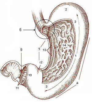
The stomach is a muscular, hollow organ in the gastrointestinal tract of humans and many other animals, including several invertebrates. The stomach has a dilated structure and functions as a vital organ in the digestive system. The stomach is involved in the gastric phase of digestion, following chewing. It performs a chemical breakdown by means of enzymes and hydrochloric acid. In humans and many other animals, the stomach is located between the oesophagus and the small intestine. The stomach secretes digestive enzymes and gastric acid to aid in food digestion. The pyloric sphincter controls the passage of partially digested food ( chyme) from the stomach into the duodenum, where peristalsis takes over to move this through the rest of intestines. Structure In the human digestive system, the stomach lies between the oesophagus and the duodenum (the first part of the small intestine). It is in the left upper quadrant of the abdominal cavity. The top of the stomach lies ag ... [...More Info...] [...Related Items...] OR: [Wikipedia] [Google] [Baidu] |
Sternocleidomastoid
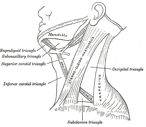
The sternocleidomastoid muscle is one of the largest and most superficial cervical muscles. The primary actions of the muscle are rotation of the head to the opposite side and flexion of the neck. The sternocleidomastoid is innervated by the accessory nerve. Etymology and location It is given the name ''sternocleidomastoid'' because it originates at the manubrium of the sternum (''sterno-'') and the clavicle (''cleido-'') and has an insertion at the mastoid process of the temporal bone of the skull. Structure The sternocleidomastoid muscle originates from two locations: the manubrium of the sternum and the clavicle. It travels obliquely across the side of the neck and inserts at the mastoid process of the temporal bone of the skull by a thin aponeurosis. The sternocleidomastoid is thick and narrow at its centre, and broader and thinner at either end. The sternal head is a round fasciculus, tendinous in front, fleshy behind, arising from the upper part of the front of the manu ... [...More Info...] [...Related Items...] OR: [Wikipedia] [Google] [Baidu] |
Cerebellopontine Angle
The cerebellopontine angle (CPA) ( la, angulus cerebellopontinus) is located between the cerebellum and the pons. The cerebellopontine angle is the site of the cerebellopontine angle cistern one of the subarachnoid cisterns that contains cerebrospinal fluid, arachnoid tissue, cranial nerves, and associated vessels. The cerebellopontine angle is also the site of a set of neurological disorders known as the cerebellopontine angle syndrome. Structure The cerebellopontine angle is formed by the cerebellopontine fissure. This fissure is made when the cerebellum folds over to the pons, creating a sharply defined angle between them. The angle formed in turn creates a subarachnoid cistern, the cerebellopontine angle cistern. The pia mater follows the outline of the fissure and the arachnoid mater continues across the divide so that the subarachnoid space is dilated at this area, forming the cerebellopontine angle cistern. The anterior inferior cerebellar artery (AICA) is the principa ... [...More Info...] [...Related Items...] OR: [Wikipedia] [Google] [Baidu] |
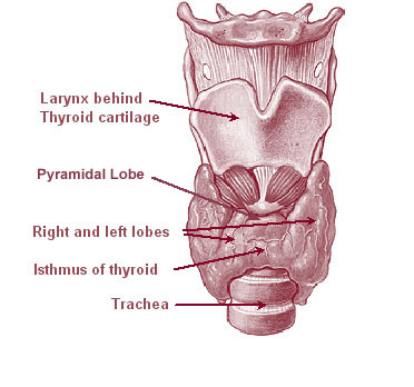
Thyroid Gland
The thyroid, or thyroid gland, is an endocrine gland in vertebrates. In humans it is in the neck and consists of two connected lobe (anatomy), lobes. The lower two thirds of the lobes are connected by a thin band of Connective tissue, tissue called the thyroid isthmus. The thyroid is located at the front of the neck, below the Adam's apple. Microscopically, the functional unit of the thyroid gland is the spherical Thyroid follicular cell#Location, thyroid follicle, lined with thyroid follicular cell, follicular cells (thyrocytes), and occasional parafollicular cells that surround a follicular lumen, lumen containing colloid. The thyroid gland secretes three hormones: the two thyroid hormonestriiodothyronine, triiodothyronine (T3) and thyroid hormone, thyroxine (T4)and a peptide hormone, calcitonin. The thyroid hormones influence the basal metabolic rate, metabolic rate and protein biosynthesis, protein synthesis, and in children, growth and development. Calcitonin plays a role in ... [...More Info...] [...Related Items...] OR: [Wikipedia] [Google] [Baidu] |
Heterotopia (medicine)
In medicine, heterotopia is the presence of a particular tissue type at a non-physiological site, but usually co-existing with original tissue in its correct anatomical location. In other words, it implies ectopic tissue, in addition to retention of the original tissue type. Examples In neuropathology, for example, gray matter heterotopia is the presence of gray matter within the cerebral white matter or ventricles. Heterotopia within the brain is often divided into three groups: subependymal heterotopia, focal cortical heterotopia and band heterotopia. Another example is a Meckel's diverticulum, which may contain heterotopic gastric or pancreatic tissue. In biology specifically, ''heterotopy'' refers to an altered location of trait expression.West-Eberhard, 2003 In her book ''Developmental Plasticity and Evolution'', Mary-Jane West Eberhard has a cover art of the sulphur crested cockatoo and comments on the back cover "Did long crest eadfeathers evolve by gradual modificat ... [...More Info...] [...Related Items...] OR: [Wikipedia] [Google] [Baidu] |
Parathyroid Gland
Parathyroid glands are small endocrine glands in the neck of humans and other tetrapods. Humans usually have four parathyroid glands, located on the back of the thyroid gland in variable locations. The parathyroid gland produces and secretes parathyroid hormone in response to a low blood calcium, which plays a key role in regulating the amount of calcium in the blood and within the bones. Parathyroid glands share a similar blood supply, venous drainage, and lymphatic drainage to the thyroid glands. Parathyroid glands are derived from the epithelial lining of the third and fourth pharyngeal pouches, with the superior glands arising from the fourth pouch and the inferior glands arising from the higher third pouch. The relative position of the inferior and superior glands, which are named according to their final location, changes because of the migration of embryological tissues. Hyperparathyroidism and hypoparathyroidism, characterized by alterations in the blood calcium levels ... [...More Info...] [...Related Items...] OR: [Wikipedia] [Google] [Baidu] |
Middle Ear
The middle ear is the portion of the ear medial to the eardrum, and distal to the oval window of the cochlea (of the inner ear). The mammalian middle ear contains three ossicles, which transfer the vibrations of the eardrum into waves in the fluid and membranes of the inner ear. The hollow space of the middle ear is also known as the tympanic cavity and is surrounded by the tympanic part of the temporal bone. The auditory tube (also known as the Eustachian tube or the pharyngotympanic tube) joins the tympanic cavity with the nasal cavity (nasopharynx), allowing pressure to equalize between the middle ear and throat. The primary function of the middle ear is to efficiently transfer acoustic energy from compression waves in air to fluid–membrane waves within the cochlea. Structure Ossicles The middle ear contains three tiny bones known as the ossicles: '' malleus'', '' incus'', and ''stapes''. The ossicles were given their Latin names for their distinctive shapes; they ar ... [...More Info...] [...Related Items...] OR: [Wikipedia] [Google] [Baidu] |
