|
Descending Aorta
In human anatomy, the descending aorta is part of the aorta, the largest artery in the body. The descending aorta begins at the aortic arch and runs down through the chest and abdomen. The descending aorta anatomically consists of two portions or segments, the thoracic and the abdominal aorta, in correspondence with the two great cavities of the trunk in which it is situated. Within the abdomen, the descending aorta branches into the two common iliac arteries which serve the pelvis and eventually legs. The ductus arteriosus connects to the junction between the pulmonary artery and the descending aorta in foetal life. This artery later regresses as the ligamentum arteriosum. See also *Abbott artery Abbott's artery describes an anomalous artery that arises from the posteromedial aspect of the proximal part of the descending aorta. Normally a minor congenital abnormality, its presence is important during surgical repair of coarctation of the ... References External links ... [...More Info...] [...Related Items...] OR: [Wikipedia] [Google] [Baidu] |
Thoracic Aorta
The descending thoracic aorta is a part of the aorta located in the thorax. It is a continuation of the aortic arch. It is located within the posterior mediastinal cavity, but frequently bulges into the left pleural cavity. The descending thoracic aorta begins at the lower border of the fourth thoracic vertebra and ends in front of the lower border of the twelfth thoracic vertebra, at the aortic hiatus in the diaphragm where it becomes the abdominal aorta. At its commencement, it is situated on the left of the vertebral column; it approaches the median line as it descends; and, at its termination, lies directly in front of the column. The descending thoracic aorta has a curved shape that faces forward, and has small branches. It has a radius of approximately 1.16 cm. Structure The descending thoracic aorta is part of the aorta, which has different parts named according to their structure or location. The descending thoracic aorta is a continuation of the descending aorta ... [...More Info...] [...Related Items...] OR: [Wikipedia] [Google] [Baidu] |
Common Iliac Artery
The common iliac artery is a large artery of the abdomen paired on each side. It originates from the aortic bifurcation at the level of the 4th lumbar vertebra. It ends in front of the sacroiliac joint, one on either side, and each bifurcates into the external and internal iliac arteries. Structure The common iliac artery are about 4 cm long in adults and more than a centimeter in diameter. It begins as a branch of the aorta. This is at the level of the 4th lumbar vertebra. It runs inferolaterally, along the medial border of the psoas muscles. It bifurcates into the external iliac artery and the internal iliac artery at the pelvic brim, in front of the sacroiliac joints. The common iliac artery, and all of its branches, exist as paired structures (that is to say, there is one on the left side and one on the right). The distribution of the common iliac artery is basically the pelvis and lower limb (as the femoral artery) on the corresponding side. Relations Both common il ... [...More Info...] [...Related Items...] OR: [Wikipedia] [Google] [Baidu] |
Mediastinum
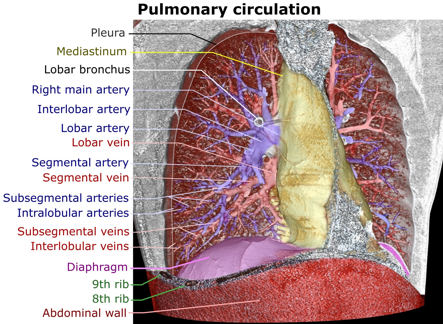
The mediastinum (from ) is the central compartment of the thoracic cavity. Surrounded by loose connective tissue, it is an undelineated region that contains a group of structures within the thorax, namely the heart and its vessels, the esophagus, the trachea, the phrenic nerve, phrenic and cardiac nerves, the thoracic duct, the thymus and the lymph nodes of the central chest. Anatomy The mediastinum lies within the thorax and is enclosed on the right and left by pulmonary pleurae, pleurae. It is surrounded by the chest wall in front, the lungs to the sides and the Spine (anatomy), spine at the back. It extends from the sternum in front to the vertebral column behind. It contains all the organs of the thorax except the lungs. It is continuous with the loose connective tissue of the neck. The mediastinum can be divided into an upper (or superior) and lower (or inferior) part: * The superior mediastinum starts at the superior thoracic aperture and ends at the #Thoracic plane, t ... [...More Info...] [...Related Items...] OR: [Wikipedia] [Google] [Baidu] |
Abbott Artery
Abbott's artery describes an anomalous artery that arises from the posteromedial aspect of the proximal part of the descending aorta. Normally a minor congenital abnormality, its presence is important during surgical repair of coarctation of the aorta Coarctation of the aorta (CoA or CoAo), also called aortic narrowing, is a congenital condition whereby the aorta is narrow, usually in the area where the ductus arteriosus (ligamentum arteriosum after regression) inserts. The word ''coarctation' .... References Arteries of the torso {{circulatory-stub ... [...More Info...] [...Related Items...] OR: [Wikipedia] [Google] [Baidu] |
Ligamentum Arteriosum
The ligamentum arteriosum (arterial ligament), also known as the Ligament of Botallo or Harvey's ligament, is a small ligament attaching the aorta to the pulmonary artery. It serves no function in adults but is the remnant of the ductus arteriosus formed within three weeks after birth. Structure At the superior end, the ligamentum attaches to the aorta—at the final part of the aortic arch (the isthmus of aorta) or the first part of the descending aorta. On the other, inferior end, the ligamentum is attached to the top of the left pulmonary artery. The ligamentum arteriosum is closely related to the left recurrent laryngeal nerve, a branch of the left vagus nerve. After splitting from the left vagus nerve, the left recurrent laryngeal loops around the aortic arch behind the ligamentum arteriosum, after which it ascends to the larynx. Function In adults, the ligamentum arteriosum has no useful function. It is a vestige of the ductus arteriosus, a temporary fetal structure tha ... [...More Info...] [...Related Items...] OR: [Wikipedia] [Google] [Baidu] |
Fetus
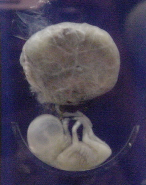
A fetus or foetus (; plural fetuses, feti, foetuses, or foeti) is the unborn offspring that develops from an animal embryo. Following embryonic development the fetal stage of development takes place. In human prenatal development, fetal development begins from the ninth week after fertilization (or eleventh week gestational age) and continues until birth. Prenatal development is a continuum, with no clear defining feature distinguishing an embryo from a fetus. However, a fetus is characterized by the presence of all the major body organs, though they will not yet be fully developed and functional and some not yet situated in their final anatomical location. Etymology The word ''fetus'' (plural ''fetuses'' or '' feti'') is related to the Latin '' fētus'' ("offspring", "bringing forth", "hatching of young") and the Greek "φυτώ" to plant. The word "fetus" was used by Ovid in Metamorphoses, book 1, line 104. The predominant British, Irish, and Commonwealth spelling is '' ... [...More Info...] [...Related Items...] OR: [Wikipedia] [Google] [Baidu] |
Pulmonary Artery
A pulmonary artery is an artery in the pulmonary circulation that carries deoxygenated blood from the right side of the heart to the lungs. The largest pulmonary artery is the ''main pulmonary artery'' or ''pulmonary trunk'' from the heart, and the smallest ones are the arterioles, which lead to the capillaries that surround the pulmonary alveoli. Structure The pulmonary arteries are blood vessels that carry systemic venous blood from the right ventricle of the heart to the microcirculation of the lungs. Unlike in other organs where arteries supply oxygenated blood, the blood carried by the pulmonary arteries is deoxygenated, as it is venous blood returning to the heart. The main pulmonary arteries emerge from the right side of the heart, and then split into smaller arteries that progressively divide and become arterioles, eventually narrowing into the capillary microcirculation of the lungs where gas exchange occurs. Pulmonary trunk In order of blood flow, the pulmonary art ... [...More Info...] [...Related Items...] OR: [Wikipedia] [Google] [Baidu] |
Ductus Arteriosus
The ''ductus arteriosus'', also called the ''ductus Botalli'', named after the Italian physiologist Leonardo Botallo, is a blood vessel in the developing fetus connecting the trunk of the pulmonary artery to the proximal descending aorta. It allows most of the blood from the right ventricle to bypass the fetus's fluid-filled non-functioning lungs. Upon closure at birth, it becomes the ''ligamentum arteriosum''. Development and structure The ''ductus arteriosus'' is formed from the left 6th aortic arch during embryonic development and attaches to the final part of the aortic arch (the isthmus of aorta) and the first part of the pulmonary artery. Disorder: Patent ductus arteriosus Consequences Failure of the ''ductus arteriosus'' to close after birth results in a condition called ''patent ductus arteriosus'', which results in the abnormal flow of blood from the aorta to the pulmonary artery: a left-to-right shunt. If left uncorrected, this usually leads to pulmonary hypertensi ... [...More Info...] [...Related Items...] OR: [Wikipedia] [Google] [Baidu] |
Pelvis
The pelvis (plural pelves or pelvises) is the lower part of the trunk, between the abdomen and the thighs (sometimes also called pelvic region), together with its embedded skeleton (sometimes also called bony pelvis, or pelvic skeleton). The pelvic region of the trunk includes the bony pelvis, the pelvic cavity (the space enclosed by the bony pelvis), the pelvic floor, below the pelvic cavity, and the perineum, below the pelvic floor. The pelvic skeleton is formed in the area of the back, by the sacrum and the coccyx and anteriorly and to the left and right sides, by a pair of hip bones. The two hip bones connect the spine with the lower limbs. They are attached to the sacrum posteriorly, connected to each other anteriorly, and joined with the two femurs at the hip joints. The gap enclosed by the bony pelvis, called the pelvic cavity, is the section of the body underneath the abdomen and mainly consists of the reproductive organs (sex organs) and the rectum, while the pelvic f ... [...More Info...] [...Related Items...] OR: [Wikipedia] [Google] [Baidu] |
Abdomen
The abdomen (colloquially called the belly, tummy, midriff, tucky or stomach) is the part of the body between the thorax (chest) and pelvis, in humans and in other vertebrates. The abdomen is the front part of the abdominal segment of the torso. The area occupied by the abdomen is called the abdominal cavity. In arthropods it is the posterior (anatomy), posterior tagma (biology), tagma of the body; it follows the thorax or cephalothorax. In humans, the abdomen stretches from the thorax at the thoracic diaphragm to the pelvis at the pelvic brim. The pelvic brim stretches from the lumbosacral joint (the intervertebral disc between Lumbar vertebrae, L5 and Vertebra#Sacrum, S1) to the pubic symphysis and is the edge of the pelvic inlet. The space above this inlet and under the thoracic diaphragm is termed the abdominal cavity. The boundary of the abdominal cavity is the abdominal wall in the front and the peritoneal surface at the rear. In vertebrates, the abdomen is a large body c ... [...More Info...] [...Related Items...] OR: [Wikipedia] [Google] [Baidu] |
Ascending Aorta
The ascending aorta (AAo) is a portion of the aorta commencing at the upper part of the base of the left ventricle, on a level with the lower border of the third costal cartilage behind the left half of the sternum. Structure It passes obliquely upward, forward, and to the right, in the direction of the heart's axis, as high as the upper border of the second right costal cartilage, describing a slight curve in its course, and being situated, about behind the posterior surface of the sternum. The total length is about . Components The aortic root is the portion of the aorta beginning at the aortic annulus and extending to the sinotubular junction. It is sometimes regarded as a part of the ascending aorta, and sometimes regarded as a separate entity from the rest of the ascending aorta. Between each commissure of the aortic valve and opposite the cusps of the aortic valve, three small dilatations called the aortic sinuses. The sinotubular junction is the point in the ascendi ... [...More Info...] [...Related Items...] OR: [Wikipedia] [Google] [Baidu] |
Chest
The thorax or chest is a part of the anatomy of humans, mammals, and other tetrapod animals located between the neck and the abdomen. In insects, crustaceans, and the extinct trilobites, the thorax is one of the three main divisions of the creature's body, each of which is in turn composed of multiple segments. The human thorax includes the thoracic cavity and the thoracic wall. It contains organs including the heart, lungs, and thymus gland, as well as muscles and various other internal structures. Many diseases may affect the chest, and one of the most common symptoms is chest pain. Etymology The word thorax comes from the Greek θώραξ ''thorax'' "breastplate, cuirass, corslet" via la, thorax. Plural: ''thoraces'' or ''thoraxes''. Human thorax Structure In humans and other hominids, the thorax is the chest region of the body between the neck and the abdomen, along with its internal organs and other contents. It is mostly protected and supported by the rib cage, spine, ... [...More Info...] [...Related Items...] OR: [Wikipedia] [Google] [Baidu] |
_Victoria_blue-HE.jpg)


