|
Duodenal
The duodenum is the first section of the small intestine in most higher vertebrates, including mammals, reptiles, and birds. In fish, the divisions of the small intestine are not as clear, and the terms anterior intestine or proximal intestine may be used instead of duodenum. In mammals the duodenum may be the principal site for iron absorption. The duodenum precedes the jejunum and ileum and is the shortest part of the small intestine. In humans, the duodenum is a hollow jointed tube about 25–38 cm (10–15 inches) long connecting the stomach to the middle part of the small intestine. It begins with the duodenal bulb and ends at the suspensory muscle of duodenum. Duodenum can be divided into four parts: the first (superior), the second (descending), the third (horizontal) and the fourth (ascending) parts. Structure The duodenum is a C-shaped structure lying adjacent to the stomach. It is divided anatomically into four sections. The first part of the duodenum lies ... [...More Info...] [...Related Items...] OR: [Wikipedia] [Google] [Baidu] |
Inferior Pancreaticoduodenal Artery
The inferior pancreaticoduodenal artery (the IPDA) is a branch of the superior mesenteric artery. It supplies the head of the pancreas, and the ascending and inferior parts of the duodenum. Rarely, it may have an aneurysm. Structure The inferior pancreaticoduodenal artery is a branch of the superior mesenteric artery. This occurs opposite the upper border of the inferior part of the duodenum. As soon as it branches, it divides into anterior and posterior branches. These run between the head of the pancreas and the lesser curvature of the duodenum. They then join (anastomose) with the anterior and posterior branches of the superior pancreaticoduodenal artery. Variation The inferior pancreaticoduodenal artery may branch from the first intestinal branch of the superior mesenteric artery rather than directly from it. Function The inferior pancreaticoduodenal artery distributes branches to the head of the pancreas and to the ascending and inferior parts of the duodenum. Clinic ... [...More Info...] [...Related Items...] OR: [Wikipedia] [Google] [Baidu] |
Superior Pancreaticoduodenal Artery
The superior pancreaticoduodenal artery is an artery that supplies blood to the duodenum and pancreas. Structure It is a branch of the gastroduodenal artery, which most commonly arises from the common hepatic artery of the celiac trunk, although there are numerous variations of the origin of the gastroduodenal artery. The pancreaticoduodenal artery divides into two branches as it descends, an anterior and posterior branch. These branches then travel around the head of the pancreas and duodenum, eventually joining with the anterior and posterior branches of the inferior pancreaticoduodenal artery. The inferior pancreaticoduodenal artery is a branch of the superior mesenteric artery. These arteries, together with the pancreatic branches of the splenic artery, form connections or anastomoses with one another, allowing blood to perfuse the pancreas and duodenum through multiple channels. The artery supplies the anterior and posterior sides of the duodenum and head of pancreas ... [...More Info...] [...Related Items...] OR: [Wikipedia] [Google] [Baidu] |
Hepatoduodenal Ligament
The hepatoduodenal ligament is the portion of the lesser omentum extending between the porta hepatis of the liver and the superior part of the duodenum. Running inside it are the following structures collectively known as the portal triad: * hepatic artery proper * portal vein * common bile duct Manual compression of the hepatoduodenal ligament during surgery is known as the Pringle manoeuvre. The cystoduodenal ligament is also found in the lesser omentum and is distinct from both the hepatoduodenal and hepatogastric ligaments. The cystoduodenal ligament is an abnormal peritoneal fold that attaches the duodenum to the gallbladder, representing a rare variation in the anatomy of the lesser sac and its foramen. Another variation sometimes present at the duodenal termination of the hepatoduodenal ligament is the duodenorenal ligament which passes to the front of the right kidney The kidneys are two reddish-brown bean-shaped organs found in vertebrates. They are located on t ... [...More Info...] [...Related Items...] OR: [Wikipedia] [Google] [Baidu] |
Pancreaticoduodenal Veins
The pancreaticoduodenal veins accompany their corresponding arteries: the superior pancreaticoduodenal artery and the inferior pancreaticoduodenal artery; the lower of the two frequently joins the right gastroepiploic vein The right gastroepiploic vein (right gastroomental vein) is a blood vessel that drains blood from the greater curvature and left part of the body of the stomach into the superior mesenteric vein. It runs from left to right along the greater curvatu .... References External links Veins of the torso {{circulatory-stub ... [...More Info...] [...Related Items...] OR: [Wikipedia] [Google] [Baidu] |
Duodenal Bulb
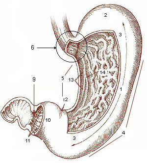
The duodenal bulb is the portion of the duodenum closest to the stomach. It normally has a length of about 5 centimeters. The duodenal bulb begins at the pylorus and ends at the neck of the gallbladder. It is located posterior to the liver and the gallbladder and superior to the pancreatic head. The gastroduodenal artery, portal vein, and common bile duct lie just behind it. The distal part of the bulb is located retroperitoneally. It is located immediately distal to the pyloric sphincter. The duodenal bulb is the place where duodenal ulcers occur. Duodenal ulcers are more common than gastric ulcers and unlike gastric ulcers, are caused by increased gastric acid secretion. Duodenal ulcers are commonly located anteriorly, and rarely posteriorly. Anterior ulcers can be complicated by perforation, while the posterior ones bleed. The reason for that is explained by their location. The peritoneal or abdominal cavity is located anterior to the duodenum. Therefore, if the ulcer grows dee ... [...More Info...] [...Related Items...] OR: [Wikipedia] [Google] [Baidu] |
Stomach
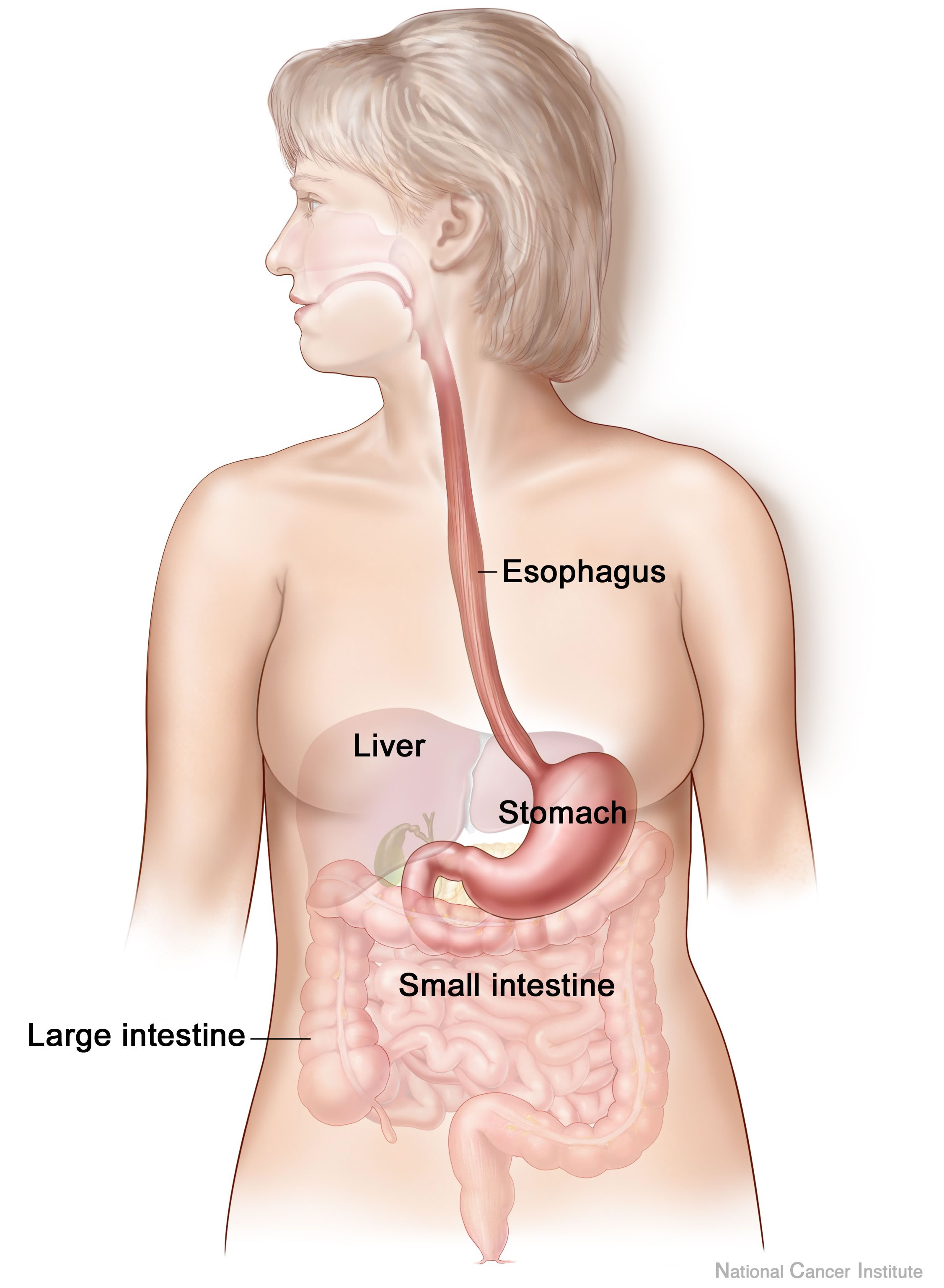
The stomach is a muscular, hollow organ in the gastrointestinal tract of humans and many other animals, including several invertebrates. The stomach has a dilated structure and functions as a vital organ in the digestive system. The stomach is involved in the gastric phase of digestion, following chewing. It performs a chemical breakdown by means of enzymes and hydrochloric acid. In humans and many other animals, the stomach is located between the oesophagus and the small intestine. The stomach secretes digestive enzymes and gastric acid to aid in food digestion. The pyloric sphincter controls the passage of partially digested food ( chyme) from the stomach into the duodenum, where peristalsis takes over to move this through the rest of intestines. Structure In the human digestive system, the stomach lies between the oesophagus and the duodenum (the first part of the small intestine). It is in the left upper quadrant of the abdominal cavity. The top of the stomach lies ag ... [...More Info...] [...Related Items...] OR: [Wikipedia] [Google] [Baidu] |
Lesser Omentum
The lesser omentum (small omentum or gastrohepatic omentum) is the double layer of peritoneum that extends from the liver to the lesser curvature of the stomach, and to the first part of the duodenum. The lesser omentum is usually divided into these two connecting parts: the hepatogastric ligament, and the hepatoduodenal ligament. Structure The lesser omentum is extremely thin, and is continuous with the two layers of peritoneum which cover respectively the antero-superior and postero-inferior surfaces of the stomach and first part of the duodenum. When these two layers reach the lesser curvature of the stomach and the upper border of the duodenum, they join and ascend as a double fold to the porta hepatis. To the left of the porta, the fold is attached to the bottom of the fossa for the ductus venosus, along which it is carried to the diaphragm, where the two layers separate to embrace the end of the esophagus. At the right border of the lesser omentum, the two layers are c ... [...More Info...] [...Related Items...] OR: [Wikipedia] [Google] [Baidu] |
Digestive System
The human digestive system consists of the gastrointestinal tract plus the accessory organs of digestion (the tongue, salivary glands, pancreas, liver, and gallbladder). Digestion involves the breakdown of food into smaller and smaller components, until they can be absorbed and assimilated into the body. The process of digestion has three stages: the cephalic phase, the gastric phase, and the intestinal phase. The first stage, the cephalic phase of digestion, begins with secretions from gastric glands in response to the sight and smell of food. This stage includes the mechanical breakdown of food by chewing, and the chemical breakdown by digestive enzymes, that takes place in the mouth. Saliva contains the digestive enzymes amylase, and lingual lipase, secreted by the salivary and serous glands on the tongue. Chewing, in which the food is mixed with saliva, begins the mechanical process of digestion. This produces a bolus which is swallowed down the esophagus to enter the st ... [...More Info...] [...Related Items...] OR: [Wikipedia] [Google] [Baidu] |
Small Intestine
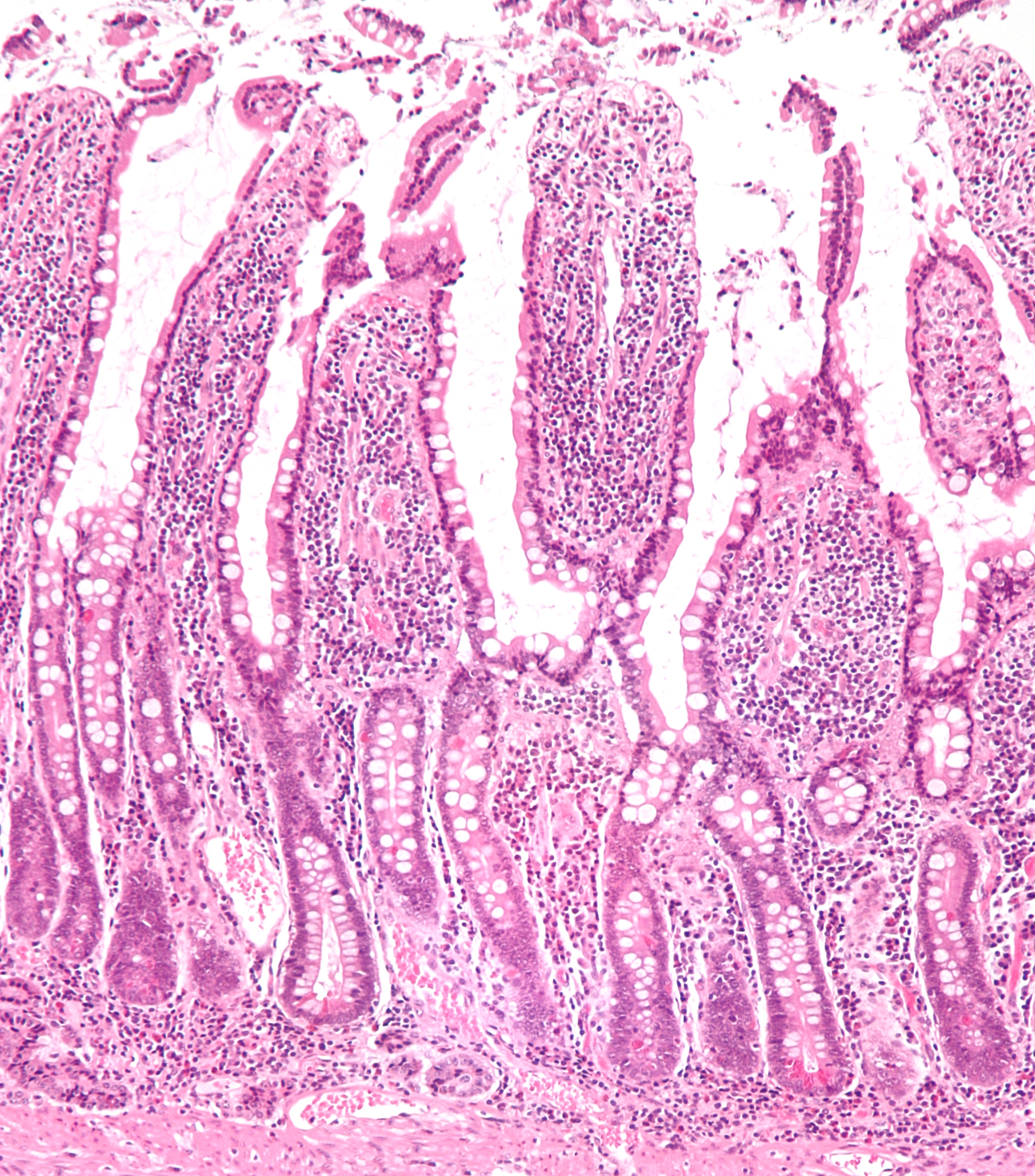
The small intestine or small bowel is an organ in the gastrointestinal tract where most of the absorption of nutrients from food takes place. It lies between the stomach and large intestine, and receives bile and pancreatic juice through the pancreatic duct to aid in digestion. The small intestine is about long and folds many times to fit in the abdomen. Although it is longer than the large intestine, it is called the small intestine because it is narrower in diameter. The small intestine has three distinct regions – the duodenum, jejunum, and ileum. The duodenum, the shortest, is where preparation for absorption through small finger-like protrusions called villi begins. The jejunum is specialized for the absorption through its lining by enterocytes: small nutrient particles which have been previously digested by enzymes in the duodenum. The main function of the ileum is to absorb vitamin B12, bile salts, and whatever products of digestion that were not absorbed by the ... [...More Info...] [...Related Items...] OR: [Wikipedia] [Google] [Baidu] |
Human Gastrointestinal Tract
The gastrointestinal tract (GI tract, digestive tract, alimentary canal) is the tract or passageway of the digestive system that leads from the mouth to the anus. The GI tract contains all the major organs of the digestive system, in humans and other animals, including the esophagus, stomach, and intestines. Food taken in through the mouth is digested to extract nutrients and absorb energy, and the waste expelled at the anus as feces. ''Gastrointestinal'' is an adjective meaning of or pertaining to the stomach and intestines. Most animals have a "through-gut" or complete digestive tract. Exceptions are more primitive ones: sponges have small pores ( ostia) throughout their body for digestion and a larger dorsal pore (osculum) for excretion, comb jellies have both a ventral mouth and dorsal anal pores, while cnidarians and acoels have a single pore for both digestion and excretion. The human gastrointestinal tract consists of the esophagus, stomach, and intestines, and is ... [...More Info...] [...Related Items...] OR: [Wikipedia] [Google] [Baidu] |
Peritoneum
The peritoneum is the serous membrane forming the lining of the abdominal cavity or coelom in amniotes and some invertebrates, such as annelids. It covers most of the intra-abdominal (or coelomic) organs, and is composed of a layer of mesothelium supported by a thin layer of connective tissue. This peritoneal lining of the cavity supports many of the abdominal organs and serves as a conduit for their blood vessels, lymphatic vessels, and nerves. The abdominal cavity (the space bounded by the vertebrae, abdominal muscles, diaphragm, and pelvic floor) is different from the intraperitoneal space (located within the abdominal cavity but wrapped in peritoneum). The structures within the intraperitoneal space are called "intraperitoneal" (e.g., the stomach and intestines), the structures in the abdominal cavity that are located behind the intraperitoneal space are called "retroperitoneal" (e.g., the kidneys), and those structures below the intraperitoneal space are called "subp ... [...More Info...] [...Related Items...] OR: [Wikipedia] [Google] [Baidu] |


