|
Dorsal Root Ganglion
A dorsal root ganglion (or spinal ganglion; also known as a posterior root ganglion) is a cluster of neurons (a ganglion) in a dorsal root of a spinal nerve. The cell bodies of sensory neurons known as first-order neurons are located in the dorsal root ganglia. The axons of dorsal root ganglion neurons are known as afferents. In the peripheral nervous system, afferents refer to the axons that relay sensory information into the central nervous system (i.e. the brain and the spinal cord). Structure The neurons comprising the dorsal root ganglion are of the pseudo-unipolar type, meaning they have a cell body (soma) with two branches that act as a single axon, often referred to as a ''distal process'' and a ''proximal process''. Unlike the majority of neurons found in the central nervous system, an action potential in posterior root ganglion neuron may initiate in the ''distal process'' in the periphery, bypass the cell body, and continue to propagate along the ''proximal p ... [...More Info...] [...Related Items...] OR: [Wikipedia] [Google] [Baidu] |
Embryo
An embryo is an initial stage of development of a multicellular organism. In organisms that reproduce sexually, embryonic development is the part of the life cycle that begins just after fertilization of the female egg cell by the male sperm cell. The resulting fusion of these two cells produces a single-celled zygote that undergoes many cell divisions that produce cells known as blastomeres. The blastomeres are arranged as a solid ball that when reaching a certain size, called a morula, takes in fluid to create a cavity called a blastocoel. The structure is then termed a blastula, or a blastocyst in mammals. The mammalian blastocyst hatches before implantating into the endometrial lining of the womb. Once implanted the embryo will continue its development through the next stages of gastrulation, neurulation, and organogenesis. Gastrulation is the formation of the three germ layers that will form all of the different parts of the body. Neurulation forms the nervous ... [...More Info...] [...Related Items...] OR: [Wikipedia] [Google] [Baidu] |
Action Potential
An action potential occurs when the membrane potential of a specific cell location rapidly rises and falls. This depolarization then causes adjacent locations to similarly depolarize. Action potentials occur in several types of animal cells, called excitable cells, which include neurons, muscle cells, and in some plant cells. Certain endocrine cells such as pancreatic beta cells, and certain cells of the anterior pituitary gland are also excitable cells. In neurons, action potentials play a central role in cell-cell communication by providing for—or with regard to saltatory conduction, assisting—the propagation of signals along the neuron's axon toward synaptic boutons situated at the ends of an axon; these signals can then connect with other neurons at synapses, or to motor cells or glands. In other types of cells, their main function is to activate intracellular processes. In muscle cells, for example, an action potential is the first step in the chain of events l ... [...More Info...] [...Related Items...] OR: [Wikipedia] [Google] [Baidu] |
Somatosensory System
In physiology, the somatosensory system is the network of neural structures in the brain and body that produce the perception of touch (haptic perception), as well as temperature (thermoception), body position (proprioception), and pain. It is a subset of the sensory nervous system, which also represents visual, auditory, olfactory, and gustatory stimuli. Somatosensation begins when mechano- and thermosensitive structures in the skin or internal organs sense physical stimuli such as pressure on the skin (see mechanotransduction, nociception). Activation of these structures, or receptors, leads to activation of peripheral sensory neurons that convey signals to the spinal cord as patterns of action potentials. Sensory information is then processed locally in the spinal cord to drive reflexes, and is also conveyed to the brain for conscious perception of touch and proprioception. Note, somatosensory information from the face and head enters the brain through periphera ... [...More Info...] [...Related Items...] OR: [Wikipedia] [Google] [Baidu] |
Ion Channel
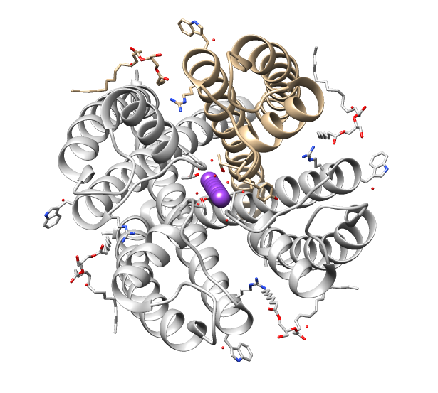
Ion channels are pore-forming membrane proteins that allow ions to pass through the channel pore. Their functions include establishing a resting membrane potential, shaping action potentials and other electrical signals by gating the flow of ions across the cell membrane, controlling the flow of ions across secretory and epithelial cells, and regulating cell volume. Ion channels are present in the membranes of all cells. Ion channels are one of the two classes of ionophoric proteins, the other being ion transporters. The study of ion channels often involves biophysics, electrophysiology, and pharmacology, while using techniques including voltage clamp, patch clamp, immunohistochemistry, X-ray crystallography, fluoroscopy, and RT-PCR. Their classification as molecules is referred to as channelomics. Basic features There are two distinctive features of ion channels that differentiate them from other types of ion transporter proteins: #The rate of ion transport through the ... [...More Info...] [...Related Items...] OR: [Wikipedia] [Google] [Baidu] |
Nociception
Nociception (also nocioception, from Latin ''nocere'' 'to harm or hurt') is the sensory nervous system's process of encoding noxious stimuli. It deals with a series of events and processes required for an organism to receive a painful stimulus, convert it to a molecular signal, and recognize and characterize the signal in order to trigger an appropriate defense response. In nociception, intense chemical (e.g., capsaicin present in Chili pepper or Cayenne pepper), mechanical (e.g., cutting, crushing), or thermal (heat and cold) stimulation of sensory neurons called nociceptors produces a signal that travels along a chain of nerve fibers via the spinal cord to the brain. Nociception triggers a variety of physiological and behavioral responses to protect the organism against an aggression and usually results in a subjective experience, or perception, of pain in sentient beings. Detection of noxious stimuli Potentially damaging mechanical, thermal, and chemical stimuli are detected ... [...More Info...] [...Related Items...] OR: [Wikipedia] [Google] [Baidu] |
Proton-sensing G Protein-coupled Receptors
Proton-sensing G protein-coupled receptors are transmembrane receptors which sense acidic pH and include GPR132 (G2A), GPR4, GPR68 (OGR1) and GPR65 (TDAG8). These G protein-coupled receptors are activated when extracellular pH falls into the range of 6.4-6.8 (typical values are above 7.0). The functional role of the low pH sensitivity of the proton-sensing G protein-coupled receptors is being studied in several tissues where cells respond to conditions of low pH including bone and inflamed tissues. The four known proton-sensing G protein-coupled receptors are Class A receptors in subfamily A15. Nociception Pain sensation can be initiated by nociceptor cells that are sensory neurons with cell bodies located in the dorsal root ganglia. Some nociceptors respond to low pH and the pH-sensitive amiloride-sensitive cation channel 3 has been described as a modulator of acid-induced pain sensation. However, results with amiloride-sensitive cation channel 3 gene knockout mice suggest ... [...More Info...] [...Related Items...] OR: [Wikipedia] [Google] [Baidu] |
Neural Tube
In the developing chordate (including vertebrates), the neural tube is the embryonic precursor to the central nervous system, which is made up of the brain and spinal cord. The neural groove gradually deepens as the neural fold become elevated, and ultimately the folds meet and coalesce in the middle line and convert the groove into the closed neural tube. In humans, neural tube closure usually occurs by the fourth week of pregnancy (the 28th day after conception). The ectodermal wall of the tube forms the rudiment of the nervous system. The centre of the tube is the ''neural canal''.It is an important structure for the development of fetus's brain and spine Development The neural tube develops in two ways: primary neurulation and secondary neurulation. Primary neurulation divides the ectoderm into three cell types: * The internally located neural tube * The externally located epidermis * The neural crest cells, which develop in the region between the neural tube and epider ... [...More Info...] [...Related Items...] OR: [Wikipedia] [Google] [Baidu] |
Neural Crest
Neural crest cells are a temporary group of cells unique to vertebrates that arise from the embryonic ectoderm germ layer, and in turn give rise to a diverse cell lineage—including melanocytes, craniofacial cartilage and bone, smooth muscle, peripheral and enteric neurons and glia. After gastrulation, neural crest cells are specified at the border of the neural plate and the non-neural ectoderm. During neurulation, the borders of the neural plate, also known as the neural folds, converge at the dorsal midline to form the neural tube. Subsequently, neural crest cells from the roof plate of the neural tube undergo an epithelial to mesenchymal transition, delaminating from the neuroepithelium and migrating through the periphery where they differentiate into varied cell types. The emergence of neural crest was important in vertebrate evolution because many of its structural derivatives are defining features of the vertebrate clade. Underlying the development of neural crest is ... [...More Info...] [...Related Items...] OR: [Wikipedia] [Google] [Baidu] |
Intervertebral Foramina
The intervertebral foramen (also called neural foramen, and often abbreviated as IV foramen or IVF) is a :wikt:foramen, foramen between two spinal vertebrae. Cervical vertebrae, Cervical, thoracic vertebrae, thoracic, and lumbar vertebrae all have intervertebral foramina. The foramina, or openings, are present between every pair of vertebrae in these areas. A number of structures pass through the foramen. These are the root of each spinal nerve, the spinal artery of the segmental artery, communicating veins between the internal and external plexuses, meningeal branches of spinal nerve, recurrent meningeal (sinu-vertebral) nerves, and transforaminal ligaments. When the spinal vertebrae are articulated with each other, the bodies form a strong pillar that supports the head and trunk, and the vertebral foramen constitutes a canal for the protection of the medulla spinalis (spinal cord). The size of the foramina is variable due to placement, pathology, spinal loading, and posture ... [...More Info...] [...Related Items...] OR: [Wikipedia] [Google] [Baidu] |
Principles Of Neural Science
First published in 1981 by Elsevier, ''Principles of Neural Science'' is an influential neuroscience textbook edited by Columbia University professors Eric R. Kandel, James H. Schwartz, and Thomas M. Jessell. The original edition was 468 pages; now on the sixth edition, the book has grown to 1646 pages. The second edition was published in 1985, third in 1991, fourth in 2000. The fifth was published on October 26, 2012 and included Steven A. Siegelbaum and A.J. Hudspeth as editors. The sixth and latest edition was published on March 8, 2021. Authors Editors * Kandel was one of the recipients of the 2000 Nobel Prize in Physiology or Medicine. He is currently a professor of biochemistry, molecular biophysics, physiology, cellular biophysics, and psychiatry at Columbia University. He is a senior investigator at the Howard Hughes Medical Institute and a recipient of the National Medal of Science. * Schwartz was a professor of physiology, cellular biophysics, neurology, and psychiatry ... [...More Info...] [...Related Items...] OR: [Wikipedia] [Google] [Baidu] |
Eric R
The given name Eric, Erich, Erikk, Erik, Erick, or Eirik is derived from the Old Norse name ''Eiríkr'' (or ''Eríkr'' in Old East Norse due to monophthongization). The first element, ''ei-'' may be derived from the older Proto-Norse ''* aina(z)'', meaning "one, alone, unique", ''as in the form'' ''Æ∆inrikr'' explicitly, but it could also be from ''* aiwa(z)'' "everlasting, eternity", as in the Gothic form ''Euric''. The second element ''- ríkr'' stems either from Proto-Germanic ''* ríks'' "king, ruler" (cf. Gothic ''reiks'') or the therefrom derived ''* ríkijaz'' "kingly, powerful, rich, prince"; from the common Proto-Indo-European root * h₃rḗǵs. The name is thus usually taken to mean "sole ruler, autocrat" or "eternal ruler, ever powerful". ''Eric'' used in the sense of a proper noun meaning "one ruler" may be the origin of ''Eriksgata'', and if so it would have meant "one ruler's journey". The tour was the medieval Swedish king's journey, when newly elected, to s ... [...More Info...] [...Related Items...] OR: [Wikipedia] [Google] [Baidu] |
Pacinian Corpuscles
Pacinian corpuscle or lamellar corpuscle or Vater-Pacini corpuscle; is one of the four major types of mechanoreceptors (specialized nerve ending with adventitious tissue for mechanical sensation) found in mammalian skin. This type of mechanoreceptor is found in both glabrous (hairless) and hirsute (hairy) skins, viscera, joints and attached to periosteum of bone, primarily responsible for sensitivity to vibration. Few of them are also sensitive to quasi-static or low frequency pressure stimulus. Most of them respond only to sudden disturbances and are especially sensitive to vibration of few hundreds of Hz. The vibrational role may be used for detecting surface texture, e.g., rough vs. smooth. Most of the Pacinian corpuscles act as rapidly adapting mechanoreceptors. Groups of corpuscles respond to pressure changes, e.g. on grasping or releasing an object. Structure Pacinian corpuscles are larger and fewer in number than tactile corpuscle, Meissner's corpuscle, Merkel cells and bul ... [...More Info...] [...Related Items...] OR: [Wikipedia] [Google] [Baidu] |


.jpg)