|
Denticulate Ligaments
Denticulate ligaments (also known as dentate ligaments) are lateral projections of the spinal pia mater forming triangular-shaped ligaments that anchor the spinal cord along its length to the dura mater on each side. There are usually 21 denticulate ligaments on each side, with the uppermost pair occurring just below the foramen magnum, and the lowest pair occurring between spinal nerve roots of T12 and L1. The denticulate ligaments are traditionally believed to provide stability for the spinal cord against motion within the vertebral column. Their tooth-like appearance originates the word which derives from Latin ''denticulatus'', from ''denticulus'' (meaning ‘small tooth’). Anatomy The bases of denticulate ligaments arise in the pia mater and are firmly attached to the arachnoid mater and dura mater at the apex. The denticulate ligaments extend across the subarachnoid space between anterior nerve roots and posterior nerve roots, piercing the intervening spinal arachnoid mat ... [...More Info...] [...Related Items...] OR: [Wikipedia] [Google] [Baidu] |
Lateral (anatomy)
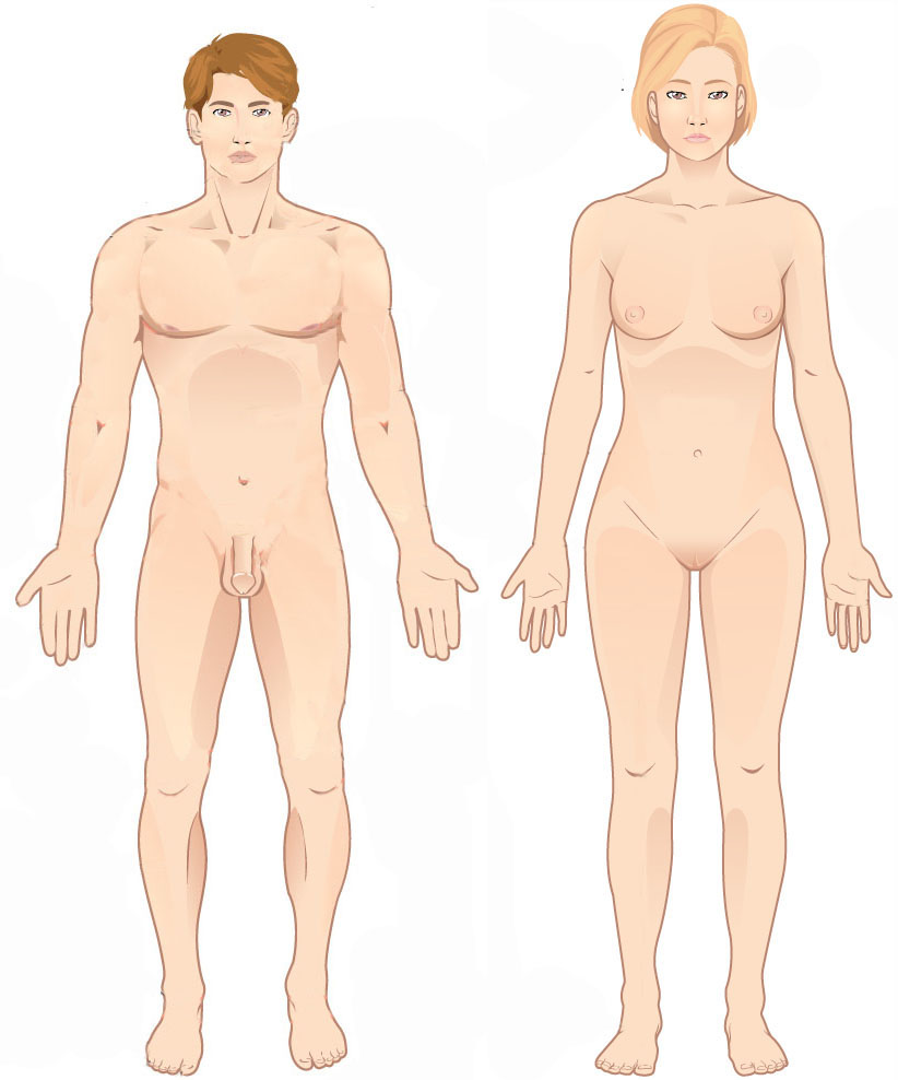
Standard anatomical terms of location are used to unambiguously describe the anatomy of animals, including humans. The terms, typically derived from Latin or Greek roots, describe something in its standard anatomical position. This position provides a definition of what is at the front ("anterior"), behind ("posterior") and so on. As part of defining and describing terms, the body is described through the use of anatomical planes and anatomical axes. The meaning of terms that are used can change depending on whether an organism is bipedal or quadrupedal. Additionally, for some animals such as invertebrates, some terms may not have any meaning at all; for example, an animal that is radially symmetrical will have no anterior surface, but can still have a description that a part is close to the middle ("proximal") or further from the middle ("distal"). International organisations have determined vocabularies that are often used as standard vocabularies for subdisciplines of anatom ... [...More Info...] [...Related Items...] OR: [Wikipedia] [Google] [Baidu] |
Pia Mater
Pia mater ( or ),Entry "pia mater" in Merriam-Webster Online Dictionary ', retrieved 2012-07-28. often referred to as simply the pia, is the delicate innermost layer of the , the membranes surrounding the and . ''Pia mater'' is medieval Latin meaning "tender mother". The other two meningeal membr ... [...More Info...] [...Related Items...] OR: [Wikipedia] [Google] [Baidu] |
Ligament
A ligament is the fibrous connective tissue that connects bones to other bones. It is also known as ''articular ligament'', ''articular larua'', ''fibrous ligament'', or ''true ligament''. Other ligaments in the body include the: * Peritoneal ligament: a fold of peritoneum or other membranes. * Fetal remnant ligament: the remnants of a fetal tubular structure. * Periodontal ligament: a group of fibers that attach the cementum of teeth to the surrounding alveolar bone. Ligaments are similar to tendons and fasciae as they are all made of connective tissue. The differences among them are in the connections that they make: ligaments connect one bone to another bone, tendons connect muscle to bone, and fasciae connect muscles to other muscles. These are all found in the skeletal system of the human body. Ligaments cannot usually be regenerated naturally; however, there are periodontal ligament stem cells located near the periodontal ligament which are involved in the adult regener ... [...More Info...] [...Related Items...] OR: [Wikipedia] [Google] [Baidu] |
Foramen Magnum
The foramen magnum ( la, great hole) is a large, oval-shaped opening in the occipital bone of the skull. It is one of the several oval or circular openings (foramina) in the base of the skull. The spinal cord, an extension of the medulla oblongata, passes through the foramen magnum as it exits the cranial cavity. Apart from the transmission of the medulla oblongata and its membranes, the foramen magnum transmits the vertebral arteries, the anterior and posterior spinal arteries, the tectorial membranes and alar ligaments. It also transmits the accessory nerve into the skull. The foramen magnum is a very important feature in bipedal mammals. One of the attributes of a biped's foramen magnum is a forward shift of the anterior border of the cerebellar tentorium; this is caused by the shortening of the cranial base. Studies on the foramen magnum position have shown a connection to the functional influences of both posture and locomotion. The forward shift of the foramen magnum i ... [...More Info...] [...Related Items...] OR: [Wikipedia] [Google] [Baidu] |
Spinal Nerve Root
Spinal nerve root may refer to: * Posterior root of spinal nerve * Anterior root of spinal nerve In anatomy and neurology, the ventral root of spinal nerve, anterior root, or motor root is the efferent motor root of a spinal nerve. At its distal end, the ventral root joins with the dorsal root The dorsal root of spinal nerve (or posterior ... Back anatomy Peripheral nervous system {{Short pages monitor ... [...More Info...] [...Related Items...] OR: [Wikipedia] [Google] [Baidu] |
Vertebral Column
The vertebral column, also known as the backbone or spine, is part of the axial skeleton. The vertebral column is the defining characteristic of a vertebrate in which the notochord (a flexible rod of uniform composition) found in all chordata, chordates has been replaced by a segmented series of bone: vertebrae separated by intervertebral discs. Individual vertebrae are named according to their region and position, and can be used as anatomical landmarks in order to guide procedures such as Lumbar puncture, lumbar punctures. The vertebral column houses the spinal canal, a cavity that encloses and protects the spinal cord. There are about 50,000 species of animals that have a vertebral column. The human vertebral column is one of the most-studied examples. Many different diseases in humans can affect the spine, with spina bifida and scoliosis being recognisable examples. The general structure of human vertebrae is fairly typical of that found in mammals, reptiles, and birds. Th ... [...More Info...] [...Related Items...] OR: [Wikipedia] [Google] [Baidu] |
Subarachnoid Space
In anatomy, the meninges (, ''singular:'' meninx ( or ), ) are the three membranes that envelop the brain and spinal cord. In mammals, the meninges are the dura mater, the arachnoid mater, and the pia mater. Cerebrospinal fluid is located in the subarachnoid space between the arachnoid mater and the pia mater. The primary function of the meninges is to protect the central nervous system. Structure Dura mater The dura mater ( la, tough mother) (also rarely called ''meninx fibrosa'' or ''pachymeninx'') is a thick, durable membrane, closest to the skull and vertebrae. The dura mater, the outermost part, is a loosely arranged, fibroelastic layer of cells, characterized by multiple interdigitating cell processes, no extracellular collagen, and significant extracellular spaces. The middle region is a mostly fibrous portion. It consists of two layers: the endosteal layer, which lies closest to the skull, and the inner meningeal layer, which lies closer to the brain. It contains large ... [...More Info...] [...Related Items...] OR: [Wikipedia] [Google] [Baidu] |
Anterior Nerve Roots
In anatomy and neurology, the ventral root of spinal nerve, anterior root, or motor root is the efferent motor root of a spinal nerve. At its distal end, the ventral root joins with the dorsal root The dorsal root of spinal nerve (or posterior root of spinal nerve or sensory root) is one of two "roots" which emerge from the spinal cord. It emerges directly from the spinal cord, and travels to the dorsal root ganglion. Nerve fibres with the v ... to form a mixed spinal nerve. Additional images Image:Cervical vertebra english.png, Cervical vertebra Image:Medulla spinalis - Section - English.svg, Medulla spinalis Image:Gray675.png, A spinal nerve with its anterior and posterior. Image:Gray764.png, The motor tract. Image:Gray770-en.svg, Diagrammatic transverse section of the medulla spinalis and its membranes. Image:Gray796.png, A portion of the spinal cord, showing its right lateral surface. The dura is opened and arranged to show the nerve roots. Image:Gray799.svg, Sche ... [...More Info...] [...Related Items...] OR: [Wikipedia] [Google] [Baidu] |
Posterior Nerve Roots
The dorsal root of spinal nerve (or posterior root of spinal nerve or sensory root) is one of two "roots" which emerge from the spinal cord. It emerges directly from the spinal cord, and travels to the dorsal root ganglion. Nerve fibres with the ventral root then combine to form a spinal nerve. The dorsal root transmits sensory information, forming the afferent sensory root of a spinal nerve. Structure The root emerges from the posterior part of the spinal cord and travels to the dorsal root ganglion. The dorsal root ganglia contain the pseudo-unipolar cell bodies of the nerve fibres which travel from the ganglia through the root into the spinal cord. The lateral division of the dorsal root contains lightly myelinated and unmyelinated fibres of small diameter. These carry pain and temperature sensation. These fibers cross through the anterior white commissure to form the anterolateral system in the lateral funiculus. The medial division of the dorsal root contains myelina ... [...More Info...] [...Related Items...] OR: [Wikipedia] [Google] [Baidu] |
Arachnoid Mater
The arachnoid mater (or simply arachnoid) is one of the three meninges, the protective membranes that cover the brain and spinal cord. It is so named because of its resemblance to a spider web. The arachnoid mater is a derivative of the neural crest mesoectoderm in the embryo. Structure It is interposed between the two other meninges, the more superficial and much thicker dura mater and the deeper pia mater, from which it is separated by the subarachnoid space. The delicate arachnoid layer is not attached to the inside of the dura but against it and surrounds the brain and spinal cord. It does not line the brain down into its sulci (folds), as does the pia mater, with the exception of the longitudinal fissure, which divides the left and right cerebral hemispheres. Cerebrospinal fluid (CSF) flows under the arachnoid in the subarachnoid space, within a meshwork of trabeculae which span between the arachnoid and the pia. The arachnoid mater makes arachnoid villi, small protrusions ... [...More Info...] [...Related Items...] OR: [Wikipedia] [Google] [Baidu] |
Nerve Root
A nerve root (Latin: ''radix nervi'') is the initial segment of a nerve leaving the central nervous system. Nerve roots can be classified as: *Cranial nerve roots: the initial or proximal segment of one of the twelve pairs of cranial nerves leaving the central nervous system from the brain stem or the highest levels of the spinal cord. *Spinal nerve roots: the initial or proximal segment of one of the thirty-one pairs of spinal nerves leaving the central nervous system from the spinal cord. Each spinal nerve is formed by the union of a sensory dorsal root and a motor ventral root, meaning that there are sixty-two dorsal/ventral root pairs, and therefore one hundred and twenty-four nerve roots in total, each of which stems from a bundle of nerve rootlets (or root filaments). Cranial nerve roots Cranial nerves originate directly from the brain's surface: two from the cerebrum and the ten others from the brain stem. Cranial roots differ from spinal roots: some of these roots do not se ... [...More Info...] [...Related Items...] OR: [Wikipedia] [Google] [Baidu] |
Caudal (anatomical Term)
Standard anatomical terms of location are used to unambiguously describe the anatomy of animals, including humans. The terms, typically derived from Latin or Greek language, Greek roots, describe something in its standard anatomical position. This position provides a definition of what is at the front ("anterior"), behind ("posterior") and so on. As part of defining and describing terms, the body is described through the use of anatomical planes and anatomical axis, anatomical axes. The meaning of terms that are used can change depending on whether an organism is bipedal or quadrupedal. Additionally, for some animals such as invertebrates, some terms may not have any meaning at all; for example, an animal that is radially symmetrical will have no anterior surface, but can still have a description that a part is close to the middle ("proximal") or further from the middle ("distal"). International organisations have determined vocabularies that are often used as standard vocabular ... [...More Info...] [...Related Items...] OR: [Wikipedia] [Google] [Baidu] |


