|
Charcot–Leyden Crystals
Charcot–Leyden crystals are microscopic crystals composed of eosinophil protein galectin-10 found in people who have allergic diseases such as asthma or parasitic infections such as parasitic pneumonia or ascariasis. Appearance Charcot–Leyden crystals are composed of an eosinophilic lysophospholipase binding protein called Galectin -10. They vary in size and may be as large as 50 µm in length. Charcot–Leyden crystals are slender and pointed at both ends, consisting of a pair of hexagonal pyramids joined at their bases. Normally colorless, they are stained purplish-red by trichrome. Clinical significance They are indicative of a disease involving eosinophilic inflammation or proliferation, such as is found in allergic reactions (asthma, bronchitis, allergic rhinitis and rhinosinusitis) and parasitic infections such as ''Entamoeba histolytica'', ''Necator americanus'', and ''Ancylostoma duodenale''. Charcot–Leyden crystals are often seen pathologically in patien ... [...More Info...] [...Related Items...] OR: [Wikipedia] [Google] [Baidu] |
Entamoeba Histolytica
''Entamoeba histolytica'' is an anaerobic parasitic amoebozoan, part of the genus ''Entamoeba''. Predominantly infecting humans and other primates causing amoebiasis, ''E. histolytica'' is estimated to infect about 35-50 million people worldwide. ''E. histolytica'' infection is estimated to kill more than 55,000 people each year. Previously, it was thought that 10% of the world population was infected, but these figures predate the recognition that at least 90% of these infections were due to a second species, ''E. dispar''. Mammals such as dogs and cats can become infected transiently, but are not thought to contribute significantly to transmission. The word '' histolysis'' literally means disintegration and dissolution of organic tissues. Transmission The active (trophozoite) stage exists only in the host and in fresh loose feces; cysts survive outside the host in water, in soils, and on foods, especially under moist conditions on the latter. The infection can occur when a ... [...More Info...] [...Related Items...] OR: [Wikipedia] [Google] [Baidu] |
Ernst Viktor Von Leyden
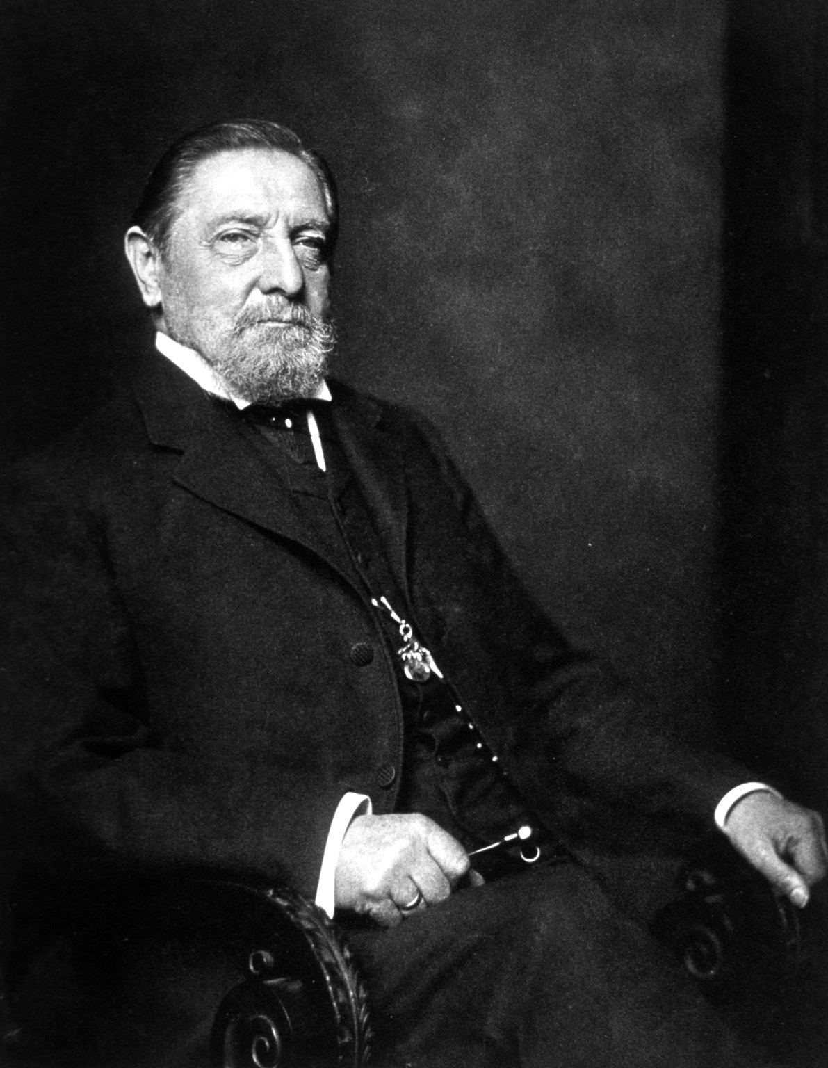
Ernst Viktor von Leyden (20 April 1832 – 5 October 1910) was a German internist from Danzig. Biography He studied medicine at the Friedrich-Wilhelms-Institut in Berlin, and was a pupil of Johann Lukas Schönlein (1793–1864) and Ludwig Traube (1818–1876). He was later a medical professor at the universities of Königsberg, Strassburg and Berlin. Leyden was an important influence to the career of Ludwig Edinger (1855–1918), and during his tenure at the University of Königsberg worked closely with Otto Spiegelberg (1830–1881) and Friedrich Daniel von Recklinghausen (1833–1910).''Ernst Viktor von Leyden'' at Among his better known students and assistants were |
Charles-Philippe Robin
Charles-Philippe Robin (4 June 1821 – 6 October 1885) was a French anatomist, biologist, and histologist born in Jasseron, département Ain. He studied medicine in Paris, and while still a student took a scientific journey with Hermann Lebert to Normandy and the Channel Islands, where they collected specimens for the Musée Orfila. In 1846 he received his medical doctorate, and at different stages of his career he was a professor of natural history, anatomy, and histology. He was a member of the Académie Nationale de Médecine (1858) and Academy of Science (1866). In 1873 he was appointed director of the marine zoology laboratory at Concarneau. Robin's contributions to medical science were many and varied. He was among the first scientists in France to use the microscope in normal and pathological anatomy. He was the first to describe the species ''Candida albicans'' (a diploid fungus), and he contributed new information on the micro-structure of ganglia and of neuroglia. He ... [...More Info...] [...Related Items...] OR: [Wikipedia] [Google] [Baidu] |
Jean-Martin Charcot
Jean-Martin Charcot (; 29 November 1825 – 16 August 1893) was a French neurology, neurologist and professor of anatomical pathology. He worked on hypnosis and hysteria, in particular with his hysteria patient Louise Augustine Gleizes. Charcot is known as "the founder of modern neurology",Lamberty (2007), p. 5 and his name has been associated with at least 15 medical eponyms, including #Eponyms, various conditions sometimes referred to as Charcot diseases. Charcot has been referred to as "the father of French neurology and one of the world's pioneers of neurology". His work greatly influenced the developing fields of neurology and psychology; modern psychiatry owes much to the work of Charcot and his direct followers.Bogousslavsky (2010), p. 7 He was the "foremost neurologist of late nineteenth-century France" and has been called "the Napoleon Bonaparte, Napoleon of the Neurosis, neuroses". Personal life Born in Paris, Charcot worked and taught at the famous Pitié-Salpêtri� ... [...More Info...] [...Related Items...] OR: [Wikipedia] [Google] [Baidu] |
Friedrich Albert Von Zenker
Friedrich Albert von Zenker (13 March 1825 – 13 June 1898) was a German pathologist and physician, celebrated for his discovery of trichinosis. He was born in Dresden, and was educated in Leipzig and Heidelberg. While in Leipzig, he worked for a while as an assistant to Justus Radius at the St. Georg Hospital. Attached to the city hospital of Dresden in 1851, he added, in 1855, the duties of professor of pathological anatomy and general pathology in the surgico-medical academy of that city. In 1862 he became professor of pathological anatomy and pharmacology at Erlangen. Three years afterwards he assumed with Hugo Wilhelm von Ziemssen the editorship of the "''Deutsches Archiv für klinische Medizin''". In 1895 he retired from active service. Zenker's diverticulum, a false pathological diverticulum of the posterior pharyngeal wall, through the thyropharyngeus and cricopharyngeus parts of the inferior constrictor muscle, is named after him. His important discovery of the dan ... [...More Info...] [...Related Items...] OR: [Wikipedia] [Google] [Baidu] |
Ancylostoma Duodenale
''Ancylostoma'' is a genus of nematodes that includes some species of hookworms. Species include: : ''Ancylostoma braziliense'', commonly infects cats, popularly known in Brazil as ''bicho-geográfico'' : ''Ancylostoma caninum'', commonly infects dogs : ''Ancylostoma ceylanicum'' : '' Ancylostoma duodenale'' : ''Ancylostoma pluridentatum'', commonly infects sylvatic cats : ''Ancylostoma tubaeforme'', infects cats along with other hosts See also * Ancylostomiasis * List of parasites (human) Endoparasites Protozoan organisms Helminths (worms) Helminth organisms (also called helminths or intestinal worms) include: Tapeworms Flukes Roundworms Other organisms Ectoparasites References {{Portal bar, Bio ... External links * Ancylostomatidae Rhabditida genera {{Rhabditida-stub ... [...More Info...] [...Related Items...] OR: [Wikipedia] [Google] [Baidu] |
Necator Americanus
''Necator americanus'' is a species of hookworm (a type of helminth) commonly known as the New World hookworm. Like other hookworms, it is a member of the phylum Nematoda. It is an obligatory parasitic nematode that lives in the small intestine of human hosts. Necatoriasis—a type of helminthiasis—is the term for the condition of being host to an infestation of a species of ''Necator''. Since ''N. americanus'' and ''Ancylostoma duodenale'' (also known as Old World hookworm) are the two species of hookworms that most commonly infest humans, they are usually dealt with under the collective heading of "hookworm infection". They differ most obviously in geographical distribution, structure of mouthparts, and relative size. ''Necator americanus'' has been proposed as an alternative to ''Trichuris suis'' in helminthic therapy. Morphology This parasite has two dorsal and two ventral cutting plates around the anterior margin of the buccal capsule. It also has a pair of subdorsal ... [...More Info...] [...Related Items...] OR: [Wikipedia] [Google] [Baidu] |
Masson's Trichrome
Masson's trichrome is a three-colour staining procedure used in histology. The recipes evolved from Claude L. Pierre Masson's (1880–1959) original formulation have different specific applications, but all are suited for distinguishing cells from surrounding connective tissue. Most recipes produce red keratin and muscle fibers, blue or green collagen and bone, light red or pink cytoplasm, and dark brown to black cell nuclei. The trichrome is applied by immersion of the fixated sample into Weigert's iron hematoxylin, and then three different solutions, labeled A, B, and C: * Weigert's hematoxylin is a sequence of three solutions: ferric chloride in diluted hydrochloric acid, hematoxylin in 95% ethanol, and potassium ferricyanide solution alkalized by sodium borate. It is used to stain the nuclei. * Solution A, also called plasma stain, contains acid fuchsin, Xylidine Ponceau, glacial acetic acid, and distilled water. Other red acid dyes can be used, e.g. the Biebrich sc ... [...More Info...] [...Related Items...] OR: [Wikipedia] [Google] [Baidu] |
Microscope
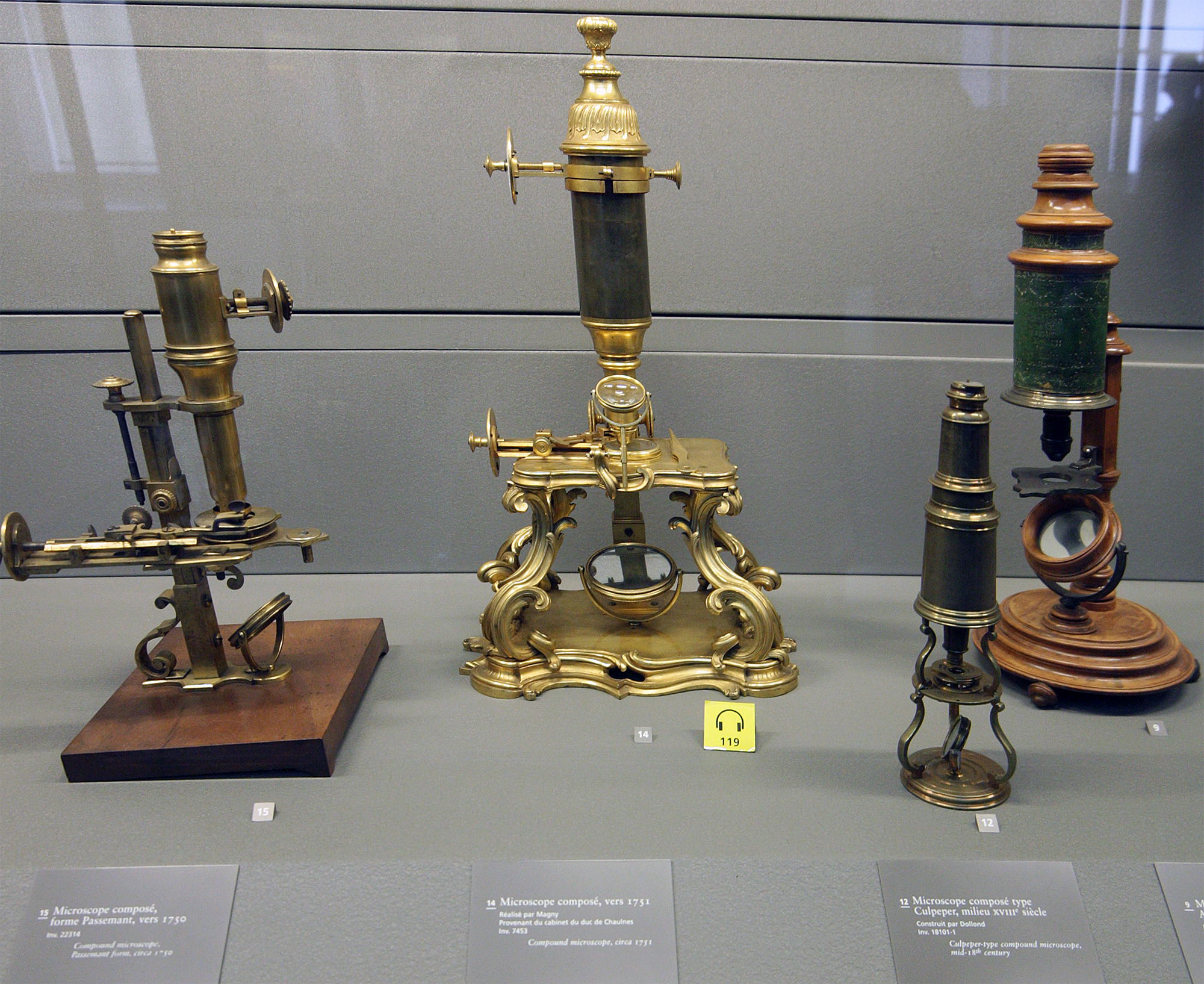
A microscope () is a laboratory instrument used to examine objects that are too small to be seen by the naked eye. Microscopy is the science of investigating small objects and structures using a microscope. Microscopic means being invisible to the eye unless aided by a microscope. There are many types of microscopes, and they may be grouped in different ways. One way is to describe the method an instrument uses to interact with a sample and produce images, either by sending a beam of light or electrons through a sample in its optical path, by detecting photon emissions from a sample, or by scanning across and a short distance from the surface of a sample using a probe. The most common microscope (and the first to be invented) is the optical microscope, which uses lenses to refract visible light that passed through a thinly sectioned sample to produce an observable image. Other major types of microscopes are the fluorescence microscope, electron microscope (both the transmi ... [...More Info...] [...Related Items...] OR: [Wikipedia] [Google] [Baidu] |
Staining (biology)
Staining is a technique used to enhance contrast in samples, generally at the microscopic level. Stains and dyes are frequently used in histology (microscopic study of biological tissues), in cytology (microscopic study of cells), and in the medical fields of histopathology, hematology, and cytopathology that focus on the study and diagnoses of diseases at the microscopic level. Stains may be used to define biological tissues (highlighting, for example, muscle fibers or connective tissue), cell populations (classifying different blood cells), or organelles within individual cells. In biochemistry, it involves adding a class-specific ( DNA, proteins, lipids, carbohydrates) dye to a substrate to qualify or quantify the presence of a specific compound. Staining and fluorescent tagging can serve similar purposes. Biological staining is also used to mark cells in flow cytometry, and to flag proteins or nucleic acids in gel electrophoresis. Light microscopes are used for viewing st ... [...More Info...] [...Related Items...] OR: [Wikipedia] [Google] [Baidu] |


