|
Cuboideonavicular Joint
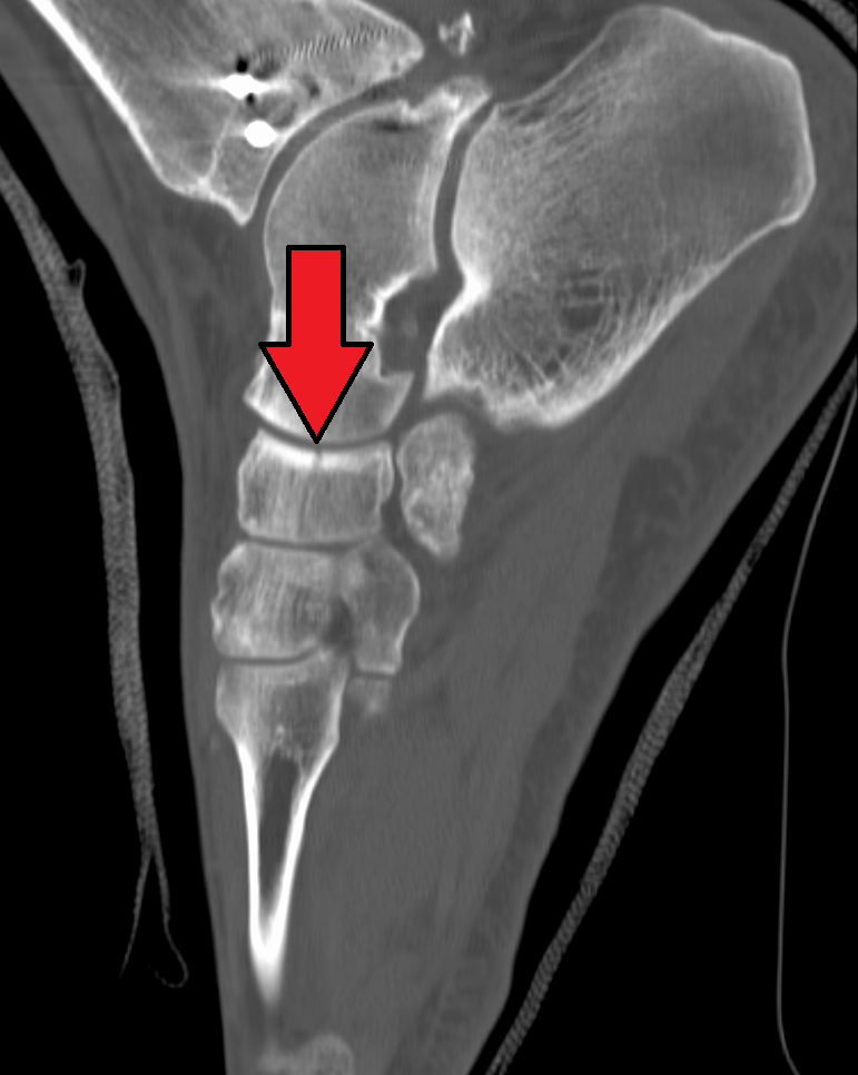
The cuboideonavicular joint is a joint (articulation) in the foot formed between the navicular bone and cuboid bone. The navicular bone is connected with the cuboid bone by the dorsal, plantar, and interosseous cuboideonavicular ligaments. It is a syndesmosis type fibrous joint.Standring, Susan. "Gray's Anatomy E-Book: The Anatomical Basis of Clinical Practice" Elsevier Health Sciences, 2015, p. 1437. The dorsal ligaments The dorsal cuboideonavicular ligament connects the lateral portion of the navicular to the posteromedial portion of the cuboid on the dorsal side. The plantar ligaments The plantar cuboideonavicular ligaments have a similar arrangement to the dorsal, and are strengthened by slips from the tendon of the tibialis posterior. Synovial membrane The synovial membrane of this joints is part of the great tarsal synovial membrane The great tarsal synovial membrane is a synovial membrane in the foot. The synovial membranes between the second and third, and the third an ... [...More Info...] [...Related Items...] OR: [Wikipedia] [Google] [Baidu] |
Plantar Ligament (other)
Plantar ligament refer to ligaments in the sole of the foot: * Plantar plates, fibrocartilaginous structures in the metatarsophalangeal and interphalangeal joints of the toes. * One of several tarsal, plantar metatarsal ligaments, metatarsal, and Plantar tarsometatarsal ligaments, tarsometatarsal ligaments: ** Long plantar ligament, that connects the calcaneus with the cuboid bone ** Plantar calcaneocuboid ligament, deep to previous ** Plantar calcaneonavicular ligament, that connects the calcaneus with the navicular bone ** Plantar cuneonavicular ligaments, that connect the navicular bone with adjacent cuneiform bones ** Plantar intercuneiform ligaments, between the cuneiform bones ** Plantar cuboideonavicular ligament, that connects the cuboid with the navicular bone ** Plantar cuneocuboid ligament, that connects the cuboid with the cuneiform bones See also * Palmar ligament (other) {{Disambig ... [...More Info...] [...Related Items...] OR: [Wikipedia] [Google] [Baidu] |
Plantar Cuboideonavicular Ligament
The plantar cuboideonavicular ligament is a fibrous band that connects the plantar surfaces of the cuboid and navicular The navicular bone is a small bone found in the feet of most mammals. Human anatomy The navicular bone in humans is one of the tarsal bones, found in the foot. Its name derives from the human bone's resemblance to a small boat, caused by th ... bones. Ligaments of the lower limb {{ligament-stub ... [...More Info...] [...Related Items...] OR: [Wikipedia] [Google] [Baidu] |
Great Tarsal Synovial Membrane
The great tarsal synovial membrane is a synovial membrane in the foot. The synovial membranes between the second and third, and the third and fourth metatarsal bones are part of the great tarsal synovial membrane; that between the fourth and fifth metatarsal bone The fifth metatarsal bone is a long bone in the foot, and is palpable along the distal outer edges of the feet. It is the second smallest of the five metatarsal bones. The fifth metatarsal is analogous to the fifth metacarpal bone in the hand. As ...s is a prolongation of the synovial membrane of the cuboideometatarsal joint. References Soft tissue {{Anatomy-stub ... [...More Info...] [...Related Items...] OR: [Wikipedia] [Google] [Baidu] |
Synovial Membrane
The synovial membrane (also known as the synovial stratum, synovium or stratum synoviale) is a specialized connective tissue that lines the inner surface of capsules of synovial joints and tendon sheath. It makes direct contact with the fibrous membrane on the outside surface and with the synovial fluid lubricant on the inside surface. In contact with the synovial fluid at the tissue surface are many rounded macrophage-like synovial cells (type A) and also type B cells, which are also known as fibroblast-like synoviocytes (FLS). Type A cells maintain the synovial fluid by removing wear-and-tear debris. As for the FLS, they produce hyaluronan, as well as other extracellular components in the synovial fluid. Structure The synovial membrane is variable but often has two layers: * The outer layer, or subintima, can be of almost any type of connective tissue – fibrous (dense collagenous type), adipose (fatty; e.g. in intra-articular fat pads) or areolar (loose collagenous typ ... [...More Info...] [...Related Items...] OR: [Wikipedia] [Google] [Baidu] |
Plantar Cuboideonavicular Ligament
The plantar cuboideonavicular ligament is a fibrous band that connects the plantar surfaces of the cuboid and navicular The navicular bone is a small bone found in the feet of most mammals. Human anatomy The navicular bone in humans is one of the tarsal bones, found in the foot. Its name derives from the human bone's resemblance to a small boat, caused by th ... bones. Ligaments of the lower limb {{ligament-stub ... [...More Info...] [...Related Items...] OR: [Wikipedia] [Google] [Baidu] |
Dorsal Cuboideonavicular Ligament
The dorsal cuboideonavicular ligament is a fibrous bundle connecting the dorsal surfaces of the cuboid and navicular The navicular bone is a small bone found in the feet of most mammals. Human anatomy The navicular bone in humans is one of the tarsal bones, found in the foot. Its name derives from the human bone's resemblance to a small boat, caused by th ... bones. Ligaments of the lower limb {{ligament-stub ... [...More Info...] [...Related Items...] OR: [Wikipedia] [Google] [Baidu] |
Syndesmosis
In anatomy, fibrous joints are joints connected by fibrous tissue, consisting mainly of collagen. These are fixed joints where bones are united by a layer of white fibrous tissue of varying thickness. In the skull the joints between the bones are called sutures. Such immovable joints are also referred to as synarthroses. Types Most fibrous joints are also called "fixed" or "immovable". These joints have no joint cavity and are connected via fibrous connective tissue. The skull bones are connected by fibrous joints called '' sutures''. In fetal skulls the sutures are wide to allow slight movement during birth. They later become rigid ( synarthrodial). Some of the long bones in the body such as the radius and ulna in the forearm are joined by a ''syndesmosis'' (along the interosseous membrane). Syndemoses are slightly moveable ( amphiarthrodial). The distal tibiofibular joint is another example. A ''gomphosis'' is a joint between the root of a tooth and the socket in the maxil ... [...More Info...] [...Related Items...] OR: [Wikipedia] [Google] [Baidu] |
Dorsal Cuboideonavicular Ligament
The dorsal cuboideonavicular ligament is a fibrous bundle connecting the dorsal surfaces of the cuboid and navicular The navicular bone is a small bone found in the feet of most mammals. Human anatomy The navicular bone in humans is one of the tarsal bones, found in the foot. Its name derives from the human bone's resemblance to a small boat, caused by th ... bones. Ligaments of the lower limb {{ligament-stub ... [...More Info...] [...Related Items...] OR: [Wikipedia] [Google] [Baidu] |
Tendon
A tendon or sinew is a tough, high-tensile-strength band of dense fibrous connective tissue that connects muscle to bone. It is able to transmit the mechanical forces of muscle contraction to the skeletal system without sacrificing its ability to withstand significant amounts of tension. Tendons are similar to ligaments; both are made of collagen. Ligaments connect one bone to another, while tendons connect muscle to bone. Structure Histologically, tendons consist of dense regular connective tissue. The main cellular component of tendons are specialized fibroblasts called tendon cells (tenocytes). Tenocytes synthesize the extracellular matrix of tendons, abundant in densely packed collagen fibers. The collagen fibers are parallel to each other and organized into tendon fascicles. Individual fascicles are bound by the endotendineum, which is a delicate loose connective tissue containing thin collagen fibrils and elastic fibres. Groups of fascicles are bounded by the epitenon, ... [...More Info...] [...Related Items...] OR: [Wikipedia] [Google] [Baidu] |
Cuboid Bone
In the human body, the cuboid bone is one of the seven tarsal bones of the foot. Structure The cuboid bone is the most lateral of the bones in the distal row of the tarsus. It is roughly cubical in shape, and presents a prominence in its inferior (or plantar) surface, the tuberosity of the cuboid. The bone provides a groove where the tendon of the peroneus longus muscle passes to reach its insertion in the first metatarsal and medial cuneiform bones. Surfaces The dorsal surface, directed upward and lateralward, is rough, for the attachment of ligaments. The plantar surface presents in front a deep groove, the peroneal sulcus, which runs obliquely forward and medialward; it lodges the tendon of the peroneus longus, and is bounded behind by a prominent ridge, to which the long plantar ligament is attached. The ridge ends laterally in an eminence, the tuberosity, the surface of which presents an oval facet; on this facet glides the sesamoid bone or cartilage frequently found ... [...More Info...] [...Related Items...] OR: [Wikipedia] [Google] [Baidu] |
Navicular Bone
The navicular bone is a small bone found in the feet of most mammals. Human anatomy The navicular bone in humans is one of the tarsal bones, found in the foot. Its name derives from the human bone's resemblance to a small boat, caused by the strongly concave proximal articular surface. The term ''navicular bone'' or ''hand navicular bone'' was formerly used for the scaphoid bone, one of the carpal bones of the wrist. The navicular bone in humans is located on the medial side of the foot, and articulates proximally with the talus, distally with the three cuneiform bones, and laterally with the cuboid. It is the last of the foot bones to start ossification and does not tend to do so until the end of the third year in girls and the beginning of the fourth year in boys, although a large range of variation has been reported. The tibialis posterior is the only muscle that attaches to the navicular bone. The main portion of the muscle inserts into the tuberosity of the navi ... [...More Info...] [...Related Items...] OR: [Wikipedia] [Google] [Baidu] |


