|
Contagious Caprine Pleuropneumonia
{{Taxobox , name = ''Mycoplasma capricolum'' subsp. ''capricolum'' , regnum = Bacteria , phylum = Mycoplasmatota , classis = Mollicutes , ordo = Mycoplasmatales , familia = Mycoplasmataceae , genus = ''Mycoplasma'' , species = ''M. capricolum'' , subspecies = ''capricolum'' Contagious caprine pleuropneumonia (CCPP) is a cause of major economic losses to goat producers in Africa, Asia and the Middle East. Disease is caused by members of the Mycoplasma genus – usually ''Mycoplasma capricolum'' subsp. ''capricolum'' but sometimes by ''M. mycoides'' subsp. ''capri'' or ''M. mycoides'' subsp. ''mycoides''. It is extremely contagious with very high morbidity and mortality rates, causing an interstitial fibrinous pleuropneumonia in infected goats. Infection is spread by close-contact aerosol, therefore overcrowding and confinement increases disease incidence. Stress factors such as malnutrition and long transport can also predispose animals to disease. Goats are the only ... [...More Info...] [...Related Items...] OR: [Wikipedia] [Google] [Baidu] |
Bacteria
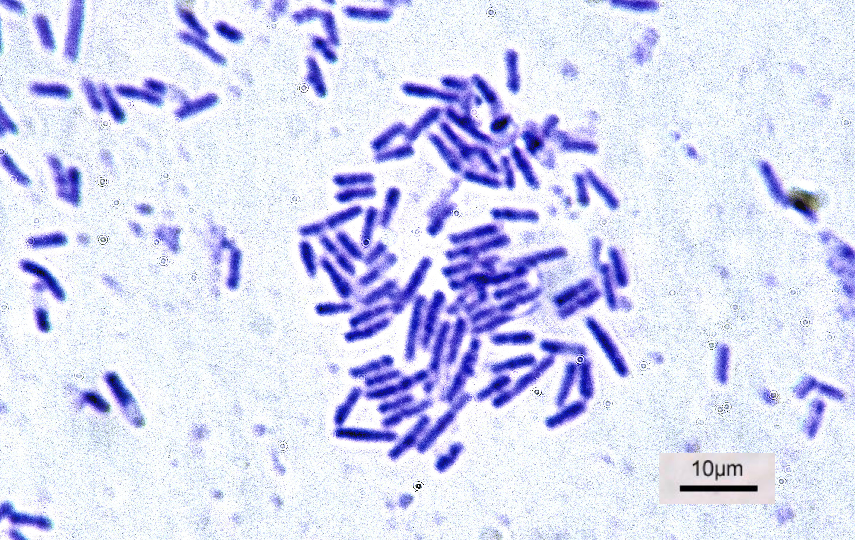
Bacteria (; singular: bacterium) are ubiquitous, mostly free-living organisms often consisting of one biological cell. They constitute a large domain of prokaryotic microorganisms. Typically a few micrometres in length, bacteria were among the first life forms to appear on Earth, and are present in most of its habitats. Bacteria inhabit soil, water, acidic hot springs, radioactive waste, and the deep biosphere of Earth's crust. Bacteria are vital in many stages of the nutrient cycle by recycling nutrients such as the fixation of nitrogen from the atmosphere. The nutrient cycle includes the decomposition of dead bodies; bacteria are responsible for the putrefaction stage in this process. In the biological communities surrounding hydrothermal vents and cold seeps, extremophile bacteria provide the nutrients needed to sustain life by converting dissolved compounds, such as hydrogen sulphide and methane, to energy. Bacteria also live in symbiotic and parasitic relationsh ... [...More Info...] [...Related Items...] OR: [Wikipedia] [Google] [Baidu] |
Tachypnoea
Tachypnea, also spelt tachypnoea, is a respiratory rate greater than normal, resulting in abnormally rapid and shallow breathing. In adult humans at rest, any respiratory rate of 1220 per minute is considered clinically normal, with tachypnea being any rate above that. Children have significantly higher resting ventilatory rates, which decline rapidly during the first three years of life and then steadily until around 18 years. Tachypnea can be an early indicator of pneumonia and other lung diseases in children, and is often an outcome of a brain injury. Distinction from other breathing terms Different sources produce different classifications for breathing terms. Some of the public describe tachypnea as any rapid breathing. Hyperventilation is then described as increased ventilation of the alveoli (which can occur through increased rate or depth of breathing, or a mix of both) where there is a smaller rise in metabolic carbon dioxide relative to this increase in ventilation. ... [...More Info...] [...Related Items...] OR: [Wikipedia] [Google] [Baidu] |
Polymerase Chain Reaction
The polymerase chain reaction (PCR) is a method widely used to rapidly make millions to billions of copies (complete or partial) of a specific DNA sample, allowing scientists to take a very small sample of DNA and amplify it (or a part of it) to a large enough amount to study in detail. PCR was invented in 1983 by the American biochemist Kary Mullis at Cetus Corporation; Mullis and biochemist Michael Smith (chemist), Michael Smith, who had developed other essential ways of manipulating DNA, were jointly awarded the Nobel Prize in Chemistry in 1993. PCR is fundamental to many of the procedures used in genetic testing and research, including analysis of Ancient DNA, ancient samples of DNA and identification of infectious agents. Using PCR, copies of very small amounts of DNA sequences are exponentially amplified in a series of cycles of temperature changes. PCR is now a common and often indispensable technique used in medical laboratory research for a broad variety of applications ... [...More Info...] [...Related Items...] OR: [Wikipedia] [Google] [Baidu] |
ELISA
The enzyme-linked immunosorbent assay (ELISA) (, ) is a commonly used analytical biochemistry assay, first described by Eva Engvall and Peter Perlmann in 1971. The assay uses a solid-phase type of enzyme immunoassay (EIA) to detect the presence of a ligand (commonly a protein) in a liquid sample using antibodies directed against the protein to be measured. ELISA has been used as a diagnostic tool in medicine, plant pathology, and biotechnology, as well as a quality control check in various industries. In the most simple form of an ELISA, antigens from the sample to be tested are attached to a surface. Then, a matching antibody is applied over the surface so it can bind the antigen. This antibody is linked to an enzyme and then any unbound antibodies are removed. In the final step, a substance containing the enzyme's substrate is added. If there was binding, the subsequent reaction produces a detectable signal, most commonly a color change. Performing an ELISA involves at least ... [...More Info...] [...Related Items...] OR: [Wikipedia] [Google] [Baidu] |
Haemagglutination Assay
The hemagglutination assay or haemagglutination assay (HA) and the hemagglutination inhibition assay (HI or HAI) were developed in 1941–42 by American virologist George Hirst as methods for quantifying the relative concentration of viruses, bacteria, or antibodies. HA and HI apply the process of hemagglutination, in which sialic acid receptors on the surface of red blood cells (RBCs) bind to the hemagglutinin glycoprotein found on the surface of influenza virus (and several other viruses) and create a network, or lattice structure, of interconnected RBCs and virus particles. The agglutinated lattice maintains the RBCs in a suspended distribution, typically viewed as a diffuse reddish solution. The formation of the lattice depends on the concentrations of the virus and RBCs, and when the relative virus concentration is too low, the RBCs are not constrained by the lattice and settle to the bottom of the well. Hemagglutination is observed in the presence of staphylococci, vibrios, ... [...More Info...] [...Related Items...] OR: [Wikipedia] [Google] [Baidu] |
Complement Fixation Test
The complement fixation test is an immunological medical test that can be used to detect the presence of either specific antibody or specific antigen in a patient's serum, based on whether complement fixation occurs. It was widely used to diagnose infections, particularly with microbes that are not easily detected by culture methods, and in rheumatic diseases. However, in clinical diagnostics labs it has been largely superseded by other serological methods such as ELISA and by DNA-based methods of pathogen detection, particularly PCR. Process The complement system is a system of serum proteins that react with antigen-antibody complexes. If this reaction occurs on a cell surface, it will result in the formation of trans-membrane pores and therefore destruction of the cell. The basic steps of a complement fixation test are as follows: # Serum is separated from the patient. # Patients naturally have different levels of complement proteins in their serum. To negate any effects thi ... [...More Info...] [...Related Items...] OR: [Wikipedia] [Google] [Baidu] |
Immunofluorescence
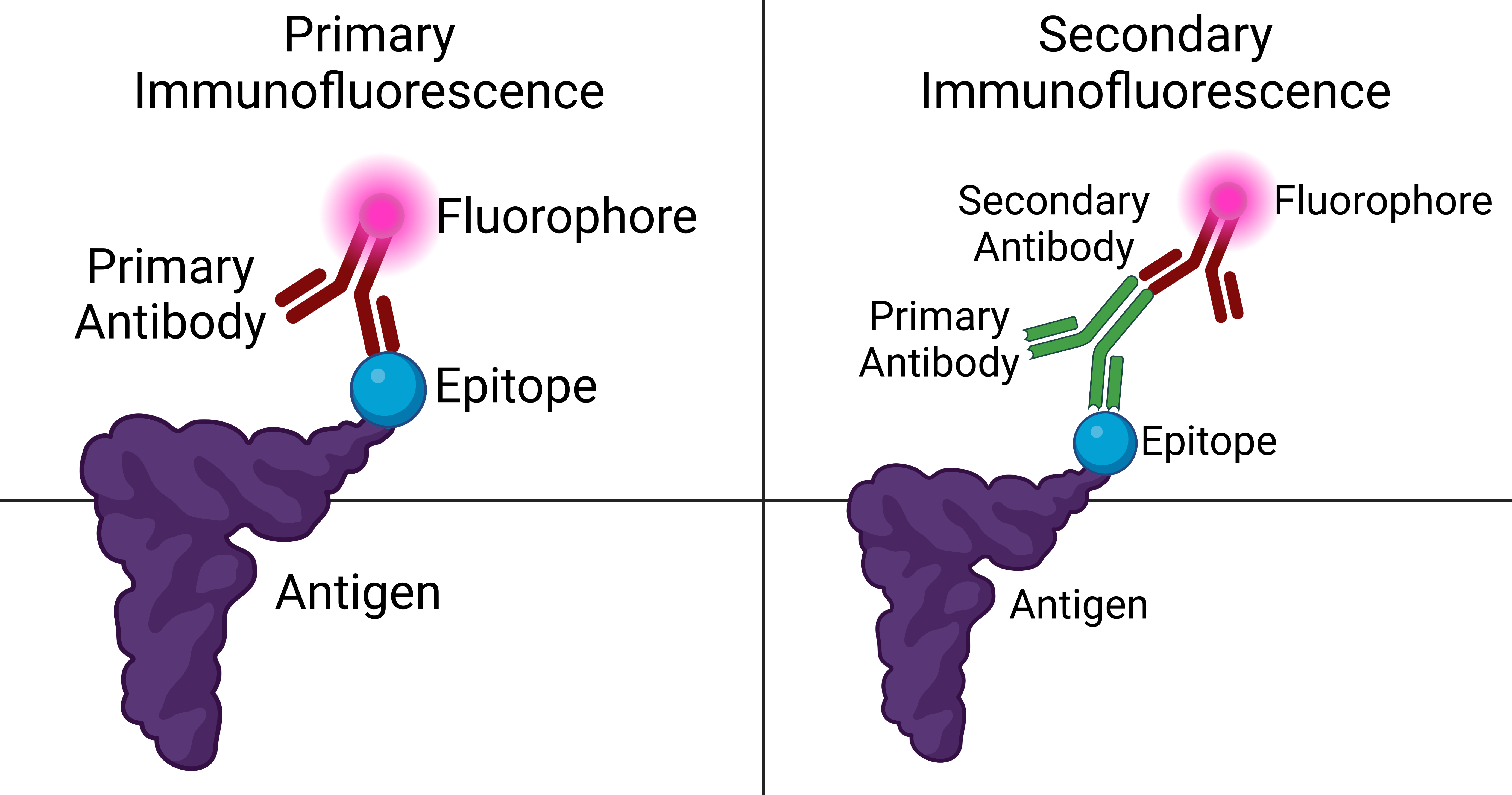
Immunofluorescence is a technique used for light microscopy with a fluorescence microscope and is used primarily on microbiological samples. This technique uses the specificity of antibodies to their antigen to target fluorescent dyes to specific biomolecule targets within a cell, and therefore allows visualization of the distribution of the target molecule through the sample. The specific region an antibody recognizes on an antigen is called an epitope. There have been efforts in epitope mapping since many antibodies can bind the same epitope and levels of binding between antibodies that recognize the same epitope can vary. Additionally, the binding of the fluorophore to the antibody itself cannot interfere with the immunological specificity of the antibody or the binding capacity of its antigen. Immunofluorescence is a widely used example of immunostaining (using antibodies to stain proteins) and is a specific example of immunohistochemistry (the use of the antibody-antigen rel ... [...More Info...] [...Related Items...] OR: [Wikipedia] [Google] [Baidu] |
Agglutination (biology)
Agglutination is the clumping of particles. The word ''agglutination'' comes from the Latin '' agglutinare'' (glueing to). Agglutination is the process that occurs if an antigen is mixed with its corresponding antibody called isoagglutinin. This term is commonly used in blood grouping. This occurs in biology in two main examples: # The clumping of cells such as bacteria or red blood cells in the presence of an antibody or complement. The antibody or other molecule binds multiple particles and joins them, creating a large complex. This increases the efficacy of microbial elimination by phagocytosis as large clumps of bacteria can be eliminated in one pass, versus the elimination of single microbial antigens. # When people are given blood transfusions of the wrong blood group, the antibodies react with the incorrectly transfused blood group and as a result, the erythrocytes clump up and stick together causing them to agglutinate. The coalescing of small particles that are suspend ... [...More Info...] [...Related Items...] OR: [Wikipedia] [Google] [Baidu] |
Bronchioles
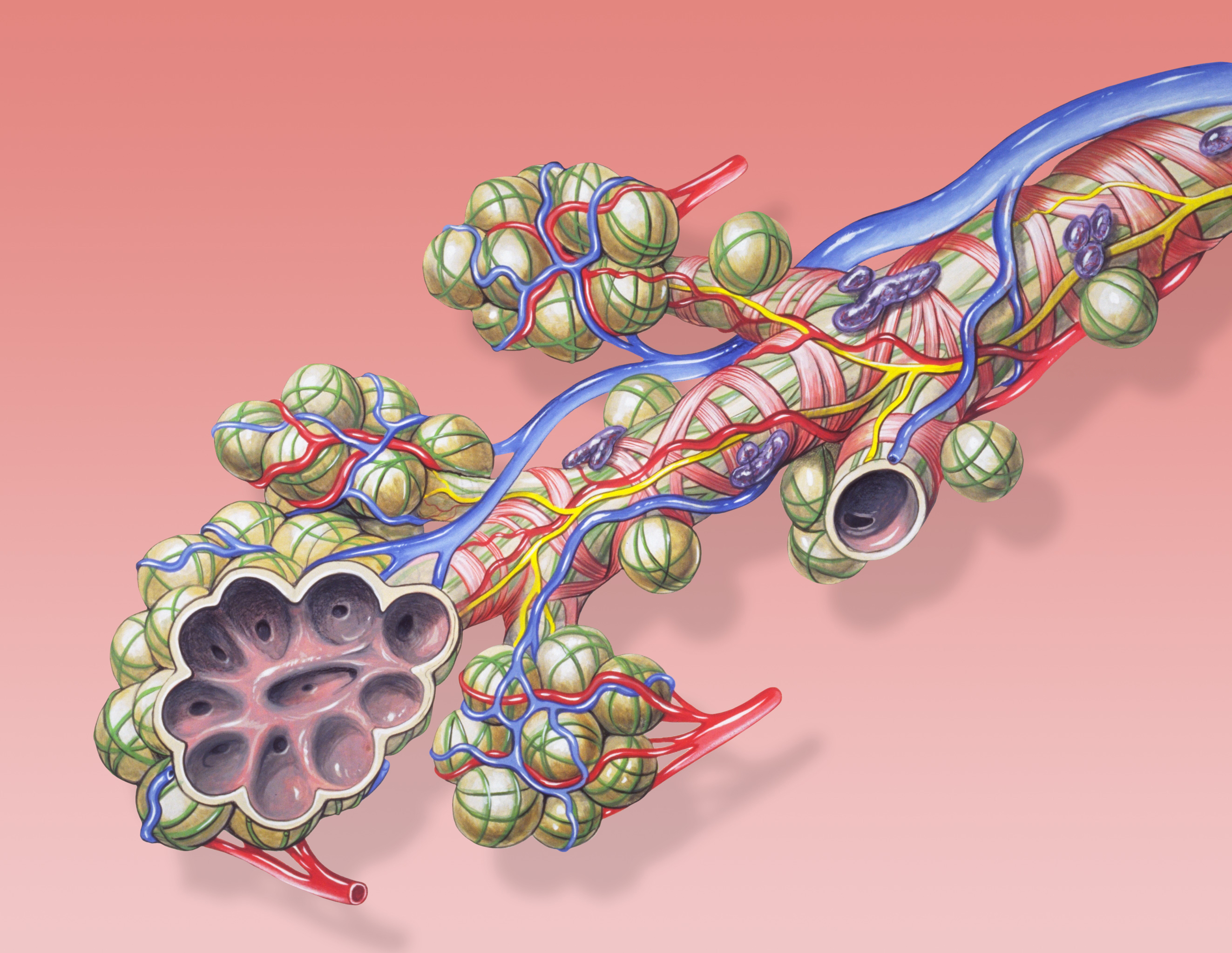
The bronchioles or bronchioli (pronounced ''bron-kee-oh-lee'') are the smaller branches of the bronchial airways in the lower respiratory tract. They include the terminal bronchioles, and finally the respiratory bronchioles that mark the start of the respiratory zone delivering air to the gas exchanging units of the alveoli. The bronchioles no longer contain the cartilage that is found in the bronchi, or glands in their submucosa. Structure The pulmonary lobule is the portion of the lung ventilated by one bronchiole. Bronchioles are approximately 1 mm or less in diameter and their walls consist of ciliated cuboidal epithelium and a layer of smooth muscle. Bronchioles divide into even smaller bronchioles, called ''terminal'', which are 0.5 mm or less in diameter. Terminal bronchioles in turn divide into smaller respiratory bronchioles which divide into alveolar ducts. Terminal bronchioles mark the end of the conducting division of air flow in the respiratory syste ... [...More Info...] [...Related Items...] OR: [Wikipedia] [Google] [Baidu] |
Pulmonary Alveolus
A pulmonary alveolus (plural: alveoli, from Latin ''alveolus'', "little cavity"), also known as an air sac or air space, is one of millions of hollow, distensible cup-shaped cavities in the lungs where oxygen is exchanged for carbon dioxide. Alveoli make up the functional tissue of the mammalian lungs known as the lung parenchyma, which takes up 90 percent of the total lung volume. Alveoli are first located in the respiratory bronchioles that mark the beginning of the respiratory zone. They are located sparsely in these bronchioles, line the walls of the alveolar ducts, and are more numerous in the blind-ended alveolar sacs. The acini are the basic units of respiration, with gas exchange taking place in all the alveoli present. The alveolar membrane is the gas exchange surface, surrounded by a network of capillaries. Across the membrane oxygen is diffused into the capillaries and carbon dioxide released from the capillaries into the alveoli to be breathed out. Alveoli are pa ... [...More Info...] [...Related Items...] OR: [Wikipedia] [Google] [Baidu] |
Pleuritis
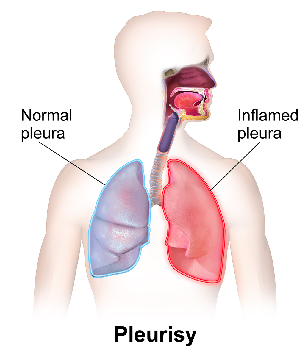
Pleurisy, also known as pleuritis, is inflammation of the membranes that surround the lungs and line the chest cavity (pleurae). This can result in a sharp chest pain while breathing. Occasionally the pain may be a constant dull ache. Other symptoms may include shortness of breath, cough, fever, or weight loss, depending on the underlying cause. The most common cause is a viral infection. Other causes include bacterial infection, pneumonia, pulmonary embolism, autoimmune disorders, lung cancer, following heart surgery, pancreatitis and asbestosis. Occasionally the cause remains unknown. The underlying mechanism involves the rubbing together of the pleurae instead of smooth gliding. Other conditions that can produce similar symptoms include pericarditis, heart attack, cholecystitis, pulmonary embolism, and pneumothorax. Diagnostic testing may include a chest X-ray, electrocardiogram (ECG), and blood tests. Treatment depends on the underlying cause. Paracetamol (acetaminophen ... [...More Info...] [...Related Items...] OR: [Wikipedia] [Google] [Baidu] |
Exudate
An exudate is a fluid emitted by an organism through pores or a wound, a process known as exuding or exudation. ''Exudate'' is derived from ''exude'' 'to ooze' from Latin ''exsūdāre'' 'to (ooze out) sweat' (''ex-'' 'out' and ''sūdāre'' 'to sweat'). Medicine An exudate is any fluid that filters from the circulatory system into lesions or areas of inflammation. It can be a pus-like or clear fluid. When an injury occurs, leaving skin exposed, it leaks out of the blood vessels and into nearby tissues. The fluid is composed of serum, fibrin, and leukocytes. Exudate may ooze from cuts or from areas of infection or inflammation. Types * Purulent or suppurative exudate consists of plasma with both active and dead neutrophils, fibrinogen, and necrotic parenchymal cells. This kind of exudate is consistent with more severe infections, and is commonly referred to as pus. * Fibrinous exudate is composed mainly of fibrinogen and fibrin. It is characteristic of rheumatic carditis, bu ... [...More Info...] [...Related Items...] OR: [Wikipedia] [Google] [Baidu] |


