|
Condylar Hyperplasia
Condylar hyperplasia (mandibular hyperplasia) is over-enlargement of the mandible bone in the skull. It was first described by Robert Adams in 1836 who related it to the overdevelopment of mandible. In humans, mandibular bone has two condyles which are known as growth centers of the mandible. When growth at the condyle exceeds its normal time span, it is referred to as condylar hyperplasia. The most common form of condylar hyperplasia is ''unilateral condylar hyperplasia'' where one condyle overgrows the other condyle leading to facial asymmetry. Hugo Obwegeser et al. classified condylar hyperplasia into two categories: ''hemimandibular hyperplasia'' and ''hemimandibular elongation''. It is estimated that about 30% of people with facial asymmetry express condylar hyperplasia. In 1986, Obwegeser and Makek specifically detailed two hemimandibular anomalies, hemimandibular hyperplasia and hemimandibular elongation. These anomalies can be clinically present in a pure form or in ... [...More Info...] [...Related Items...] OR: [Wikipedia] [Google] [Baidu] |
Facial Asymmetry
Facial symmetry is one specific measure of bodily symmetry. Along with traits such as averageness and youthfulness it influences judgments of aesthetic traits of physical attractiveness and beauty. For instance, in mate selection, people have been shown to have a preference for symmetry. Facial bilateral symmetry is typically defined as fluctuating asymmetry of the face comparing random differences in facial features of the two sides of the face. The human face also has systematic, directional asymmetry: on average, the face (mouth, nose and eyes) sits systematically to the left with respect to the axis through the ears, the so-called ''aurofacial asymmetry''. Directional asymmetry Directional asymmetry is a systematic asymmetry of some parts of the face across the population. A theory of directional asymmetries in the human body is the axial twist hypothesis. As predicted by this theory, the eyes, nose and mouth are, on average, located slightly to the left of the axis through ... [...More Info...] [...Related Items...] OR: [Wikipedia] [Google] [Baidu] |
Hugo Obwegeser
Hugo Obwegeser (21 October 1920 – 2 September 2017) was an Austrian Oral and Maxillo-Facial Surgeon and Plastic Surgeon who is known as the father of the modern orthognathic surgery. In his publication of 1970, he was the first surgeon to describe the simultaneous procedure which involved surgeries of both Maxilla and Mandible involving Le Fort I and Bilateral Sagittal Split Osteotomy technique. Career In 1945, Obwegeser attended the Rockitansky Institute of Pathological Anatomy at University of Vienna after he consulted his uncle who was a physician. He received 1 year of general surgery training, 2 years of pathology training. Then he went to University of Graz, Graz University to train in Oral and Maxillofacial Surgery under Richard Trauner. He trained there for 6 years and performed many surgeries related to war injuries. Hugo then left to train under Harold Gillies, who was known as the founder of modern plastic and reconstructive surgery. He worked with him from 1951 to 195 ... [...More Info...] [...Related Items...] OR: [Wikipedia] [Google] [Baidu] |
Temporomandibular Joint
In anatomy, the temporomandibular joints (TMJ) are the two joints connecting the jawbone to the skull. It is a bilateral synovial articulation between the temporal bone of the skull above and the mandible below; it is from these bones that its name is derived. This joint is unique in that it is a bilateral joint that functions as one unit. Since the TMJ is connected to the mandible, the right and left joints must function together and therefore are not independent of each other. Structure The main components are the joint capsule, articular disc, mandibular condyles, articular surface of the temporal bone, temporomandibular ligament, stylomandibular ligament, sphenomandibular ligament, and lateral pterygoid muscle. Capsule The articular capsule (capsular ligament) is a thin, loose envelope, attached above to the circumference of the mandibular fossa and the articular tubercle immediately in front; below, to the neck of the condyle of the mandible. Its loose attachment to th ... [...More Info...] [...Related Items...] OR: [Wikipedia] [Google] [Baidu] |
Nuclear Imaging
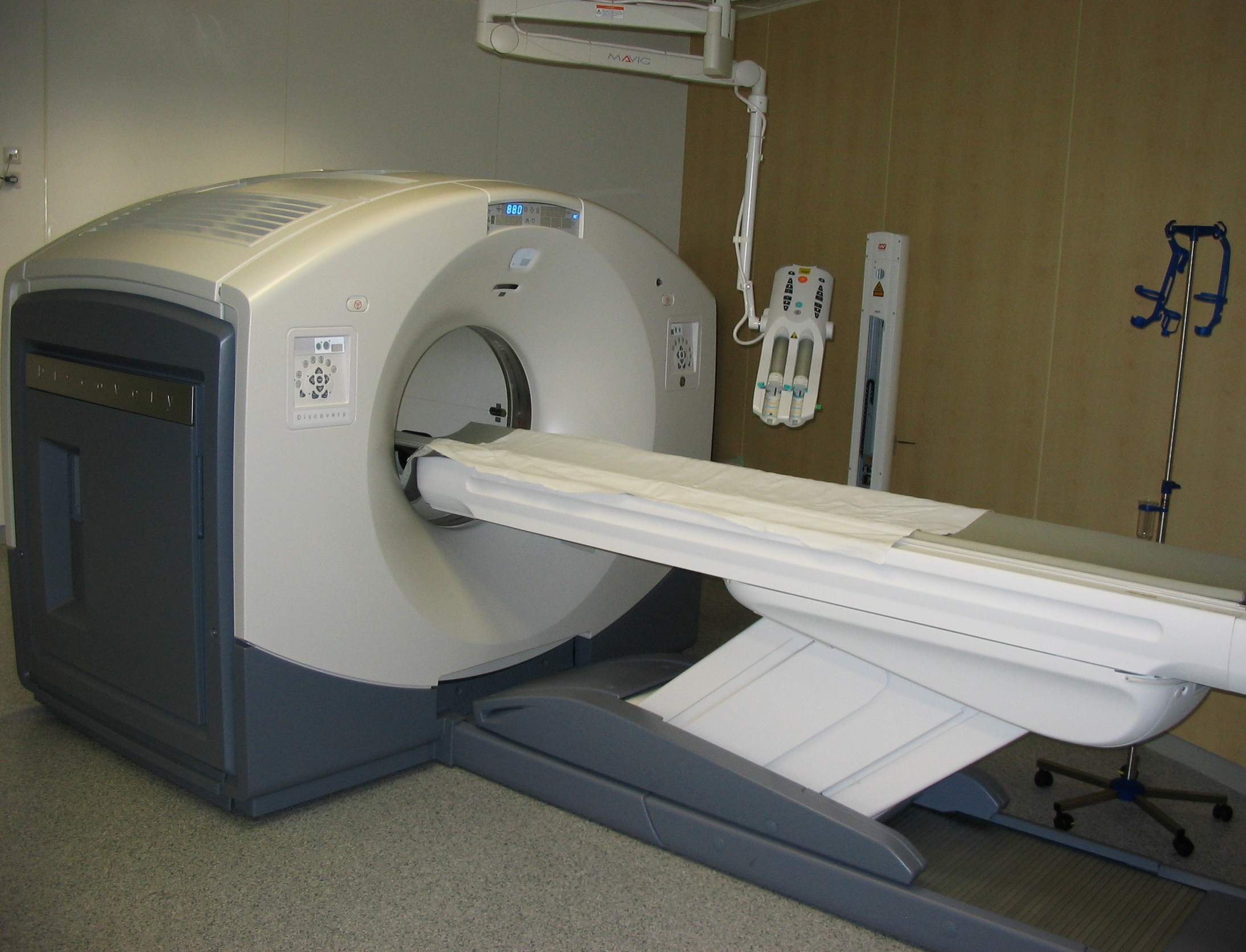
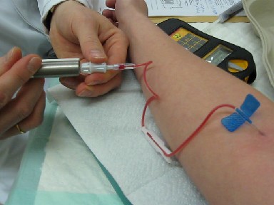
Nuclear medicine or nucleology is a medical specialty involving the application of radioactive substances in the diagnosis and treatment of disease. Nuclear imaging, in a sense, is " radiology done inside out" because it records radiation emitting from within the body rather than radiation that is generated by external sources like X-rays. In addition, nuclear medicine scans differ from radiology, as the emphasis is not on imaging anatomy, but on the function. For such reason, it is called a physiological imaging modality. Single photon emission computed tomography (SPECT) and positron emission tomography (PET) scans are the two most common imaging modalities in nuclear medicine. Diagnostic medical imaging Diagnostic In nuclear medicine imaging, radiopharmaceuticals are taken internally, for example, through inhalation, intravenously or orally. Then, external detectors ( gamma cameras) capture and form images from the radiation emitted by the radiopharmaceuticals. Thi ... [...More Info...] [...Related Items...] OR: [Wikipedia] [Google] [Baidu] |
Single-photon Emission Computed Tomography
Single-photon emission computed tomography (SPECT, or less commonly, SPET) is a nuclear medicine tomographic imaging technique using gamma rays. It is very similar to conventional nuclear medicine planar imaging using a gamma camera (that is, scintigraphy), but is able to provide true 3D information. This information is typically presented as cross-sectional slices through the patient, but can be freely reformatted or manipulated as required. The technique needs delivery of a gamma-emitting radioisotope (a radionuclide) into the patient, normally through injection into the bloodstream. On occasion, the radioisotope is a simple soluble dissolved ion, such as an isotope of gallium(III). Most of the time, though, a marker radioisotope is attached to a specific ligand to create a radioligand, whose properties bind it to certain types of tissues. This marriage allows the combination of ligand and radiopharmaceutical to be carried and bound to a place of interest in the body, where ... [...More Info...] [...Related Items...] OR: [Wikipedia] [Google] [Baidu] |
Positron Emission Tomography
Positron emission tomography (PET) is a functional imaging technique that uses radioactive substances known as radiotracers to visualize and measure changes in Metabolism, metabolic processes, and in other physiological activities including blood flow, regional chemical composition, and absorption. Different tracers are used for various imaging purposes, depending on the target process within the body. For example, 18F-FDG, -FDG is commonly used to detect cancer, Sodium fluoride#Medical imaging, NaF is widely used for detecting bone formation, and Isotopes of oxygen#Oxygen-15, oxygen-15 is sometimes used to measure blood flow. PET is a common medical imaging, imaging technique, a Scintigraphy#Process, medical scintillography technique used in nuclear medicine. A radiopharmaceutical, radiopharmaceutical — a radioisotope attached to a drug — is injected into the body as a radioactive tracer, tracer. When the radiopharmaceutical undergoes beta plus decay, a positron is ... [...More Info...] [...Related Items...] OR: [Wikipedia] [Google] [Baidu] |
Bone Scintigraphy
A bone scan or bone scintigraphy is a nuclear medicine imaging technique of the bone. It can help diagnose a number of bone conditions, including cancer of the bone or metastasis, location of bone inflammation and fractures (that may not be visible in traditional X-ray images), and bone infection (osteomyelitis). Nuclear medicine provides functional imaging and allows visualisation of bone metabolism or bone remodeling, which most other imaging techniques (such as X-ray computed tomography, CT) cannot. Bone scintigraphy competes with positron emission tomography (PET) for imaging of abnormal metabolism in bones, but is considerably less expensive. Bone scintigraphy has higher sensitivity but lower specificity than CT or MRI for diagnosis of scaphoid fractures following negative plain radiography. History Some of the earliest investigations into skeletal metabolism were carried out by George de Hevesy in the 1930s, using phosphorus-32 and by Charles Pecher in the 1940s. In t ... [...More Info...] [...Related Items...] OR: [Wikipedia] [Google] [Baidu] |
Technetium-99m
Technetium-99m (99mTc) is a metastable nuclear isomer of technetium-99 (itself an isotope of technetium), symbolized as 99mTc, that is used in tens of millions of medical diagnostic procedures annually, making it the most commonly used medical radioisotope in the world. Technetium-99m is used as a radioactive tracer and can be detected in the body by medical equipment (gamma cameras). It is well suited to the role, because it emits readily detectable gamma rays with a photon energy of 140 keV (these 8.8 pm photons are about the same wavelength as emitted by conventional X-ray diagnostic equipment) and its half-life for gamma emission is 6.0058 hours (meaning 93.7% of it decays to 99Tc in 24 hours). The relatively "short" physical half-life of the isotope and its biological half-life of 1 day (in terms of human activity and metabolism) allows for scanning procedures which collect data rapidly but keep total patient radiation exposure low. The same characteristics make the ... [...More Info...] [...Related Items...] OR: [Wikipedia] [Google] [Baidu] |
Orthognathic Surgery
Orthognathic surgery (), also known as corrective jaw surgery or simply jaw surgery, is surgery designed to correct conditions of the jaw and lower face related to structure, growth, airway issues including sleep apnea, TMJ disorders, malocclusion problems primarily arising from skeletal disharmonies, other orthodontic dental bite problems that cannot be easily treated with braces, as well as the broad range of facial imbalances, disharmonies, asymmetries and malproportions where correction can be considered to improve facial aesthetics and self esteem. The origins of orthognathic surgery belong in oral surgery, and the basic operations related to the surgical removal of impacted or displaced teeth – especially where indicated by orthodontics to enhance dental treatments of malocclusion and dental crowding. One of the first published cases of orthognathic surgery was the one from Dr. Simon P. Hullihen in 1849. Originally coined by Harold Hargis, it was more widely popularised ... [...More Info...] [...Related Items...] OR: [Wikipedia] [Google] [Baidu] |
Articular Disk
The articular disk (or disc) is a thin, oval plate of fibrocartilage present in several joints which separates synovial cavities. This separation of the cavity space allows for separate movements to occur in each space. The presence of an articular disk also permits a more even distribution of forces between the articulating surfaces of bones, increases the stability of the joint, and aids in directing the flow of synovial fluid to areas of the articular cartilage that experience the most friction. The term "meniscus" has a very similar meaning. Additional images File:Gray325.png, Sternoclavicular articulation. Anterior view. File:Gray300.png , Diagrammatic section of a diarthrodial joint, with an articular disk. See also * Triangular fibrocartilage ("articular disk of the distal radioulnar articulation") * Articular disk of the temporomandibular joint * Articular disk of sternoclavicular articulation The articular disc of the sternoclavicular joint is flat and nearly circula ... [...More Info...] [...Related Items...] OR: [Wikipedia] [Google] [Baidu] |
.jpg)


