|
Articular Disk
The articular disk (or disc) is a thin, oval plate of fibrocartilage present in several joints which separates synovial cavities. This separation of the cavity space allows for separate movements to occur in each space. The presence of an articular disk also permits a more even distribution of forces between the articulating surfaces of bones, increases the stability of the joint, and aids in directing the flow of synovial fluid to areas of the articular cartilage that experience the most friction. The term "meniscus Meniscus may refer to: * Meniscus (anatomy), crescent-shaped fibrocartilaginous structure that partly divides a joint cavity * Meniscus (liquid), a curve in the upper surface of liquid contained in an object *Meniscus (optics) A lens is a ..." has a very similar meaning. Additional images File:Gray325.png, Sternoclavicular articulation. Anterior view. File:Gray300.png , Diagrammatic section of a diarthrodial joint, with an articular disk. See also * Tr ... [...More Info...] [...Related Items...] OR: [Wikipedia] [Google] [Baidu] |
Fibrocartilage
Fibrocartilage consists of a mixture of white fibrous tissue and cartilaginous tissue in various proportions. It owes its inflexibility and toughness to the former of these constituents, and its elasticity to the latter. It is the only type of cartilage that contains type I collagen in addition to the normal type II. Structure The extracellular matrix of fibrocartilage is mainly made from type I collagen secreted by chondroblasts. Locations of fibrocartilage in the human body * secondary cartilaginous joints: ** pubic symphysis ** annulus fibrosis of intervertebral discs ** manubriosternal joint * glenoid labrum of shoulder joint * acetabular labrum of hip joint * medial and lateral menisci of the knee joint * location where tendons and ligaments A ligament is the fibrous connective tissue that connects bones to other bones. It is also known as ''articular ligament'', ''articular larua'', ''fibrous ligament'', or ''true ligament''. Other ligaments in the bo ... [...More Info...] [...Related Items...] OR: [Wikipedia] [Google] [Baidu] |
Joints
A joint or articulation (or articular surface) is the connection made between bones, ossicles, or other hard structures in the body which link an animal's skeletal system into a functional whole.Saladin, Ken. Anatomy & Physiology. 7th ed. McGraw-Hill Connect. Webp.274/ref> They are constructed to allow for different degrees and types of movement. Some joints, such as the knee, elbow, and shoulder, are self-lubricating, almost frictionless, and are able to withstand compression and maintain heavy loads while still executing smooth and precise movements. Other joints such as sutures between the bones of the skull permit very little movement (only during birth) in order to protect the brain and the sense organs. The connection between a tooth and the jawbone is also called a joint, and is described as a fibrous joint known as a gomphosis. Joints are classified both structurally and functionally. Classification The number of joints depends on if sesamoids are included, age of the h ... [...More Info...] [...Related Items...] OR: [Wikipedia] [Google] [Baidu] |
Meniscus (anatomy)
A meniscus is a crescent-shaped fibrocartilaginous anatomical structure that, in contrast to an articular disc, only partly divides a joint cavity.Platzer (2004), p 208 In humans they are present in the knee, wrist, acromioclavicular, sternoclavicular, and temporomandibular joints; in other animals they may be present in other joints. Generally, the term "meniscus" is used to refer to the cartilage of the knee, either to the lateral or medial meniscus. Both are cartilaginous tissues that provide structural integrity to the knee when it undergoes tension and torsion. The menisci are also known as "semi-lunar" cartilages, referring to their half-moon, crescent shape. The term "meniscus" is from the Ancient Greek word (), meaning "crescent". Structure The menisci of the knee are two pads of fibrocartilaginous tissue which serve to disperse friction in the knee joint between the lower leg (tibia) and the thigh (femur). They are concave on the top and flat on the bott ... [...More Info...] [...Related Items...] OR: [Wikipedia] [Google] [Baidu] |
Triangular Fibrocartilage
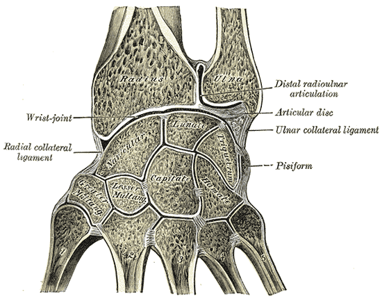
The triangular fibrocartilage complex (TFCC) is formed by the triangular fibrocartilage discus (TFC), the radioulnar ligaments (RULs) and the ulnocarpal ligaments (UCLs). Structure Triangular fibrocartilage disc The triangular fibrocartilage disc (TFC) is an articular discus that lies on the pole of the distal ulna. It has a triangular shape and a biconcave body; the periphery is thicker than its center. The central portion of the TFC is thin and consists of chondroid fibrocartilage; this type of tissue is often seen in structures that can bear compressive loads. This central area is often so thin that it is translucent and in some cases it is even absent. The peripheral portion of the TFC is well vascularized, while the central portion has no blood supply. This discus is attached by thick tissue to the base of the ulnar styloid and by thinner tissue to the edge of the radius just proximal to the radiocarpal articular surface. Radioulnar ligaments The radioulnar ligaments ... [...More Info...] [...Related Items...] OR: [Wikipedia] [Google] [Baidu] |
Articular Disk Of The Temporomandibular Joint
The articular disk of the temporomandibular joint is a thin, oval plate made of non-vascular fibrous connective tissue located between the mandible's condyloid process and the cranium's mandibular fossa. Its ''upper surface'' is concavo-convex from before backward, to accommodate itself to the form of the mandibular fossa and the articular tubercle. Its lower surface, in contact with the condyle, is concave. Its circumference is connected to the articular capsule, and in front to the tendon of the lateral pterygoid muscle. It is thicker at its periphery, especially behind, than at its center. The fibers of which the disc is composed have a concentric arrangement, more apparent at the circumference than at the center. It divides the joint into two cavities, each of which is furnished with a synovial membrane. It is attached as follows. * The anterior portion of the disc attaches inferiorly to the anterior condyle and superiorly to the eminence Eminence may refer to: Places * ... [...More Info...] [...Related Items...] OR: [Wikipedia] [Google] [Baidu] |
Articular Disk Of Sternoclavicular Articulation
The articular disc of the sternoclavicular joint is flat and nearly circular, interposed between the articulating surfaces of the sternum and clavicle. It is attached, above, to the upper and posterior border of the articular surface of the clavicle; below, to the cartilage of the first rib, near its junction with the sternum; and by its circumference to the interclavicular and anterior and posterior sternoclavicular ligaments. It is thicker at the circumference, especially its upper and back part, than at its center. It divides the joint into two cavities, each of which is furnished with a synovial membrane. See also * Articular disc The articular disk (or disc) is a thin, oval plate of fibrocartilage present in several joints which separates synovial cavities. This separation of the cavity space allows for separate movements to occur in each space. The presence of an articula ... References Upper limb anatomy {{Portal bar, Anatomy ... [...More Info...] [...Related Items...] OR: [Wikipedia] [Google] [Baidu] |


