|
Computed Tomography Laser Mammography
Computed tomography laser mammography (CTLM) is the trademark of Imaging Diagnostic Systems, Inc. (IDSI, United States) for its optical tomographic technique for female breast imaging. This medical imaging technique uses laser energy in the near infrared region of the spectrum, to detect angiogenesis in the breast tissue. It is optical molecular imaging for hemoglobin both oxygenated and deoxygenated. The technology uses laser in the same way computed tomography A computed tomography scan (CT scan; formerly called computed axial tomography scan or CAT scan) is a medical imaging technique used to obtain detailed internal images of the body. The personnel that perform CT scans are called radiographers ... uses X-Rays, these beams travel through tissue and suffer attenuation. A laser detector measures the intensity drop and the data is collected as the laser detector moves across the breast creating a tomography image. CTLM images show hemoglobin distribution in a tissu ... [...More Info...] [...Related Items...] OR: [Wikipedia] [Google] [Baidu] |
Mammography
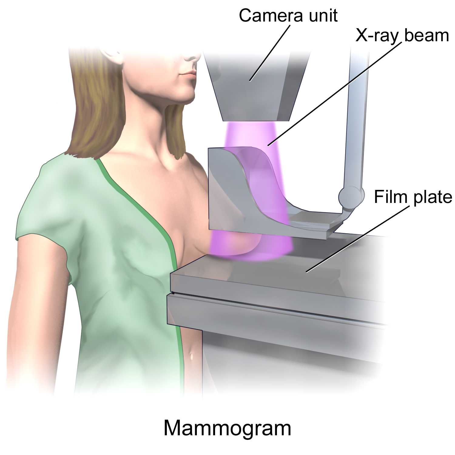
Mammography (also called mastography) is the process of using low-energy X-rays (usually around 30 kVp) to examine the human breast for diagnosis and screening. The goal of mammography is the early detection of breast cancer, typically through detection of characteristic masses or microcalcifications. As with all X-rays, mammograms use doses of ionizing radiation to create images. These images are then analyzed for abnormal findings. It is usual to employ lower-energy X-rays, typically Mo (K-shell X-ray energies of 17.5 and 19.6 keV) and Rh (20.2 and 22.7 keV) than those used for radiography of bones. Mammography may be 2D or 3D ( tomosynthesis), depending on the available equipment and/or purpose of the examination. Ultrasound, ductography, positron emission mammography (PEM), and magnetic resonance imaging (MRI) are adjuncts to mammography. Ultrasound is typically used for further evaluation of masses found on mammography or palpable masses that may or may not be seen on ma ... [...More Info...] [...Related Items...] OR: [Wikipedia] [Google] [Baidu] |
Trademark
A trademark (also written trade mark or trade-mark) is a type of intellectual property consisting of a recognizable sign, design, or expression that identifies products or services from a particular source and distinguishes them from others. The trademark owner can be an individual, business organization, or any legal entity. A trademark may be located on a package, a label, a voucher, or on the product itself. Trademarks used to identify services are sometimes called service marks. The first legislative act concerning trademarks was passed in 1266 under the reign of Henry III of England, requiring all bakers to use a distinctive mark for the bread they sold. The first modern trademark laws emerged in the late 19th century. In France, the first comprehensive trademark system in the world was passed into law in 1857. The Trade Marks Act 1938 of the United Kingdom changed the system, permitting registration based on "intent-to-use", creating an examination based pro ... [...More Info...] [...Related Items...] OR: [Wikipedia] [Google] [Baidu] |
Imaging Diagnostic Systems, Inc
Imaging is the representation or reproduction of an object's form; especially a visual representation (i.e., the formation of an image). Imaging technology is the application of materials and methods to create, preserve, or duplicate images. Imaging science is a multidisciplinary field concerned with the generation, collection, duplication, analysis, modification, and visualization of images,Joseph P. Hornak, ''Encyclopedia of Imaging Science and Technology'' (John Wiley & Sons, 2002) including imaging things that the human eye cannot detect. As an evolving field it includes research and researchers from physics, mathematics, electrical engineering, computer vision, computer science, and perceptual psychology. ''Imager'' are imaging sensors. Imaging chain The foundation of imaging science as a discipline is the "imaging chain" – a conceptual model describing all of the factors which must be considered when developing a system for creating visual renderings (images). In g ... [...More Info...] [...Related Items...] OR: [Wikipedia] [Google] [Baidu] |
Optical Tomography
Optical tomography is a form of computed tomography that creates a digital volumetric model of an object by reconstructing images made from light transmitted and scattered through an object. Optical tomography is used mostly in medical imaging research. Optical tomography in industry is used as a sensor of thickness and internal structure of semiconductors. Principle Optical tomography relies on the object under study being at least partially light-transmitting or translucent, so it works best on soft tissue, such as breast and brain tissue. The high scatter-based attenuation involved is generally dealt with by using intense, often pulsed or intensity modulated, light sources, and highly sensitive light sensors, and the use of infrared light at frequencies where body tissues are most transmissive. Soft tissues are highly scattering but weakly absorbing in the near-infrared and red parts of the spectrum, so that this is the wavelength range usually used. Types Diffuse optical t ... [...More Info...] [...Related Items...] OR: [Wikipedia] [Google] [Baidu] |
Medical Imaging
Medical imaging is the technique and process of imaging the interior of a body for clinical analysis and medical intervention, as well as visual representation of the function of some organs or tissues ( physiology). Medical imaging seeks to reveal internal structures hidden by the skin and bones, as well as to diagnose and treat disease. Medical imaging also establishes a database of normal anatomy and physiology to make it possible to identify abnormalities. Although imaging of removed organs and tissues can be performed for medical reasons, such procedures are usually considered part of pathology instead of medical imaging. Measurement and recording techniques that are not primarily designed to produce images, such as electroencephalography (EEG), magnetoencephalography (MEG), electrocardiography (ECG), and others, represent other technologies that produce data susceptible to representation as a parameter graph versus time or maps that contain data about the measurement ... [...More Info...] [...Related Items...] OR: [Wikipedia] [Google] [Baidu] |
Laser
A laser is a device that emits light through a process of optical amplification based on the stimulated emission of electromagnetic radiation. The word "laser" is an acronym for "light amplification by stimulated emission of radiation". The first laser was built in 1960 by Theodore H. Maiman at Hughes Research Laboratories, based on theoretical work by Charles Hard Townes and Arthur Leonard Schawlow. A laser differs from other sources of light in that it emits light which is coherence (physics), ''coherent''. Spatial coherence allows a laser to be focused to a tight spot, enabling applications such as laser cutting and Photolithography#Light sources, lithography. Spatial coherence also allows a laser beam to stay narrow over great distances (collimated light, collimation), enabling applications such as laser pointers and lidar (light detection and ranging). Lasers can also have high temporal coherence, which allows them to emit light with a very narrow frequency spectrum, spectru ... [...More Info...] [...Related Items...] OR: [Wikipedia] [Google] [Baidu] |
Angiogenesis
Angiogenesis is the physiological process through which new blood vessels form from pre-existing vessels, formed in the earlier stage of vasculogenesis. Angiogenesis continues the growth of the vasculature by processes of sprouting and splitting. Vasculogenesis is the embryonic formation of endothelial cells from mesoderm cell precursors, and from neovascularization, although discussions are not always precise (especially in older texts). The first vessels in the developing embryo form through vasculogenesis, after which angiogenesis is responsible for most, if not all, blood vessel growth during development and in disease. Angiogenesis is a normal and vital process in growth and development, as well as in wound healing and in the formation of granulation tissue. However, it is also a fundamental step in the transition of tumors from a benign state to a malignant one, leading to the use of angiogenesis inhibitors in the treatment of cancer. The essential role of angi ... [...More Info...] [...Related Items...] OR: [Wikipedia] [Google] [Baidu] |
Hemoglobin
Hemoglobin (haemoglobin BrE) (from the Greek word αἷμα, ''haîma'' 'blood' + Latin ''globus'' 'ball, sphere' + ''-in'') (), abbreviated Hb or Hgb, is the iron-containing oxygen-transport metalloprotein present in red blood cells (erythrocytes) of almost all vertebrates (the exception being the fish family Channichthyidae) as well as the tissues of some invertebrates. Hemoglobin in blood carries oxygen from the respiratory organs (''e.g.'' lungs or gills) to the rest of the body (''i.e.'' tissues). There it releases the oxygen to permit aerobic respiration to provide energy to power functions of an organism in the process called metabolism. A healthy individual human has 12to 20grams of hemoglobin in every 100mL of blood. In mammals, the chromoprotein makes up about 96% of the red blood cells' dry content (by weight), and around 35% of the total content (including water). Hemoglobin has an oxygen-binding capacity of 1.34mL O2 per gram, which increases the total blood oxygen ca ... [...More Info...] [...Related Items...] OR: [Wikipedia] [Google] [Baidu] |
Computed Tomography
A computed tomography scan (CT scan; formerly called computed axial tomography scan or CAT scan) is a medical imaging technique used to obtain detailed internal images of the body. The personnel that perform CT scans are called radiographers or radiology technologists. CT scanners use a rotating X-ray tube and a row of detectors placed in a gantry to measure X-ray attenuations by different tissues inside the body. The multiple X-ray measurements taken from different angles are then processed on a computer using tomographic reconstruction algorithms to produce tomographic (cross-sectional) images (virtual "slices") of a body. CT scans can be used in patients with metallic implants or pacemakers, for whom magnetic resonance imaging (MRI) is contraindicated. Since its development in the 1970s, CT scanning has proven to be a versatile imaging technique. While CT is most prominently used in medical diagnosis, it can also be used to form images of non-living objects. The 1979 N ... [...More Info...] [...Related Items...] OR: [Wikipedia] [Google] [Baidu] |
Dense Breast Tissue
Dense breast tissue, also known as dense breasts, is a condition of the breasts where a higher proportion of the breasts are made up of glandular tissue and fibrous tissue than fatty tissue. Around 40–50% of women have dense breast tissue and one of the main medical components of the condition is that mammograms are unable to differentiate tumorous tissue from the surrounding dense tissue. This increases the risk of late diagnosis of breast cancer in women with dense breast tissue. Additionally, women with such tissue have a higher likelihood of developing breast cancer in general, though the reasons for this are poorly understood. Definition Dense breast tissue is defined based on the amount of glandular and fibrous tissue as compared to the percentage of fatty tissue. The current mammography classifications split up the density of breasts into four categories. Approximately 10% of women have almost entirely fatty breasts, 40% with small pockets of dense tissue, 40% with even ... [...More Info...] [...Related Items...] OR: [Wikipedia] [Google] [Baidu] |
Operation Of Computed Tomography
X-ray computed tomography operates by using an X-ray generator that rotates around the object; X-ray detectors are positioned on the opposite side of the circle from the X-ray source. A visual representation of the raw data obtained is called a ''sinogram'', yet it is not sufficient for interpretation. Once the scan data has been acquired, the data must be processed using a form of tomographic reconstruction, which produces a series of cross-sectional images. In terms of mathematics, the raw data acquired by the scanner consists of multiple "projections" of the object being scanned. These projections are effectively the Radon transformation of the structure of the object. Reconstruction essentially involves solving the inverse Radon transformation. Structure In conventional CT machines, an X-ray tube and detector are physically rotated behind a circular shroud (see the image above right). An alternative, short lived design, known as electron beam tomography (EBT), used electrom ... [...More Info...] [...Related Items...] OR: [Wikipedia] [Google] [Baidu] |


