|
Caudal Duplication
Caudal duplication, (or caudal duplication syndrome) is a rare congenital disorder in which various structures of the caudal region, embryonic cloaca, and neural tube exhibit a spectrum of abnormalities such as duplication and malformations. The exact causes of the condition is unknown, though there are several theories implicating abnormal embryological development as a cause for the condition. Diagnosis is often made during prenatal development of the second trimester through anomaly scans or immediately after birth. However, rare cases of adulthood diagnosis has also been observed. Treatment is often required to correct such abnormalities according to the range of symptoms present, whilst treatment options vary from conservative expectant management to resection of caudal tissue to restore normal function or appearance. As a rare congenital disorder, the prevalence at birth is less than 1 per 100,000 with less than 100 cases reported worldwide. The term "caudal duplication synd ... [...More Info...] [...Related Items...] OR: [Wikipedia] [Google] [Baidu] |
Birth Defect
A birth defect is an abnormal condition that is present at birth, regardless of its cause. Birth defects may result in disabilities that may be physical, intellectual, or developmental. The disabilities can range from mild to severe. Birth defects are divided into two main types: structural disorders in which problems are seen with the shape of a body part and functional disorders in which problems exist with how a body part works. Functional disorders include metabolic and degenerative disorders. Some birth defects include both structural and functional disorders. Birth defects may result from genetic or chromosomal disorders, exposure to certain medications or chemicals, or certain infections during pregnancy. Risk factors include folate deficiency, drinking alcohol or smoking during pregnancy, poorly controlled diabetes, and a mother over the age of 35 years old. Many birth defects are believed to involve multiple factors. Birth defects may be visible at birth or dia ... [...More Info...] [...Related Items...] OR: [Wikipedia] [Google] [Baidu] |
Meckel's Diverticulum
A Meckel's diverticulum, a true congenital diverticulum, is a slight bulge in the small intestine present at birth and a vestigial remnant of the vitelline duct. It is the most common malformation of the Human gastrointestinal tract, gastrointestinal tract and is present in approximately 2% of the population, with males more frequently experiencing symptoms. Meckel's diverticulum was first explained by Fabricius Hildanus in the sixteenth century and later named after Johann Friedrich Meckel, who described the embryological origin of this type of diverticulum in 1809. Signs and symptoms The majority of people with a Meckel's diverticulum are asymptomatic. An asymptomatic Meckel's diverticulum is called a ''silent'' Meckel's diverticulum. If symptoms do occur, they typically appear before the age of two years. The most common presenting symptom is painless rectal bleeding such as melaena-like black offensive stools, followed by intestinal obstruction, volvulus and intussusception (m ... [...More Info...] [...Related Items...] OR: [Wikipedia] [Google] [Baidu] |
Spina Bifida
Spina bifida (SB; ; Latin for 'split spine') is a birth defect in which there is incomplete closing of the vertebral column, spine and the meninges, membranes around the spinal cord during embryonic development, early development in pregnancy. There are three main types: spina bifida occulta, meningocele and myelomeningocele. Meningocele and myelomeningocele may be grouped as spina bifida cystica. The most common location is the Lumbar vertebrae, lower back, but in rare cases it may be in the Thoracic vertebrae, middle back or Cervical vertebrae, neck. Occulta has no or only mild signs, which may include a hairy patch, dimple, dark spot or swelling on the back at the site of the gap in the spine. Meningocele typically causes mild problems, with a sac of fluid present at the gap in the spine. Myelomeningocele, also known as open spina bifida, is the most severe form. Problems associated with this form include poor ability to walk, impaired Neurogenic bladder dysfunction, bladder o ... [...More Info...] [...Related Items...] OR: [Wikipedia] [Google] [Baidu] |
Cryptorchidism
Cryptorchidism, also known as undescended testis, is the failure of one or both testes to descend into the scrotum. The word is . It is the most common birth defect of the male genital tract. About 3% of full-term and 30% of premature infant boys are born with at least one undescended testis. However, about 80% of cryptorchid testes descend by the first year of life (the majority within three months), making the true incidence of cryptorchidism around 1% overall. Cryptorchidism may develop after infancy, sometimes as late as young adulthood, but that is exceptional. Cryptorchidism is distinct from monorchism, the condition of having only one testicle. Though the condition may occur on one or both sides, it more commonly affects the right testis. A testis absent from the normal scrotal position may be: # Anywhere along the "path of descent" from high in the posterior (retroperitoneal) abdomen, just below the kidney, to the inguinal ring # In the inguinal canal # Ectopic, havin ... [...More Info...] [...Related Items...] OR: [Wikipedia] [Google] [Baidu] |
Bladder Exstrophy
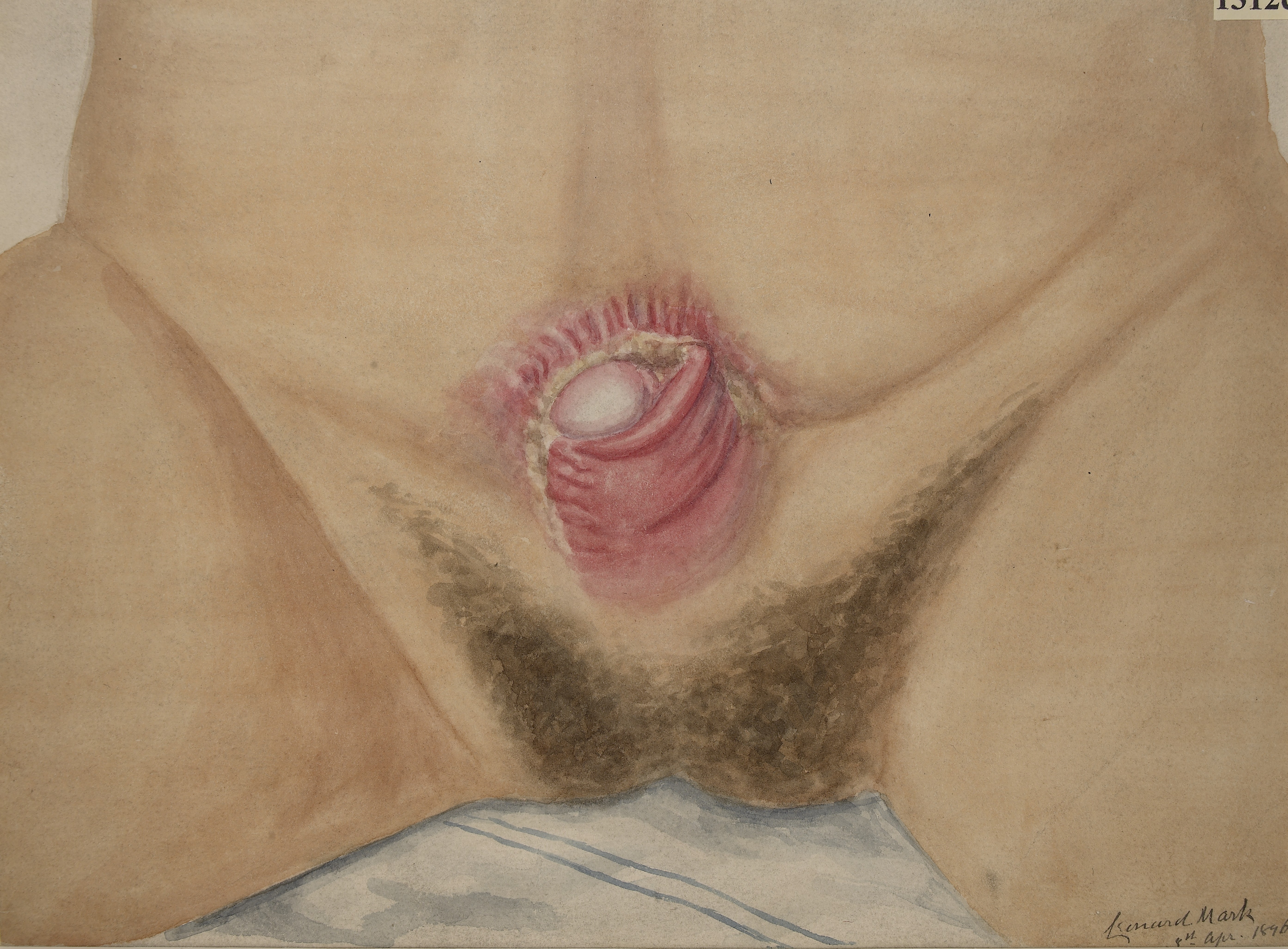
Bladder exstrophy is a congenital anomaly that exists along the spectrum of the exstrophy-epispadias complex, and most notably involves protrusion of the urinary bladder through a defect in the abdominal wall. Its presentation is variable, often including abnormalities of the bony pelvis, pelvic floor, and genitalia. The underlying embryologic mechanism leading to bladder exstrophy is unknown, though it is thought to be in part due to failed reinforcement of the cloacal membrane by underlying mesoderm. Exstrophy means the inversion of a hollow organ. Signs and symptoms The classic manifestation of bladder exstrophy presents with: * A defect in the abdominal wall occupied by both the exstrophied bladder as well as a portion of the urethra * A flattened puborectal sling * Separation of the pubic symphysis * Shortening of a pubic rami * External rotation of the pelvis. Females frequently have a displaced and narrowed vaginal orifice, a bifid clitoris, and divergent labia ... [...More Info...] [...Related Items...] OR: [Wikipedia] [Google] [Baidu] |
Testicle
A testicle or testis ( testes) is the gonad in all male bilaterians, including humans, and is Homology (biology), homologous to the ovary in females. Its primary functions are the production of sperm and the secretion of Androgen, androgens, primarily testosterone. The release of testosterone is regulated by luteinizing hormone (LH) from the anterior pituitary gland. Sperm production is controlled by follicle-stimulating hormone (FSH) from the anterior pituitary gland and by testosterone produced within the gonads. Structure Appearance Males have two testicles of similar size contained within the scrotum, which is an extension of the abdominal wall. Scrotal asymmetry, in which one testicle extends farther down into the scrotum than the other, is common. This is because of the differences in the vasculature's anatomy. For 85% of men, the right testis hangs lower than the left one. Measurement and volume The volume of the testicle can be estimated by palpating it and compari ... [...More Info...] [...Related Items...] OR: [Wikipedia] [Google] [Baidu] |
Scrotum
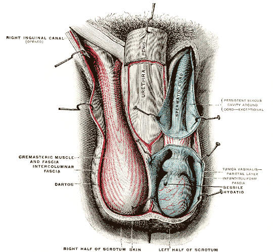
In most terrestrial mammals, the scrotum (: scrotums or scrota; possibly from Latin ''scortum'', meaning "hide" or "skin") or scrotal sac is a part of the external male genitalia located at the base of the penis. It consists of a sac of skin containing the external spermatic fascia, testicles, epididymides, and vasa deferentia. The scrotum will usually tighten when exposed to cold temperatures. The scrotum is homologous to the labia majora in females. Structure In regards to humans, the scrotum is a suspended two-chambered sac of skin and muscular tissue containing the testicles and the lower part of the spermatic cords. It is located behind the penis and above the perineum. The perineal raphe is a small, vertical ridge of skin that expands from the anus and runs through the middle of the scrotum front to back. The scrotum is also a distention of the perineum and carries some abdominal tissues into its cavity including the testicular artery, testicular vein, and ... [...More Info...] [...Related Items...] OR: [Wikipedia] [Google] [Baidu] |
Ureter
The ureters are tubes composed of smooth muscle that transport urine from the kidneys to the urinary bladder. In an adult human, the ureters typically measure 20 to 30 centimeters in length and about 3 to 4 millimeters in diameter. They are lined with urothelial cells, a form of transitional epithelium, and feature an extra layer of smooth muscle in the lower third to aid in peristalsis. The ureters can be affected by a number of diseases, including urinary tract infections and kidney stone. is when a ureter is narrowed, due to for example chronic inflammation. Congenital abnormalities that affect the ureters can include the development of two ureters on the same side or abnormally placed ureters. Additionally, reflux of urine from the bladder back up the ureters is a condition commonly seen in children. The ureters have been identified for at least two thousand years, with the word "ureter" stemming from the stem relating to urinating and seen in written records since at ... [...More Info...] [...Related Items...] OR: [Wikipedia] [Google] [Baidu] |
Uterus
The uterus (from Latin ''uterus'', : uteri or uteruses) or womb () is the hollow organ, organ in the reproductive system of most female mammals, including humans, that accommodates the embryonic development, embryonic and prenatal development, fetal development of one or more Fertilized egg, fertilized eggs until birth. The uterus is a hormone-responsive sex organ that contains uterine gland, glands in its endometrium, lining that secrete uterine milk for embryonic nourishment. (The term ''uterus'' is also applied to analogous structures in some non-mammalian animals.) In humans, the lower end of the uterus is a narrow part known as the Uterine isthmus, isthmus that connects to the cervix, the anterior gateway leading to the vagina. The upper end, the body of the uterus, is connected to the fallopian tubes at the uterine horns; the rounded part, the fundus, is above the openings to the fallopian tubes. The connection of the uterine cavity with a fallopian tube is called the utero ... [...More Info...] [...Related Items...] OR: [Wikipedia] [Google] [Baidu] |
Urethra
The urethra (: urethras or urethrae) is the tube that connects the urinary bladder to the urinary meatus, through which Placentalia, placental mammals Urination, urinate and Ejaculation, ejaculate. The external urethral sphincter is a striated muscle that allows voluntary control over urination. The Internal urethral sphincter, internal sphincter, formed by the involuntary smooth muscles lining the bladder neck and urethra, receives its nerve supply by the Sympathetic nervous system, sympathetic division of the autonomic nervous system. The internal sphincter is present both in males and females. Structure The urethra is a fibrous and muscular tube which connects the urinary bladder to the external urethral meatus. Its length differs between the sexes, because it passes through the penis in males. Male In the human male, the urethra is on average long and opens at the end of the external urethral meatus. The urethra is divided into four parts in men, named after the lo ... [...More Info...] [...Related Items...] OR: [Wikipedia] [Google] [Baidu] |
Urinary Bladder
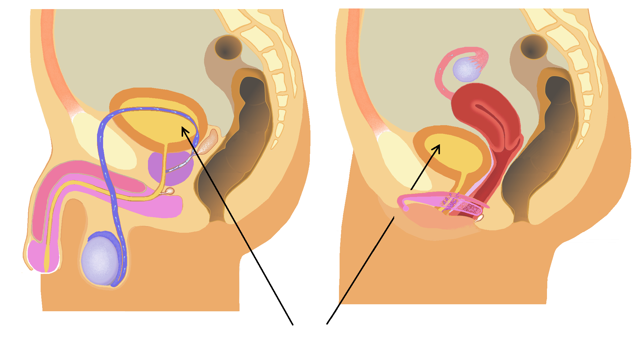
The bladder () is a hollow organ in humans and other vertebrates that stores urine from the Kidney (vertebrates), kidneys. In placental mammals, urine enters the bladder via the ureters and exits via the urethra during urination. In humans, the bladder is a distensible organ that sits on the pelvic floor. The typical adult human bladder will hold between 300 and (10 and ) before the urge to empty occurs, but can hold considerably more. The Latin phrase for "urinary bladder" is ''vesica urinaria'', and the term ''vesical'' or prefix ''vesico-'' appear in connection with associated structures such as vesical veins. The modern Latin word for "bladder" – ''cystis'' – appears in associated terms such as cystitis (inflammation of the bladder). Structure In humans, the bladder is a hollow muscular organ situated at the base of the pelvis. In gross anatomy, the bladder can be divided into a broad (base), a body, an apex, and a neck. The apex (also called the vertex) is directed ... [...More Info...] [...Related Items...] OR: [Wikipedia] [Google] [Baidu] |
Cervix
The cervix (: cervices) or cervix uteri is a dynamic fibromuscular sexual organ of the female reproductive system that connects the vagina with the uterine cavity. The human female cervix has been documented anatomically since at least the time of Hippocrates, over 2,000 years ago. The cervix is approximately 4 cm long with a diameter of approximately 3 cm and tends to be described as a cylindrical shape, although the front and back walls of the cervix are contiguous. The size of the cervix changes throughout a woman's life cycle. For example, women in the fertile years of their reproductive cycle tend to have larger cervixes than postmenopausal women; likewise, women who have produced offspring have a larger cervix than those who have not. In relation to the vagina, the part of the cervix that opens to the uterus is called the ''internal os'' and the opening of the cervix in the vagina is called the ''external os''. Between them is a conduit commonly called the cervic ... [...More Info...] [...Related Items...] OR: [Wikipedia] [Google] [Baidu] |


