|
CDH15
Cadherin-15 is a protein that in humans is encoded by the ''CDH15'' gene. Function This gene is a member of the cadherin superfamily of genes, encoding calcium-dependent intercellular adhesion glycoproteins. Cadherins consist of an extracellular domain containing 5 cadherin domains, a transmembrane region, and a conserved cytoplasmic domain. Transcripts from this particular cadherin are expressed in myoblasts and upregulated in myotubule-forming cells. The protein is thought to be essential for the control of morphogenetic processes, specifically myogenesis, and may provide a trigger for terminal muscle cell differentiation. Interactions CDH15 has been shown to interact with ARVCF Armadillo repeat protein deleted in velo-cardio-facial syndrome is a protein that in humans is encoded by the ''ARVCF'' gene. Function Armadillo repeat gene deleted in Velo-Cardio-Facial syndrome (ARVCF) is a member of the catenin family wh .... References Further reading * ... [...More Info...] [...Related Items...] OR: [Wikipedia] [Google] [Baidu] |
Cadherin
Cadherins (named for "calcium-dependent adhesion") are a type of cell adhesion molecule (CAM) that is important in the formation of adherens junctions to allow cells to adhere to each other . Cadherins are a class of type-1 transmembrane proteins, and they are dependent on calcium (Ca2+) ions to function, hence their name. Cell-cell adhesion is mediated by extracellular cadherin domains, whereas the intracellular cytoplasmic tail associates with numerous adaptors and signaling proteins, collectively referred to as the cadherin adhesome. The cadherin family is essential in maintaining the cell-cell contact and regulating cytoskeletal complexes. The cadherin superfamily includes cadherins, protocadherins, desmogleins, desmocollins, and more. In structure, they share ''cadherin repeats'', which are the extracellular Ca2+-binding domains. There are multiple classes of cadherin molecules, each designated with a prefix (in general, noting the types of tissue with which it is associated). ... [...More Info...] [...Related Items...] OR: [Wikipedia] [Google] [Baidu] |
Protein
Proteins are large biomolecules and macromolecules that comprise one or more long chains of amino acid residues. Proteins perform a vast array of functions within organisms, including catalysing metabolic reactions, DNA replication, responding to stimuli, providing structure to cells and organisms, and transporting molecules from one location to another. Proteins differ from one another primarily in their sequence of amino acids, which is dictated by the nucleotide sequence of their genes, and which usually results in protein folding into a specific 3D structure that determines its activity. A linear chain of amino acid residues is called a polypeptide. A protein contains at least one long polypeptide. Short polypeptides, containing less than 20–30 residues, are rarely considered to be proteins and are commonly called peptides. The individual amino acid residues are bonded together by peptide bonds and adjacent amino acid residues. The sequence of amino acid residue ... [...More Info...] [...Related Items...] OR: [Wikipedia] [Google] [Baidu] |
Gene
In biology, the word gene (from , ; "...Wilhelm Johannsen coined the word gene to describe the Mendelian units of heredity..." meaning ''generation'' or ''birth'' or ''gender'') can have several different meanings. The Mendelian gene is a basic unit of heredity and the molecular gene is a sequence of nucleotides in DNA that is transcribed to produce a functional RNA. There are two types of molecular genes: protein-coding genes and noncoding genes. During gene expression, the DNA is first copied into RNA. The RNA can be directly functional or be the intermediate template for a protein that performs a function. The transmission of genes to an organism's offspring is the basis of the inheritance of phenotypic traits. These genes make up different DNA sequences called genotypes. Genotypes along with environmental and developmental factors determine what the phenotypes will be. Most biological traits are under the influence of polygenes (many different genes) as well as gen ... [...More Info...] [...Related Items...] OR: [Wikipedia] [Google] [Baidu] |
Glycoprotein
Glycoproteins are proteins which contain oligosaccharide chains covalently attached to amino acid side-chains. The carbohydrate is attached to the protein in a cotranslational or posttranslational modification. This process is known as glycosylation. Secreted extracellular proteins are often glycosylated. In proteins that have segments extending extracellularly, the extracellular segments are also often glycosylated. Glycoproteins are also often important integral membrane proteins, where they play a role in cell–cell interactions. It is important to distinguish endoplasmic reticulum-based glycosylation of the secretory system from reversible cytosolic-nuclear glycosylation. Glycoproteins of the cytosol and nucleus can be modified through the reversible addition of a single GlcNAc residue that is considered reciprocal to phosphorylation and the functions of these are likely to be an additional regulatory mechanism that controls phosphorylation-based signalling. In contrast, ... [...More Info...] [...Related Items...] OR: [Wikipedia] [Google] [Baidu] |
Myoblast
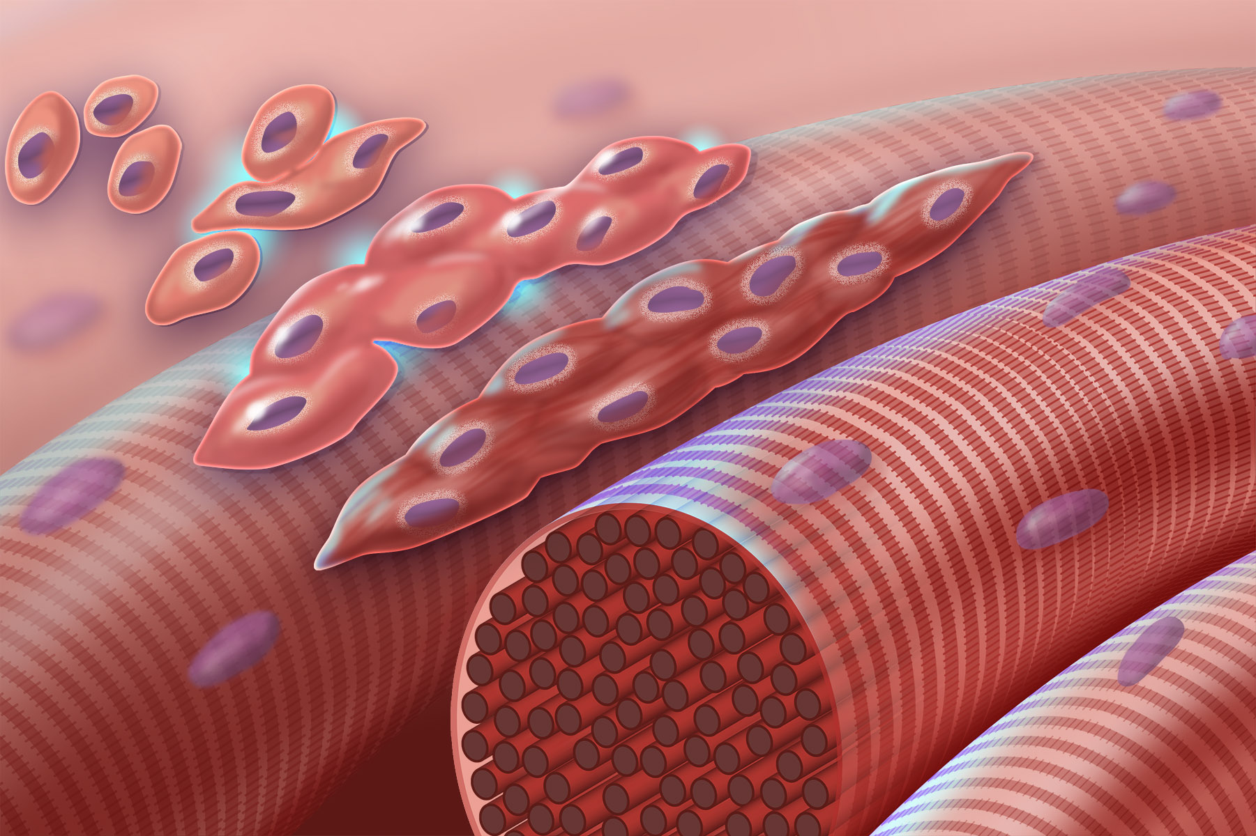
Myogenesis is the formation of skeletal muscular tissue, particularly during embryonic development. Muscle fibers generally form through the fusion of precursor myoblasts into multinucleated fibers called ''myotubes''. In the early development of an embryo, myoblasts can either proliferate, or differentiate into a myotube. What controls this choice in vivo is generally unclear. If placed in cell culture, most myoblasts will proliferate if enough fibroblast growth factor (FGF) or another growth factor is present in the medium surrounding the cells. When the growth factor runs out, the myoblasts cease division and undergo terminal differentiation into myotubes. Myoblast differentiation proceeds in stages. The first stage, involves cell cycle exit and the commencement of expression of certain genes. The second stage of differentiation involves the alignment of the myoblasts with one another. Studies have shown that even rat and chick myoblasts can recognise and align with one an ... [...More Info...] [...Related Items...] OR: [Wikipedia] [Google] [Baidu] |
Morphogenetic
Morphogenesis (from the Greek ''morphê'' shape and ''genesis'' creation, literally "the generation of form") is the biological process that causes a cell, tissue or organism to develop its shape. It is one of three fundamental aspects of developmental biology along with the control of tissue growth and patterning of cellular differentiation. The process controls the organized spatial distribution of cells during the embryonic development of an organism. Morphogenesis can take place also in a mature organism, such as in the normal maintenance of tissue by stem cells or in regeneration of tissues after damage. Cancer is an example of highly abnormal and pathological tissue morphogenesis. Morphogenesis also describes the development of unicellular life forms that do not have an embryonic stage in their life cycle. Morphogenesis is essential for the evolution of new forms. Morphogenesis is a mechanical process involving forces that generate mechanical stress, strain, and mo ... [...More Info...] [...Related Items...] OR: [Wikipedia] [Google] [Baidu] |
Myogenesis
Myogenesis is the formation of skeletal muscular tissue, particularly during embryonic development. Muscle fibers generally form through the fusion of precursor myoblasts into multinucleated fibers called ''myotubes''. In the early development of an embryo, myoblasts can either proliferate, or differentiate into a myotube. What controls this choice in vivo is generally unclear. If placed in cell culture, most myoblasts will proliferate if enough fibroblast growth factor (FGF) or another growth factor is present in the medium surrounding the cells. When the growth factor runs out, the myoblasts cease division and undergo terminal differentiation into myotubes. Myoblast differentiation proceeds in stages. The first stage, involves cell cycle exit and the commencement of expression of certain genes. The second stage of differentiation involves the alignment of the myoblasts with one another. Studies have shown that even rat and chick myoblasts can recognise and align with one a ... [...More Info...] [...Related Items...] OR: [Wikipedia] [Google] [Baidu] |


.jpg)