|
Antigen Presenting Cells
An antigen-presenting cell (APC) or accessory cell is a cell that displays antigen bound by major histocompatibility complex (MHC) proteins on its surface; this process is known as antigen presentation. T cells may recognize these complexes using their T cell receptors (TCRs). APCs process antigens and present them to T-cells. Almost all cell types can present antigens in some way. They are found in a variety of tissue types. Professional antigen-presenting cells, including macrophages, B cells and dendritic cells, present foreign antigens to helper T cells, while virus-infected cells (or cancer cells) can present antigens originating inside the cell to cytotoxic T cells. In addition to the MHC family of proteins, antigen presentation relies on other specialized signaling molecules on the surfaces of both APCs and T cells. Antigen-presenting cells are vital for effective adaptive immune response, as the functioning of both cytotoxic and helper T cells is dependent on APCs. Antigen ... [...More Info...] [...Related Items...] OR: [Wikipedia] [Google] [Baidu] |
Antigen Presentation
Antigen presentation is a vital immune process that is essential for T cell immune response triggering. Because T cells recognize only fragmented antigens displayed on cell surfaces, antigen processing must occur before the antigen fragment, now bound to the major histocompatibility complex (MHC), is transported to the surface of the cell, a process known as presentation, where it can be recognized by a T-cell receptor. If there has been an infection with viruses or bacteria, the cell will present an endogenous or exogenous peptide fragment derived from the antigen by MHC molecules. There are two types of MHC molecules which differ in the behaviour of the antigens: MHC class I molecules (MHC-I) bind peptides from the cell cytosol, while peptides generated in the endocytic vesicles after internalisation are bound to MHC class II (MHC-II). Cellular membranes separate these two cellular environments - intracellular and extracellular. Each T cell can only recognize tens to hundreds ... [...More Info...] [...Related Items...] OR: [Wikipedia] [Google] [Baidu] |
Cytotoxic T Cell
A cytotoxic T cell (also known as TC, cytotoxic T lymphocyte, CTL, T-killer cell, cytolytic T cell, CD8+ T-cell or killer T cell) is a T lymphocyte (a type of white blood cell) that kills cancer cells, cells that are infected by intracellular pathogens (such as viruses or bacteria), or cells that are damaged in other ways. Most cytotoxic T cells express T-cell receptors (TCRs) that can recognize a specific antigen. An antigen is a molecule capable of stimulating an immune response and is often produced by cancer cells, viruses, bacteria or intracellular signals. Antigens inside a cell are bound to class I MHC molecules, and brought to the surface of the cell by the class I MHC molecule, where they can be recognized by the T cell. If the TCR is specific for that antigen, it binds to the complex of the class I MHC molecule and the antigen, and the T cell destroys the cell. In order for the TCR to bind to the class I MHC molecule, the former must be accompanied by a glycoprotein ... [...More Info...] [...Related Items...] OR: [Wikipedia] [Google] [Baidu] |
Monocyte
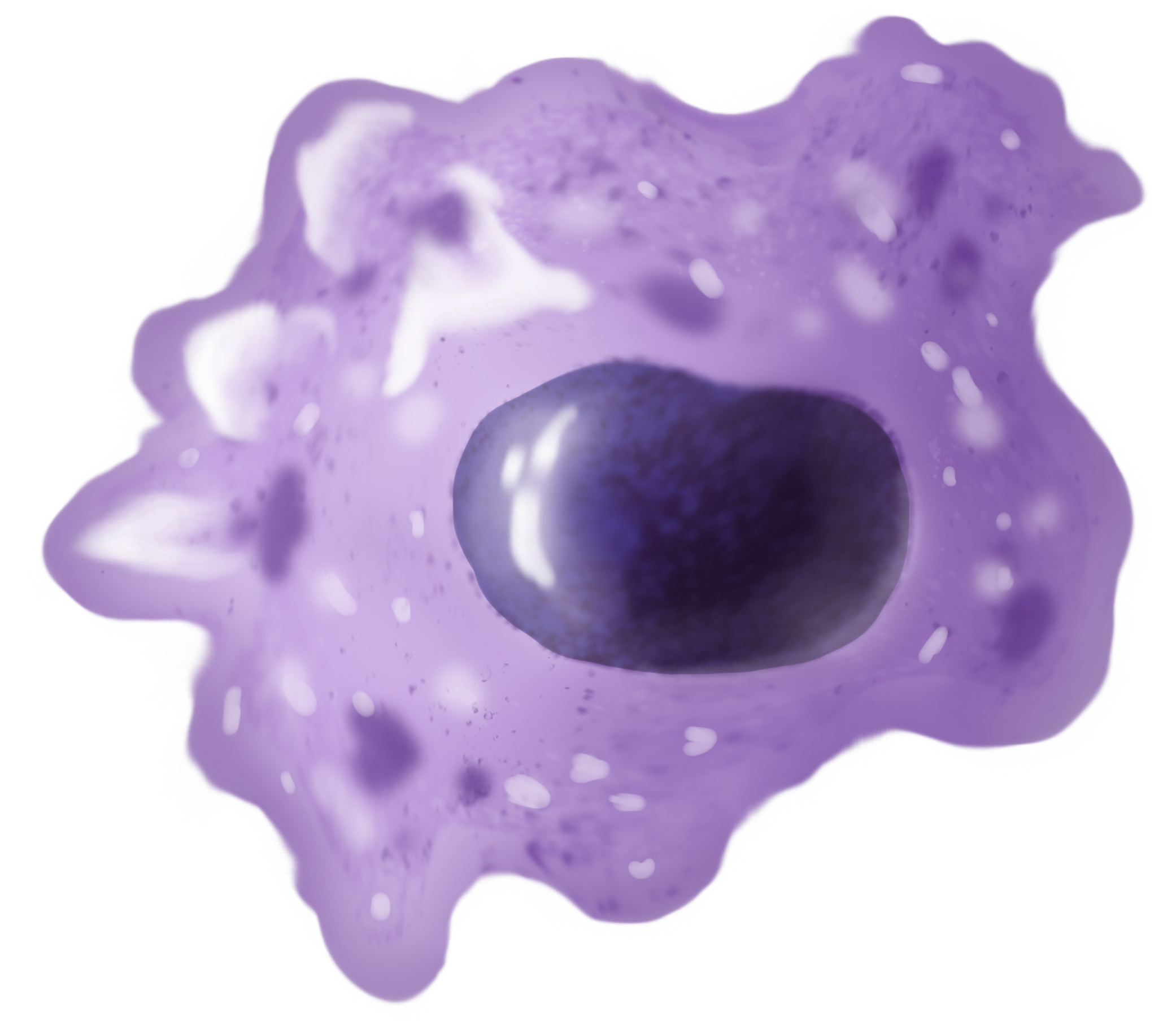
Monocytes are a type of leukocyte or white blood cell. They are the largest type of leukocyte in blood and can differentiate into macrophages and conventional dendritic cells. As a part of the vertebrate innate immune system monocytes also influence adaptive immune responses and exert tissue repair functions. There are at least three subclasses of monocytes in human blood based on their phenotypic receptors. Structure Monocytes are amoeboid in appearance, and have nongranulated cytoplasm. Thus they are classified as agranulocytes, although they might occasionally display some azurophil granules and/or vacuoles. With a diameter of 15–22 μm, monocytes are the largest cell type in peripheral blood. Monocytes are mononuclear cells and the ellipsoidal nucleus is often lobulated/indented, causing a bean-shaped or kidney-shaped appearance. Monocytes compose 2% to 10% of all leukocytes in the human body. Development Monocytes are produced by the bone marrow from precursors ca ... [...More Info...] [...Related Items...] OR: [Wikipedia] [Google] [Baidu] |
Interferon
Interferons (IFNs, ) are a group of signaling proteins made and released by host cells in response to the presence of several viruses. In a typical scenario, a virus-infected cell will release interferons causing nearby cells to heighten their anti-viral defenses. IFNs belong to the large class of proteins known as cytokines, molecules used for communication between cells to trigger the protective defenses of the immune system that help eradicate pathogens. Interferons are named for their ability to "interfere" with viral replication by protecting cells from virus infections. However, virus-encoded genetic elements have the ability to antagonize the IFN response contributing to viral pathogenesis and viral diseases. IFNs also have various other functions: they activate immune cells, such as natural killer cells and macrophages, and they increase host defenses by up-regulating antigen presentation by virtue of increasing the expression of major histocompatibility complex (M ... [...More Info...] [...Related Items...] OR: [Wikipedia] [Google] [Baidu] |
Macrophage
Macrophages (abbreviated as M φ, MΦ or MP) ( el, large eaters, from Greek ''μακρός'' (') = large, ''φαγεῖν'' (') = to eat) are a type of white blood cell of the immune system that engulfs and digests pathogens, such as cancer cells, microbes, cellular debris, and foreign substances, which do not have proteins that are specific to healthy body cells on their surface. The process is called phagocytosis, which acts to defend the host against infection and injury. These large phagocytes are found in essentially all tissues, where they patrol for potential pathogens by amoeboid movement. They take various forms (with various names) throughout the body (e.g., histiocytes, Kupffer cells, alveolar macrophages, microglia, and others), but all are part of the mononuclear phagocyte system. Besides phagocytosis, they play a critical role in nonspecific defense (innate immunity) and also help initiate specific defense mechanisms (adaptive immunity) by recruiting other immune ... [...More Info...] [...Related Items...] OR: [Wikipedia] [Google] [Baidu] |
CD28
CD28 (Cluster of Differentiation 28) is one of the proteins expressed on T cells that provide co-stimulatory signals required for T cell activation and survival. T cell stimulation through CD28 in addition to the T-cell receptor ( TCR) can provide a potent signal for the production of various interleukins ( IL-6 in particular). CD28 is the receptor for CD80 (B7.1) and CD86 (B7.2) proteins. When activated by Toll-like receptor ligands, the CD80 expression is upregulated in antigen-presenting cells (APCs). The CD86 expression on antigen-presenting cells is constitutive (expression is independent of environmental factors). CD28 is the only B7 receptor constitutively expressed on naive T cells. Association of the TCR of a naive T cell with MHC:antigen complex without CD28:B7 interaction results in a T cell that is anergic. Furthermore, CD28 was also identified on bone marrow stromal cells, plasma cells, neutrophils and eosinophils, but the functional importance of CD28 on these ... [...More Info...] [...Related Items...] OR: [Wikipedia] [Google] [Baidu] |
CD40 (protein)
Cluster of differentiation 40, CD40 is a costimulatory protein found on antigen-presenting cells and is required for their activation. The binding of CD154 (CD40L) on TH cells to CD40 activates antigen presenting cells and induces a variety of downstream effects. Deficiency can cause Hyper-IgM syndrome type 3. Function The protein receptor encoded by this gene is a member of the TNF-receptor superfamily. This receptor has been found to be essential in mediating a broad variety of immune and inflammatory responses including T cell-dependent immunoglobulin class switching, memory B cell development, and germinal center formation. AT-hook transcription factor AKNA is reported to coordinately regulate the expression of this receptor and its ligand, which may be important for homotypic cell interactions. The interaction of this receptor and its ligand is found to be necessary for amyloid-beta-induced microglial activation, and thus is thought to be an early event in Alzheimer disea ... [...More Info...] [...Related Items...] OR: [Wikipedia] [Google] [Baidu] |
Autoimmune Disease
An autoimmune disease is a condition arising from an abnormal immune response to a functioning body part. At least 80 types of autoimmune diseases have been identified, with some evidence suggesting that there may be more than 100 types. Nearly any body part can be involved. Common symptoms can be diverse and transient, ranging from mild to severe, and generally include low grade fever and feeling tired. The cause is unknown. Some autoimmune diseases such as lupus run in families, and certain cases may be triggered by infections or other environmental factors. Some common diseases that are generally considered autoimmune include celiac disease, diabetes mellitus type 1, graves' disease, inflammatory bowel disease, multiple sclerosis, alopecia areata, addison’s disease, pernicious anemia, psoriasis, rheumatoid arthritis, and systemic lupus erythematosus. The diagnosis can be difficult to determine. Treatment depends on the type and severity of the condition. Nonsteroidal ant ... [...More Info...] [...Related Items...] OR: [Wikipedia] [Google] [Baidu] |
Peripheral Tolerance
In immunology, peripheral tolerance is the second branch of immunological tolerance, after central tolerance. It takes place in the immune periphery (after T and B cells egress from primary lymphoid organs). Its main purpose is to ensure that self-reactive T and B cells which escaped central tolerance do not cause autoimmune disease. Peripheral tolerance prevents immune response to harmless food antigens and allergens, too. Deletion of self-reactive T cells in the thymus is only 60-70% efficient, and naive T cell repertoire contains a significant portion of low-avidity self-reactive T cells. These cells can trigger an autoimmune response, and there are several mechanisms of peripheral tolerance to prevent their activation. Antigen-specific mechanisms of peripheral tolerance include persistent of T cell in quiescence, ignorance of antigen and direct inactivation of effector T cells by either clonal deletion, conversion to regulatory T cells (Tregs) or induction of anergy. Tre ... [...More Info...] [...Related Items...] OR: [Wikipedia] [Google] [Baidu] |
Cross-presentation
Cross-presentation is the ability of certain professional antigen-presenting cells (mostly dendritic cells) to take up, process and present ''extracellular'' antigens with MHC class I molecules to CD8 T cells (cytotoxic T cells). Cross-priming, the result of this process, describes the stimulation of naive cytotoxic CD8+ T cells into activated cytotoxic CD8+ T cells. This process is necessary for immunity against most tumors and against viruses that infect dendritic cells and sabotage their presentation of virus antigens. Cross presentation is also required for the induction of cytotoxic immunity by vaccination with protein antigens, for example, tumour vaccination. Cross-presentation is of particular importance, because it permits the presentation of exogenous antigens, which are normally presented by MHC II on the surface of dendritic cells, to also be presented through the MHC I pathway. The MHC I pathway is normally used to present endogenous antigens that have infected a parti ... [...More Info...] [...Related Items...] OR: [Wikipedia] [Google] [Baidu] |
Dendritic Cell
Dendritic cells (DCs) are antigen-presenting cells (also known as ''accessory cells'') of the mammalian immune system. Their main function is to process antigen material and present it on the cell surface to the T cells of the immune system. They act as messengers between the innate and the adaptive immune systems. Dendritic cells are present in those tissues that are in contact with the external environment, such as the skin (where there is a specialized dendritic cell type called the Langerhans cell) and the inner lining of the nose, lungs, stomach and intestines. They can also be found in an immature state in the blood. Once activated, they migrate to the lymph nodes where they interact with T cells and B cells to initiate and shape the adaptive immune response. At certain development stages they grow branched projections, the ''dendrites'' that give the cell its name (δένδρον or déndron being Greek for 'tree'). While similar in appearance, these are structures ... [...More Info...] [...Related Items...] OR: [Wikipedia] [Google] [Baidu] |
Receptor-mediated Endocytosis
Receptor-mediated endocytosis (RME), also called clathrin-mediated endocytosis, is a process by which cells absorb metabolites, hormones, proteins – and in some cases viruses – by the inward budding of the plasma membrane (invagination). This process forms vesicles containing the absorbed substances and is strictly mediated by receptors on the surface of the cell. Only the receptor-specific substances can enter the cell through this process. Process Although receptors and their ligands can be brought into the cell through a few mechanisms (e.g. caveolin and lipid raft), clathrin-mediated endocytosis remains the best studied. Clathrin-mediated endocytosis of many receptor types begins with the ligands binding to receptors on the cell plasma membrane. The ligand and receptor will then recruit adaptor proteins and clathrin triskelions to the plasma membrane around where invagination will take place. Invagination of the plasma membrane then occurs, forming a clathrin-coated pit. ... [...More Info...] [...Related Items...] OR: [Wikipedia] [Google] [Baidu] |

.png)


