|
BCL6
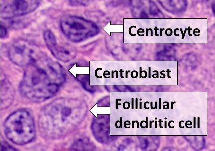
Bcl-6 (B-cell lymphoma 6) is a protein that in humans is encoded by the ''BCL6'' gene. BCL6 is a master transcription factor for regulation of T follicular helper cells (TFH cells) proliferation. BCL6 has three evolutionary conserved structural domains. The interaction of these domains with corepressors allows for germinal center development and leads to B cell proliferation. The ''deletion'' of BCL6 is known to lead to failure to germinal center formation in the follicles of the lymph nodes, preventing B cells from undergoing somatic hypermutation. ''Mutations'' in BCL6 can lead to B cell lymphomas because it promotes unchecked B cell growth. Clinically, BCL6 can be used to diagnose B cell lymphomas and is shown to be upregulated in a number of cancers. Other BCL genes, including BCL2, BCL3, BCL5, BCL7A, BCL9, and BCL10, also have clinical significance in lymphoma. Normal Physiological Function Structure The protein encoded by the BCL6 gene is a zinc finger transcription f ... [...More Info...] [...Related Items...] OR: [Wikipedia] [Google] [Baidu] |
Corepressor
In the field of molecular biology, a corepressor is a molecule that represses the expression of genes. In prokaryotes, corepressors are small molecules whereas in eukaryotes, corepressors are proteins. A corepressor does not directly bind to DNA, but instead indirectly regulates gene expression by binding to repressors. A corepressor downregulates (or represses) the expression of genes by binding to and activating a repressor transcription factor. The repressor in turn binds to a gene's operator sequence (segment of DNA to which a transcription factor binds to regulate gene expression), thereby blocking transcription of that gene. Function Prokaryotes In prokaryotes, the term corepressor is used to denote the activating ligand of a repressor protein. For example, the '' E. coli'' tryptophan repressor (TrpR) is only able to bind to DNA and repress transcription of the ''trp'' operon when its corepressor tryptophan is bound to it. TrpR in the absence of tryptophan is ... [...More Info...] [...Related Items...] OR: [Wikipedia] [Google] [Baidu] |
BCL2
Bcl-2 (B-cell lymphoma 2), encoded in humans by the ''BCL2'' gene, is the founding member of the Bcl-2 family of regulator proteins that regulate cell death (apoptosis), by either inhibiting (anti-apoptotic) or inducing (pro-apoptotic) apoptosis. It was the first apoptosis regulator identified in any organism. Bcl-2 derives its name from ''B-cell lymphoma 2'', as it is the second member of a range of proteins initially described in chromosomal translocations involving chromosomes 14 and 18 in follicular lymphomas. Orthologs (such as ''Bcl2'' in mice) have been identified in numerous mammals for which complete genome data are available. Like BCL3, BCL5, BCL6, BCL7A, BCL9, and BCL10, it has clinical significance in lymphoma. Isoforms The two isoforms of Bcl-2, Isoform 1, and Isoform 2, exhibit a similar fold. However, results in the ability of these isoforms to bind to the BAD and BAK proteins, as well as in the structural topology and electrostatic potential of the binding g ... [...More Info...] [...Related Items...] OR: [Wikipedia] [Google] [Baidu] |
BCL9
B-cell CLL/lymphoma 9 protein is a protein that in humans is encoded by the ''BCL9'' gene. Function BCL9, together with its paralogue gene BCL9L (BCL9 like or BCL9.2), have been extensively studied for their role as transcriptional beta-catenin cofactors, fundamental for the transcription of Wnt target genes. Recent work, using the mouse (Mus musculus) and Zebrafish (Danio rerio) as model organisms, identified an ancient role of BCL9 and BCL9L as key factors required for cardiac development. This work emphasises the tissue-specific nature of the Wnt/β-catenin mechanism of action, and implicates alterations in BCL9 and BCL9L in human congenital heart defects. BCL9 and BCL9L have been shown to take part in other tissue-specific molecular mechanisms, showing that their role in the Wnt signaling cascade is only one aspect of their mode of action. The conserved homology domain HD1 of BCL9 (and BCL9L) has recently been shown to be interacting with TBX3 in the context of intestin ... [...More Info...] [...Related Items...] OR: [Wikipedia] [Google] [Baidu] |
Follicular B Helper T Cells
Follicular helper T cells (also known as follicular B helper T cells and abbreviated as TFH), are antigen-experienced CD4+ T cells found in the periphery within B cell follicles of secondary lymphoid organs such as lymph nodes, spleen and Peyer's patches, and are identified by their constitutive expression of the B cell follicle homing receptor CXCR5. Upon cellular interaction and cross-signaling with their cognate follicular (Fo B) B cells, TFH cells trigger the formation and maintenance of germinal centers through the expression of CD40 ligand (CD40L) and the secretion of IL-21 and IL-4. TFH cells also migrate from T cell zones into these seeded germinal centers, predominantly composed of rapidly dividing B cells mutating their Ig genes. Within germinal centers, TFH cells play a critical role in mediating the selection and survival of B cells that go on to differentiate either into long-lived plasma cells capable of producing high affinity antibodies against foreign antigen ... [...More Info...] [...Related Items...] OR: [Wikipedia] [Google] [Baidu] |
BTB/POZ Domain
The BTB/POZ domain is a common structural domain contained within some proteins. The BTB (for BR-C, ttk and bab) or POZ (for Pox virus and Zinc finger) domain is present near the N-terminus of a fraction of zinc finger proteins and in proteins that contain the Kelch motif and a family of pox virus proteins. The BTB/POZ domain mediates homomeric dimerisation and in some instances heteromeric dimerisation. The structure of the dimerised PLZF BTB/POZ domain has been solved and consists of a tightly intertwined homodimer. The central scaffolding of the protein is made up of a cluster of alpha-helices flanked by short beta-sheets at both the top and bottom of the molecule. BTB/POZ domains from several zinc finger proteins have been shown to mediate transcriptional repression and to interact with components of histone deacetylase Histone deacetylases (, HDAC) are a class of enzymes that remove acetyl groups (O=C-CH3) from an ε-N-acetyl lysine amino acid on a histone, allowing the hi ... [...More Info...] [...Related Items...] OR: [Wikipedia] [Google] [Baidu] |
Blimp-1
PR domain zinc finger protein 1, or B lymphocyte-induced maturation protein-1 (BLIMP-1), is a protein in humans encoded by the gene ''PRDM1'' located on chromosome 6q21. BLIMP-1 is considered a 'master regulator' of hematopoietic stem cells, and plays a critical role in the development of plasma B cells, T cells, dendritic cells (DCs), macrophages, and osteoclasts. Pattern Recognition Receptors (PRRs) can activate BLIMP-1, both as a direct target and through downstream activation. BLIMP-1 is a transcription factor that triggers expression of many downstream signaling cascades. As a fine-tuned and contextual rheostat of the immune system, BLIMP-1 up- or down-regulates immune responses depending on the precise scenarios. BLIMP-1 is highly expressed in exhausted T-cells – clones of dysfunctional T-cells with diminished functions due to chronic immune response against cancer, viral infections, or organ transplant. Function As a potent repressor of beta-interferon (IFN-β), B ... [...More Info...] [...Related Items...] OR: [Wikipedia] [Google] [Baidu] |
PRDM1
PR domain zinc finger protein 1, or B lymphocyte-induced maturation protein-1 (BLIMP-1), is a protein in humans encoded by the gene ''PRDM1'' located on chromosome 6q21. BLIMP-1 is considered a 'master regulator' of Hematopoietic stem cell, hematopoietic stem cells, and plays a critical role in the development of Plasma cell, plasma B cells, T cell, T cells, Dendritic cell, dendritic cells (DCs), Macrophage, macrophages, and Osteoclast, osteoclasts. Pattern recognition receptor, Pattern Recognition Receptors (PRRs) can activate BLIMP-1, both as a direct target and through downstream activation. BLIMP-1 is a transcription factor that triggers expression of many downstream signaling cascades. As a fine-tuned and contextual rheostat of the immune system, BLIMP-1 up- or down-regulates immune responses depending on the precise scenarios. BLIMP-1 is highly expressed in T cell#Exhaustion, exhausted T-cells – clones of dysfunctional T-cells with diminished functions due to chronic immune ... [...More Info...] [...Related Items...] OR: [Wikipedia] [Google] [Baidu] |
BCL3
B-cell lymphoma 3-encoded protein is a protein that in humans is encoded by the ''BCL3'' gene. This gene is a proto-oncogene candidate. It is identified by its translocation into the immunoglobulin alpha-locus in some cases of B-cell leukemia. The protein encoded by this gene contains seven ankyrin repeats, which are most closely related to those found in I kappa B proteins. This protein functions as a transcriptional coactivator that activates through its association with NF-kappa B homodimers. The expression of this gene can be induced by NF-kappa B, which forms a part of the autoregulatory loop that controls the nuclear residence of p50 NF-kappa B. Like BCL2, BCL5, BCL6, BCL7A, BCL9, and BCL10, it has clinical significance in lymphoma. Interactions BCL3 has been shown to interact with: * BARD1, * C-Fos, * C-jun, * C22orf25, * COPS5, * EP300, * HTATIP, * NFKB1, * NFKB2, * PIR, and * NR2B1. Clinical significance Genetic variations in ''BCL3'' gene ha ... [...More Info...] [...Related Items...] OR: [Wikipedia] [Google] [Baidu] |
BCL10
B-cell lymphoma/leukemia 10 is a protein that in humans is encoded by the ''BCL10'' gene. Like BCL2, BCL3, BCL5, BCL6, BCL7A, and BCL9, it has clinical significance in lymphoma. Function Bcl10 was identified by its translocation in a case of mucosa-associated lymphoid tissue (MALT) lymphoma. The protein encoded by this gene contains a caspase recruitment domain (CARD), and has been shown to induce apoptosis and to activate NF-κB. This protein is reported to interact with other CARD and coiled coil domain containing proteins including CARD9, -10, -11 and -14, which are thought to function as upstream regulators in NF-κB signaling. This protein is found to form a complex with the paracaspase MALT1, a protein encoded by another gene known to be translocated in MALT lymphoma. MALT1 and Bcl10 thought to synergize in the activation of NF-κB, and the deregulation of either of them may contribute to the same pathogenetic process that leads to the malignancy. Bcl10 is evolution ... [...More Info...] [...Related Items...] OR: [Wikipedia] [Google] [Baidu] |
Interleukin 21
Interleukin 21 (IL-21) is a protein that in humans is encoded by the ''IL21'' gene. Interleukin-21 is a cytokine that has potent regulatory effects on cells of the immune system, including natural killer (NK) cells and cytotoxic T cells that can destroy virally infected or cancerous cells. This cytokine induces cell division/ proliferation in its target cells. Gene The human IL-21 gene is about 8.43kb, mapped to chromosome 4 and 180kb from IL-2 gene, and the mRNA product is 616 nucleotides long. Tissue and cell distribution IL-21 is expressed in activated human CD4+ T cells but not in most other tissues. In addition, IL-21 expression is up-regulated in Th2 and Th17 subsets of T helper cells, as well as T follicular cells. In fact, it was shown that IL-21 can be used to identify peripheral T follicular helper cells. Furthermore, IL-21 is expressed in NK T cells regulating the function of these cells. Interleukin-21 is also produced by Hodgkin's lymphoma (HL) cancer ce ... [...More Info...] [...Related Items...] OR: [Wikipedia] [Google] [Baidu] |
Dendritic Cell
Dendritic cells (DCs) are antigen-presenting cells (also known as ''accessory cells'') of the mammalian immune system. Their main function is to process antigen material and present it on the cell surface to the T cells of the immune system. They act as messengers between the innate and the adaptive immune systems. Dendritic cells are present in those tissues that are in contact with the external environment, such as the skin (where there is a specialized dendritic cell type called the Langerhans cell) and the inner lining of the nose, lungs, stomach and intestines. They can also be found in an immature state in the blood. Once activated, they migrate to the lymph nodes where they interact with T cells and B cells to initiate and shape the adaptive immune response. At certain development stages they grow branched projections, the ''dendrites'' that give the cell its name (δένδρον or déndron being Greek for 'tree'). While similar in appearance, these are structures ... [...More Info...] [...Related Items...] OR: [Wikipedia] [Google] [Baidu] |
Macrophage
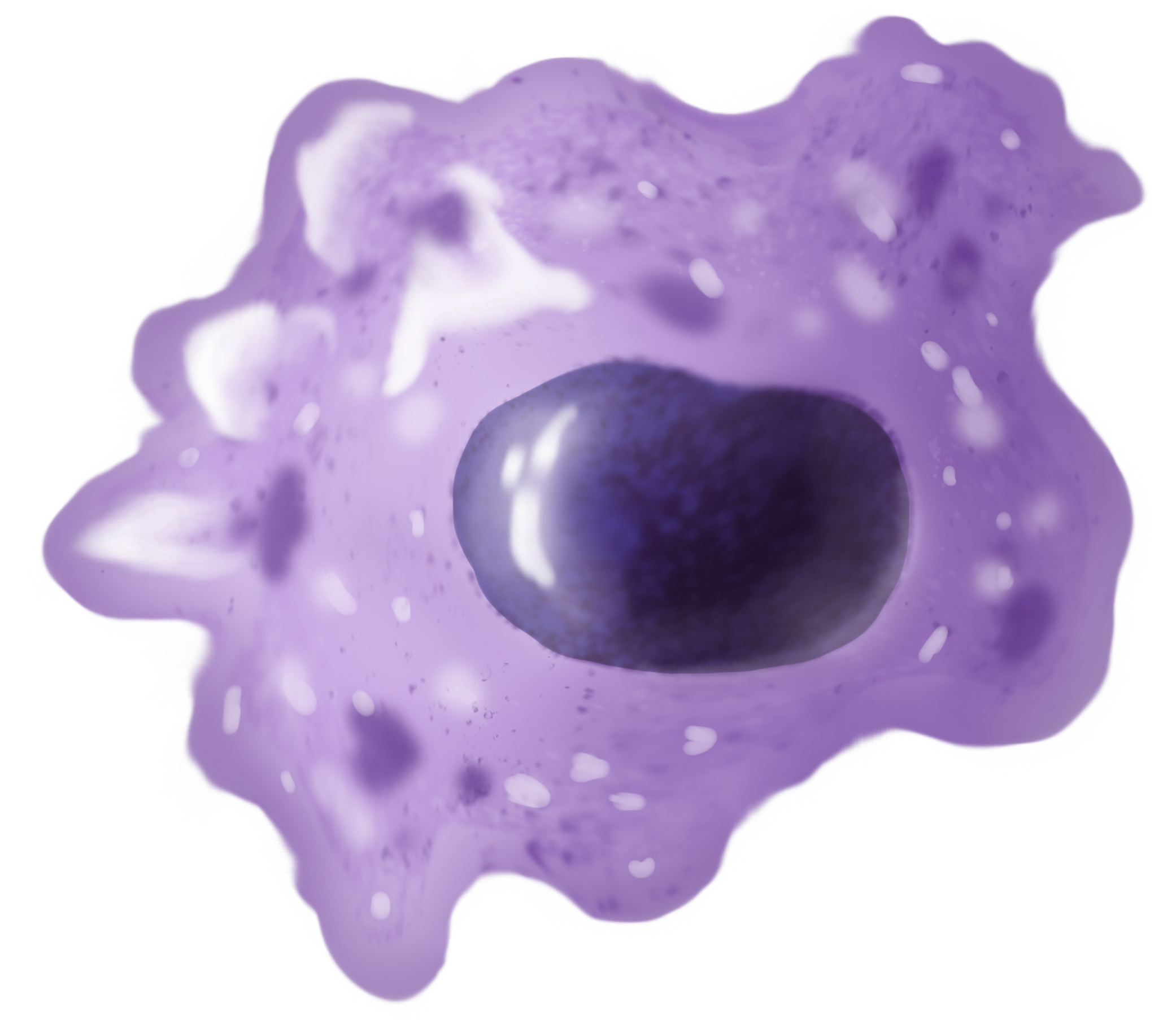
Macrophages (abbreviated as M φ, MΦ or MP) ( el, large eaters, from Greek ''μακρός'' (') = large, ''φαγεῖν'' (') = to eat) are a type of white blood cell of the immune system that engulfs and digests pathogens, such as cancer cells, microbes, cellular debris, and foreign substances, which do not have proteins that are specific to healthy body cells on their surface. The process is called phagocytosis, which acts to defend the host against infection and injury. These large phagocytes are found in essentially all tissues, where they patrol for potential pathogens by amoeboid movement. They take various forms (with various names) throughout the body (e.g., histiocytes, Kupffer cells, alveolar macrophages, microglia, and others), but all are part of the mononuclear phagocyte system. Besides phagocytosis, they play a critical role in nonspecific defense (innate immunity) and also help initiate specific defense mechanisms (adaptive immunity) by recruiting other immune ... [...More Info...] [...Related Items...] OR: [Wikipedia] [Google] [Baidu] |


