|
Bitewing
Dental radiographs, commonly known as X-rays, are radiographs used to diagnose hidden dental structures, malignant or benign masses, bone loss, and cavities. A radiographic image is formed by a controlled burst of X-ray radiation which penetrates oral structures at different levels, depending on varying anatomical densities, before striking the film or sensor. Teeth appear lighter because less radiation penetrates them to reach the film. Dental caries, infections and other changes in the bone density, and the periodontal ligament, appear darker because X-rays readily penetrate these less dense structures. Dental restorations (fillings, crowns) may appear lighter or darker, depending on the density of the material. The dosage of X-ray radiation received by a dental patient is typically small (around 0.150 mSv for a full mouth series), equivalent to a few days' worth of background environmental radiation exposure, or similar to the dose received during a cross-country airplane fl ... [...More Info...] [...Related Items...] OR: [Wikipedia] [Google] [Baidu] |
X-rays
An X-ray, or, much less commonly, X-radiation, is a penetrating form of high-energy electromagnetic radiation. Most X-rays have a wavelength ranging from 10 Picometre, picometers to 10 Nanometre, nanometers, corresponding to frequency, frequencies in the range 30 Hertz, petahertz to 30 Hertz, exahertz ( to ) and energies in the range 145 electronvolt, eV to 124 keV. X-ray wavelengths are shorter than those of ultraviolet, UV rays and typically longer than those of gamma rays. In many languages, X-radiation is referred to as Röntgen radiation, after the German scientist Wilhelm Röntgen, Wilhelm Conrad Röntgen, who discovered it on November 8, 1895. He named it ''X-radiation'' to signify an unknown type of radiation.Novelline, Robert (1997). ''Squire's Fundamentals of Radiology''. Harvard University Press. 5th edition. . Spellings of ''X-ray(s)'' in English include the variants ''x-ray(s)'', ''xray(s)'', and ''X ray(s)''. The most familiar use of X-ra ... [...More Info...] [...Related Items...] OR: [Wikipedia] [Google] [Baidu] |
Human Mandible
In anatomy, the mandible, lower jaw or jawbone is the largest, strongest and lowest bone in the human facial skeleton. It forms the lower jaw and holds the lower tooth, teeth in place. The mandible sits beneath the maxilla. It is the only movable bone of the skull (discounting the ossicles of the middle ear). It is connected to the temporal bones by the temporomandibular joints. The bone is formed prenatal development, in the fetus from a fusion of the left and right mandibular prominences, and the point where these sides join, the mandibular symphysis, is still visible as a faint ridge in the midline. Like other symphyses in the body, this is a midline articulation where the bones are joined by fibrocartilage, but this articulation fuses together in early childhood.Illustrated Anatomy of the Head and Neck, Fehrenbach and Herring, Elsevier, 2012, p. 59 The word "mandible" derives from the Latin word ''mandibula'', "jawbone" (literally "one used for chewing"), from ''wikt:mandere ... [...More Info...] [...Related Items...] OR: [Wikipedia] [Google] [Baidu] |
Cephalogram
A cephalogram is an X-ray of the craniofacial area. A cephalometric analysis Cephalometric analysis is the clinical application of cephalometry. It is analysis of the dental and skeletal relationships of a human skull. It is frequently used by dentists, orthodontists, and oral and maxillofacial surgeons as a treatment ... could be used as means for measuring growth in children. The lateral cephalogram is a profile x-ray of the skull and soft tissues and is used to assess the relation of the teeth in the jaws, the relation of the jaws to the skull and the relation of the soft tissues to the teeth and jaws. In children, growth predictions can be made and we can also determine the changes that have occurred with treatment. In adults, treatment can be predicted with varying degrees of accuracy and results quantified. References Further reading * * Human head and neck Radiography {{med-diagnostic-stub ... [...More Info...] [...Related Items...] OR: [Wikipedia] [Google] [Baidu] |
Evidence-based Medicine
Evidence-based medicine (EBM) is "the conscientious, explicit and judicious use of current best evidence in making decisions about the care of individual patients". The aim of EBM is to integrate the experience of the clinician, the values of the patient, and the best available scientific information to guide decision-making about clinical management. The term was originally used to describe an approach to teaching the practice of medicine and improving decisions by individual physicians about individual patients. Background, history and definition Medicine has a long history of scientific inquiry about the prevention, diagnosis, and treatment of human disease. The concept of a controlled clinical trial was first described in 1662 by Jan Baptist van Helmont in reference to the practice of bloodletting. Wrote Van Helmont: The first published report describing the conduct and results of a controlled clinical trial was by James Lind, a Scottish naval surgeon who conducted rese ... [...More Info...] [...Related Items...] OR: [Wikipedia] [Google] [Baidu] |
Selection Criteria In Dental Radiography
Selection may refer to: Science * Selection (biology), also called natural selection, selection in evolution ** Sex selection, in genetics ** Mate selection, in mating ** Sexual selection in humans, in human sexuality ** Human mating strategies, in human sexuality * Social selection, within social groups * Selection (linguistics), the ability of predicates to determine the semantic content of their arguments * Selection in schools, the admission of students on the basis of selective criteria * Selection effect, a distortion of data arising from the way that the data are collected * A selection, or choice function, a function that selects an element from a set Religion * Divine selection, selection by God * Papal selection, selection by clergy Computing * Selection (user interface) ** X Window selection * Selection (genetic algorithm) * Selection (relational algebra) * Selection-based search, a search engine system in which the user invokes a search query using only the mo ... [...More Info...] [...Related Items...] OR: [Wikipedia] [Google] [Baidu] |
Royal College Of Surgeons Of England
The Royal College of Surgeons of England (RCS England) is an independent professional body and registered charity that promotes and advances standards of surgical care for patients, and regulates surgery and dentistry in England and Wales. The College is located at Lincoln's Inn Fields in London. It publishes multiple medical journals including the ''Annals of the Royal College of Surgeons of England'', the '' Faculty Dental Journal'', and the '' Bulletin of the Royal College of Surgeons of England''. History The origins of the college date to the fourteenth century with the foundation of the "Guild of Surgeons Within the City of London". Certain sources date this as occurring in 1368. There was ongoing dispute between the surgeons and barber surgeons until an agreement was signed between them in 1493, giving the fellowship of surgeons the power of incorporation. This union was formalised further in 1540 by Henry VIII between the Worshipful Company of Barbers (incorporated 14 ... [...More Info...] [...Related Items...] OR: [Wikipedia] [Google] [Baidu] |
Faculty Of General Dental Practice
The Faculty of General Dental Practice (UK) (FGDP(UK)) is an independent UK professional body for general dental practitioners. It was established in 1992 as a faculty of the Royal College of Surgeons of England and is located in the College’s headquarters in Lincoln’s Inn Fields, London. Membership of the FGDP(UK) is open to all dentists, dental surgeons and dental care professionals registered with the General Dental Council. It opened its membership to dental care professionals (DCPs) in 2005 and to students in years 4 or 5 of their Bachelor of Dental Surgery undergraduate degree in 2011. Education FGDP(UK) is involved in many educational aspects of dental care in general practice and primary dental care. It runs a programme of training courses and continuous professional development for the whole dental team. It also provides the continuing professional development (CPD) and training needs of both dentists and dental care professionals working in primary care. The Fac ... [...More Info...] [...Related Items...] OR: [Wikipedia] [Google] [Baidu] |
Premolar
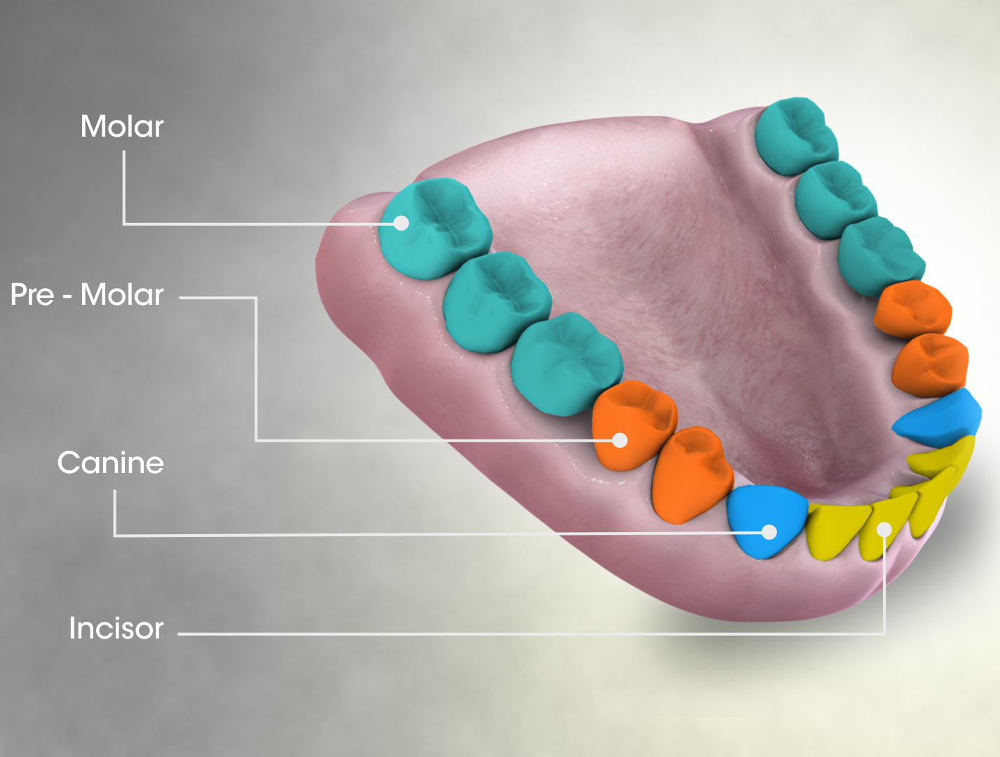
The premolars, also called premolar teeth, or bicuspids, are transitional teeth located between the canine and molar teeth. In humans, there are two premolars per quadrant in the permanent set of teeth, making eight premolars total in the mouth. They have at least two cusps. Premolars can be considered transitional teeth during chewing, or mastication. They have properties of both the canines, that lie anterior and molars that lie posterior, and so food can be transferred from the canines to the premolars and finally to the molars for grinding, instead of directly from the canines to the molars. Human anatomy The premolars in humans are the maxillary first premolar, maxillary second premolar, mandibular first premolar, and the mandibular second premolar. Premolar teeth by definition are permanent teeth distal to the canines, preceded by deciduous molars. Morphology There is always one large buccal cusp, especially so in the mandibular first premolar. The lower second ... [...More Info...] [...Related Items...] OR: [Wikipedia] [Google] [Baidu] |
Molar (tooth)
The molars or molar teeth are large, flat teeth at the back of the mouth. They are more developed in mammals. They are used primarily to grind food during chewing. The name ''molar'' derives from Latin, ''molaris dens'', meaning "millstone tooth", from ''mola'', millstone and ''dens'', tooth. Molars show a great deal of diversity in size and shape across mammal groups. The third molar of humans is sometimes vestigial. Human anatomy In humans, the molar teeth have either four or five cusps. Adult humans have 12 molars, in four groups of three at the back of the mouth. The third, rearmost molar in each group is called a wisdom tooth. It is the last tooth to appear, breaking through the front of the gum at about the age of 20, although this varies from individual to individual. Race can also affect the age at which this occurs, with statistical variations between groups. In some cases, it may not even erupt at all. The human mouth contains upper (maxillary) and lower (mandib ... [...More Info...] [...Related Items...] OR: [Wikipedia] [Google] [Baidu] |
Parotid Gland
The parotid gland is a major salivary gland in many animals. In humans, the two parotid glands are present on either side of the mouth and in front of both ears. They are the largest of the salivary glands. Each parotid is wrapped around the mandibular ramus, and secretes serous saliva through the parotid duct into the mouth, to facilitate mastication and swallowing and to begin the digestion of starches. There are also two other types of salivary glands; they are submandibular and sublingual glands. Sometimes accessory parotid glands are found close to the main parotid glands. Etymology The word ''parotid'' literally means "beside the ear". From Greek παρωτίς (stem παρωτιδ-) : (gland) behind the ear < παρά - pará : in front, and οὖς - ous (stem ὠτ-, ōt-) : ear. Structure The parotid glands are a pair of mainly |
Saliva
Saliva (commonly referred to as spit) is an extracellular fluid produced and secreted by salivary glands in the mouth. In humans, saliva is around 99% water, plus electrolytes, mucus, white blood cells, epithelial cells (from which DNA can be extracted), enzymes (such as lipase and amylase), antimicrobial agents (such as secretory IgA, and lysozymes). The enzymes found in saliva are essential in beginning the process of digestion of dietary starches and fats. These enzymes also play a role in breaking down food particles entrapped within dental crevices, thus protecting teeth from bacterial decay. Saliva also performs a lubricating function, wetting food and permitting the initiation of swallowing, and protecting the oral mucosa from drying out. Various animal species have special uses for saliva that go beyond predigestion. Some swifts use their gummy saliva to build nests. ''Aerodramus'' nests form the basis of bird's nest soup. Cobras, vipers, and certain other membe ... [...More Info...] [...Related Items...] OR: [Wikipedia] [Google] [Baidu] |
Sialolithiasis
Sialolithiasis (also termed salivary calculi, or salivary stones) is a crystallopathy where a calcified mass or ''sialolith'' forms within a salivary gland, usually in the duct of the submandibular gland (also termed "Wharton's duct"). Less commonly the parotid gland or rarely the sublingual gland or a minor salivary gland may develop salivary stones. The usual symptoms are pain and swelling of the affected salivary gland, both of which get worse when salivary flow is stimulated, e.g. with the sight, thought, smell or taste of food, or with hunger or chewing. This is often termed "mealtime syndrome". Inflammation or infection of the gland may develop as a result. Sialolithiasis may also develop because of the presence of existing chronic infection of the glands, dehydration (e.g. use of phenothiazines), Sjögren's syndrome and/or increased local levels of calcium, but in many instances the cause is idiopathic (unknown). The condition is usually managed by removing the stone, and ... [...More Info...] [...Related Items...] OR: [Wikipedia] [Google] [Baidu] |


