|
Biarticular Muscle
Biarticular muscles are muscles that cross two joints rather than just one, such as the hamstrings which cross both the hip and the knee. The function of these muscles is complex and often depends upon both their anatomy and the activity of other muscles at the joints in question. Their role in movement is currently poorly understood. Anatomy Biarticular muscles cross two joints in series, usually in a limb, but the details of the origin (proximal attachment) and insertion (distal attachment) can play a large role in determining muscle function. For instance, the human gastrocnemius technically spans both the knee and ankle joints. However, the origin point of the muscle is so close to the axis of rotation of the knee joint that the muscle's effective lever arm would be very small, especially compared to its large lever arm at the ankle. As a result, even though it spans two joints, the strong bias in lever arms allows it to function primarily as an ankle plantar flexor. Other m ... [...More Info...] [...Related Items...] OR: [Wikipedia] [Google] [Baidu] |
Muscle
Skeletal muscles (commonly referred to as muscles) are organs of the vertebrate muscular system and typically are attached by tendons to bones of a skeleton. The muscle cells of skeletal muscles are much longer than in the other types of muscle tissue, and are often known as muscle fibers. The muscle tissue of a skeletal muscle is striated – having a striped appearance due to the arrangement of the sarcomeres. Skeletal muscles are voluntary muscles under the control of the somatic nervous system. The other types of muscle are cardiac muscle which is also striated and smooth muscle which is non-striated; both of these types of muscle tissue are classified as involuntary, or, under the control of the autonomic nervous system. A skeletal muscle contains multiple fascicles – bundles of muscle fibers. Each individual fiber, and each muscle is surrounded by a type of connective tissue layer of fascia. Muscle fibers are formed from the fusion of developmental myoblasts in ... [...More Info...] [...Related Items...] OR: [Wikipedia] [Google] [Baidu] |
Joint
A joint or articulation (or articular surface) is the connection made between bones, ossicles, or other hard structures in the body which link an animal's skeletal system into a functional whole.Saladin, Ken. Anatomy & Physiology. 7th ed. McGraw-Hill Connect. Webp.274/ref> They are constructed to allow for different degrees and types of movement. Some joints, such as the knee, elbow, and shoulder, are self-lubricating, almost frictionless, and are able to withstand compression and maintain heavy loads while still executing smooth and precise movements. Other joints such as sutures between the bones of the skull permit very little movement (only during birth) in order to protect the brain and the sense organs. The connection between a tooth and the jawbone is also called a joint, and is described as a fibrous joint known as a gomphosis. Joints are classified both structurally and functionally. Classification The number of joints depends on if sesamoids are included, age of the ... [...More Info...] [...Related Items...] OR: [Wikipedia] [Google] [Baidu] |
Hamstring
In human anatomy, a hamstring () is any one of the three posterior thigh muscles in between the hip and the knee (from medial to lateral: semimembranosus, semitendinosus and biceps femoris). The hamstrings are susceptible to injury. In quadrupeds, the hamstring is the single large tendon found behind the knee or comparable area. Criteria The common criteria of any hamstring muscles are: # Muscles should originate from ischial tuberosity. # Muscles should be inserted over the knee joint, in the tibia or in the fibula. # Muscles will be innervated by the tibial branch of the sciatic nerve. # Muscle will participate in flexion of the knee joint and extension of the hip joint. Those muscles which fulfill all of the four criteria are called true hamstrings. The adductor magnus reaches only up to the adductor tubercle of the femur, but it is included amongst the hamstrings because the tibial collateral ligament of the knee joint morphologically is the degenerated tendon of this muscl ... [...More Info...] [...Related Items...] OR: [Wikipedia] [Google] [Baidu] |
Knee
In humans and other primates, the knee joins the thigh with the leg and consists of two joints: one between the femur and tibia (tibiofemoral joint), and one between the femur and patella (patellofemoral joint). It is the largest joint in the human body. The knee is a modified hinge joint, which permits flexion and extension as well as slight internal and external rotation. The knee is vulnerable to injury and to the development of osteoarthritis. It is often termed a ''compound joint'' having tibiofemoral and patellofemoral components. (The fibular collateral ligament is often considered with tibiofemoral components.) Structure The knee is a modified hinge joint, a type of synovial joint, which is composed of three functional compartments: the patellofemoral articulation, consisting of the patella, or "kneecap", and the patellar groove on the front of the femur through which it slides; and the medial and lateral tibiofemoral articulations linking the femur, or thigh bone ... [...More Info...] [...Related Items...] OR: [Wikipedia] [Google] [Baidu] |
Gastrocnemius
The gastrocnemius muscle (plural ''gastrocnemii'') is a superficial two-headed muscle that is in the back part of the lower leg of humans. It runs from its two heads just above the knee to the heel, a three joint muscle (knee, ankle and subtalar joints). The muscle is named via Latin, from Greek γαστήρ (''gaster'') 'belly' or 'stomach' and κνήμη (''knḗmē'') 'leg', meaning 'stomach of the leg' (referring to the bulging shape of the calf). Structure The gastrocnemius is located with the soleus in the posterior (back) compartment of the leg. The lateral head originates from the lateral condyle of the femur, while the medial head originates from the medial condyle of the femur. Its other end forms a common tendon with the soleus muscle; this tendon is known as the calcaneal tendon or Achilles tendon and inserts onto the posterior surface of the calcaneus, or heel bone. It is considered a superficial muscle as it is located directly under skin, and its shape may often b ... [...More Info...] [...Related Items...] OR: [Wikipedia] [Google] [Baidu] |
Lever Arm
In physics and mechanics, torque is the rotational equivalent of linear force. It is also referred to as the moment of force (also abbreviated to moment). It represents the capability of a force to produce change in the rotational motion of the body. The concept originated with the studies by Archimedes of the usage of levers, which is reflected in his famous quote: "''Give me a lever and a place to stand and I will move the Earth''". Just as a linear force is a push or a pull, a torque can be thought of as a twist to an object around a specific axis. Torque is defined as the product of the magnitude of the perpendicular component of the force and the distance of the line of action of a force from the point around which it is being determined. The law of conservation of energy can also be used to understand torque. The symbol for torque is typically \boldsymbol\tau, the lowercase Greek alphabet, Greek letter ''tau''. When being referred to as moment (physics), moment of force, it ... [...More Info...] [...Related Items...] OR: [Wikipedia] [Google] [Baidu] |
Muscle Contraction
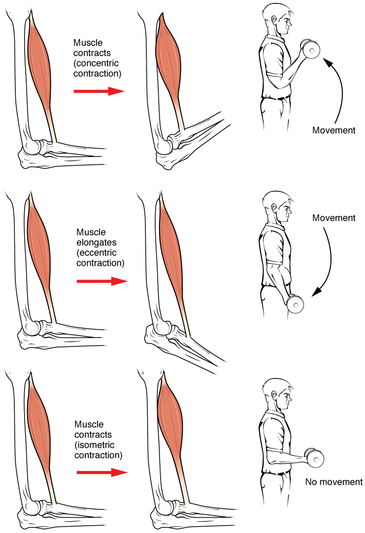
Muscle contraction is the activation of tension-generating sites within muscle cells. In physiology, muscle contraction does not necessarily mean muscle shortening because muscle tension can be produced without changes in muscle length, such as when holding something heavy in the same position. The termination of muscle contraction is followed by muscle relaxation, which is a return of the muscle fibers to their low tension-generating state. For the contractions to happen, the muscle cells must rely on the interaction of two types of filaments which are the thin and thick filaments. Thin filaments are two strands of actin coiled around each, and thick filaments consist of mostly elongated proteins called myosin. Together, these two filaments form myofibrils which are important organelles in the skeletal muscle system. Muscle contraction can also be described based on two variables: length and tension. A muscle contraction is described as isometric if the muscle tension changes ... [...More Info...] [...Related Items...] OR: [Wikipedia] [Google] [Baidu] |
Four-bar Linkage
In the study of mechanisms, a four-bar linkage, also called a four-bar, is the simplest closed- chain movable linkage. It consists of four bodies, called ''bars'' or ''links'', connected in a loop by four joints. Generally, the joints are configured so the links move in parallel planes, and the assembly is called a ''planar four-bar linkage''. Spherical and spatial four-bar linkages also exist and are used in practice. Planar four-bar linkage Planar four-bar linkages are constructed from four links connected in a loop by four one- degree-of-freedom joints. A joint may be either a revolute joint – also known as a pin joint or hinged joint – denoted by R, or a prismatic joint – also known as a sliding pair – denoted by P. A link that are fixed in place relative to the viewer is called a ''ground link.'' A link connecting to the ground by a revolute joint that can perform a complete revolution is called a '' crank link.'' A link connecting to the ground by a revolute join ... [...More Info...] [...Related Items...] OR: [Wikipedia] [Google] [Baidu] |
Inverse Dynamics
Inverse dynamics is an inverse problem. It commonly refers to either inverse rigid body dynamics or inverse structural dynamics. Inverse rigid-body dynamics is a method for computing forces and/or moment of force, moments of force (torques) based on the kinematics (motion) of a body and the body's inertial properties (mass and moment of inertia). Typically it uses link-segment models to represent the mechanical behaviour of interconnected segments, such as the Limb (anatomy), limbs of humans or animals or the joint extensions of robots, where given the kinematics of the various parts, inverse dynamics derives the minimum forces and moments responsible for the individual movements. In practice, inverse dynamics computes these internal moments and forces from measurements of the motion of limbs and external forces such as ground reaction forces, under a special set of assumptions.Robertson DGE, et al., Research Methods in Biomechanics, Champaign IL:Human Kinetics Pubs., 2004. Applicati ... [...More Info...] [...Related Items...] OR: [Wikipedia] [Google] [Baidu] |


