|
B-cell Prolymphocytic Leukemia
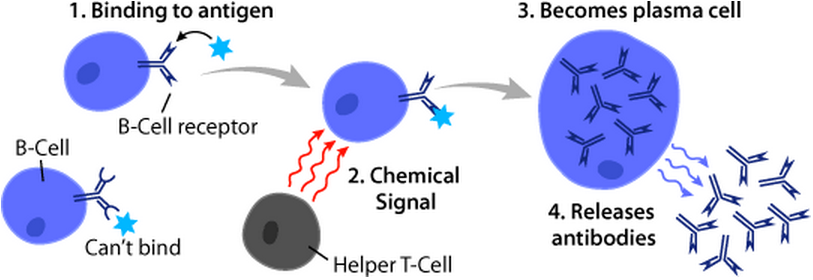
B-cell prolymphocytic leukemia, referred to as B-PLL, is a rare blood cancer. It is a more aggressive, but still treatable, form of leukemia. Specifically, B-PLL is a prolymphocytic leukemia (PLL) that affects prolymphocytes – immature forms of B-lymphocytes and T-lymphocytes – in the peripheral blood, bone marrow, and spleen. It is an aggressive cancer that presents poor response to treatment. Mature lymphocytes are infection-fighting immune system cells. B-lymphocytes have two responsibilities: # Production of antibodies – In response to antigens, B-lymphocytes produce and release antibodies specific to foreign substances in order to aid in their identification and elimination phagocytes # Generation of memory cells – Interactions between antibodies and antigens allow B-lymphocytes to establish cellular memories, otherwise known as immunities that allow the body to respond more rapidly and efficiently to previously encountered species Classification It is categorize ... [...More Info...] [...Related Items...] OR: [Wikipedia] [Google] [Baidu] |
Prolymphocyte
A prolymphocyte is a white blood cell with a certain state of cellular differentiation in lymphocytopoiesis. In the 20th century it was believed that a sequence of general maturation changed cells from lymphoblasts to prolymphocytes and then to lymphocytes (the lymphocytic series), with each being a precursor of the last. Today it is believed that the differentiation of cells in the lymphocyte line is not always simply chronologic but rather depends on antigen exposure, such that, for example, lymphocytes can become lymphoblasts. The size is between 10 and 18 μm. See also * Pluripotential hemopoietic stem cell Hematopoietic stem cells (HSCs) are the stem cells that give rise to other blood cells. This process is called haematopoiesis. In vertebrates, the very first definitive HSCs arise from the ventral endothelial wall of the embryonic aorta within t ... * Prolymphocytic leukemia References External links Histology at hematologyatlas.com (found in sixth row) Lymph ... [...More Info...] [...Related Items...] OR: [Wikipedia] [Google] [Baidu] |
Splenomegaly
Splenomegaly is an enlargement of the spleen. The spleen usually lies in the left upper quadrant (LUQ) of the human abdomen. Splenomegaly is one of the four cardinal signs of ''hypersplenism'' which include: some reduction in number of circulating blood cells affecting granulocytes, erythrocytes or platelets in any combination; a compensatory proliferative response in the bone marrow; and the potential for correction of these abnormalities by splenectomy. Splenomegaly is usually associated with increased workload (such as in hemolytic anemias), which suggests that it is a response to hyperfunction. It is therefore not surprising that splenomegaly is associated with any disease process that involves abnormal red blood cells being destroyed in the spleen. Other common causes include congestion due to portal hypertension and infiltration by leukemias and lymphomas. Thus, the finding of an enlarged spleen, along with caput medusae, is an important sign of portal hypertension. Definiti ... [...More Info...] [...Related Items...] OR: [Wikipedia] [Google] [Baidu] |
CD22
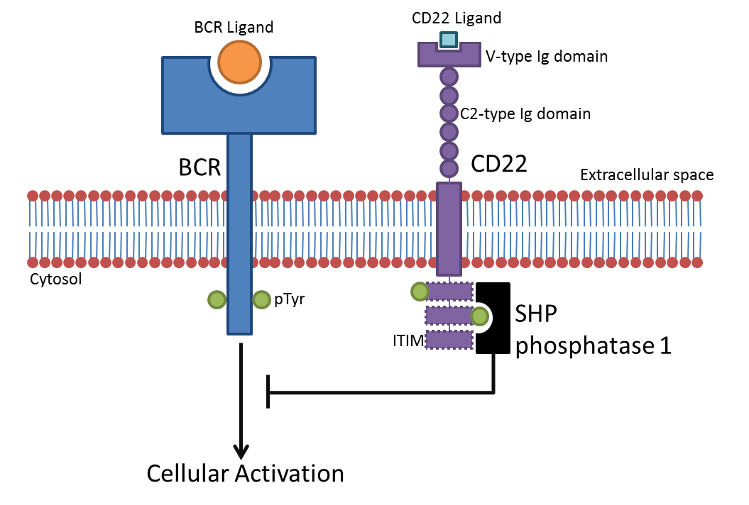
CD22, or cluster of differentiation-22, is a molecule belonging to the SIGLEC family of lectins. It is found on the surface of mature B cells and to a lesser extent on some immature B cells. Generally speaking, CD22 is a regulatory molecule that prevents the overactivation of the immune system and the development of autoimmune diseases. CD22 is a sugar binding transmembrane protein, which specifically binds sialic acid with an immunoglobulin (Ig) domain located at its N-terminus. The presence of Ig domains makes CD22 a member of the immunoglobulin superfamily. CD22 functions as an inhibitory receptor for B cell receptor (BCR) signaling. It is also involved in the B cell trafficking to Peyer's patches in mice. In mice, it has been shown that CD22 blockade restores homeostatic microglial phagocytosis in aging brains. Structure CD22 is a transmembrane protein with a molecular weight of 140 kDa. The extracellular part of CD22 consists of seven immunoglobulin domains and the intr ... [...More Info...] [...Related Items...] OR: [Wikipedia] [Google] [Baidu] |
CD20
B-lymphocyte antigen CD20 or CD20 is expressed on the surface of all B-cells beginning at the pro-B phase (CD45R+, CD117+) and progressively increasing in concentration until maturity. In humans CD20 is encoded by the ''MS4A1'' gene. This gene encodes a member of the membrane-spanning 4A gene family. Members of this nascent protein family are characterized by common structural features and similar intron/exon splice boundaries and display unique expression patterns among hematopoietic cells and nonlymphoid tissues. This gene encodes a B-lymphocyte surface molecule that plays a role in the development and differentiation of B-cells into plasma cells. This family member is localized to 11q12, among a cluster of family members. Alternative splicing of this gene results in two transcript variants that encode the same protein. Function The protein has no known natural Ligand (biochemistry), ligand and its function is to enable optimal B-cell immune response, specifically against T-i ... [...More Info...] [...Related Items...] OR: [Wikipedia] [Google] [Baidu] |
CD19
B-lymphocyte antigen CD19, also known as CD19 molecule ( Cluster of Differentiation 19), B-Lymphocyte Surface Antigen B4, T-Cell Surface Antigen Leu-12 and CVID3 is a transmembrane protein that in humans is encoded by the gene ''CD19''. In humans, CD19 is expressed in all B lineage cells. Contrary to some early doubts, human plasma cells do express CD19, as confirmed by others. CD19 plays two major roles in human B cells: on the one hand, it acts as an adaptor protein to recruit cytoplasmic signaling proteins to the membrane; on the other, it works within the CD19/CD21 complex to decrease the threshold for B cell receptor signaling pathways. Due to its presence on all B cells, it is a biomarker for B lymphocyte development, lymphoma diagnosis and can be utilized as a target for leukemia immunotherapies. Structure In humans, CD19 is encoded by the 7.41 kilobase ''CD19'' gene located on the short arm of chromosome 16. It contains at least fifteen exons, four that encode extrac ... [...More Info...] [...Related Items...] OR: [Wikipedia] [Google] [Baidu] |
NC Ratio
The nuclear-cytoplasmic ratio (also variously known as the nucleus:cytoplasm ratio, nucleus-cytoplasm ratio, N:C ratio, or N/C) is a measurement used in cell biology. It is a ratio of the size (i.e., volume) of the nucleus of a cell to the size of the cytoplasm of that cell. The N:C ratio indicates the maturity of a cell, because as a cell matures the size of its nucleus generally decreases. For example, "blast" forms of erythrocytes, leukocytes, and megakaryocytes start with an N:C ratio of 4:1, which decreases as they mature to 2:1 or even 1:1 (with exceptions for mature thrombocytes and erythrocytes, which are anuclear cells, and mature lymphocytes, which only decrease to a 3:1 ratio and often retain the original 4:1 ratio). An increased N:C ratio is commonly associated with precancerous dysplasia as well as with malignant cells. See also * *Cytopathology *Nuclear atypia Nuclear atypia refers to abnormal appearance of cell nuclei. It is a term used in cytopathology and his ... [...More Info...] [...Related Items...] OR: [Wikipedia] [Google] [Baidu] |
Chromatin
Chromatin is a complex of DNA and protein found in eukaryotic cells. The primary function is to package long DNA molecules into more compact, denser structures. This prevents the strands from becoming tangled and also plays important roles in reinforcing the DNA during cell division, preventing DNA damage, and regulating gene expression and DNA replication. During mitosis and meiosis, chromatin facilitates proper segregation of the chromosomes in anaphase; the characteristic shapes of chromosomes visible during this stage are the result of DNA being coiled into highly condensed chromatin. The primary protein components of chromatin are histones. An octamer of two sets of four histone cores (Histone H2A, Histone H2B, Histone H3, and Histone H4) bind to DNA and function as "anchors" around which the strands are wound.Maeshima, K., Ide, S., & Babokhov, M. (2019). Dynamic chromatin organization without the 30-nm fiber. ''Current opinion in cell biology, 58,'' 95–104. https://doi.o ... [...More Info...] [...Related Items...] OR: [Wikipedia] [Google] [Baidu] |
Nucleolus
The nucleolus (, plural: nucleoli ) is the largest structure in the nucleus of eukaryotic cells. It is best known as the site of ribosome biogenesis, which is the synthesis of ribosomes. The nucleolus also participates in the formation of signal recognition particles and plays a role in the cell's response to stress. Nucleoli are made of proteins, DNA and RNA, and form around specific chromosomal regions called nucleolar organizing regions. Malfunction of nucleoli can be the cause of several human conditions called "nucleolopathies" and the nucleolus is being investigated as a target for cancer chemotherapy. History The nucleolus was identified by bright-field microscopy during the 1830s. Little was known about the function of the nucleolus until 1964, when a study of nucleoli by John Gurdon and Donald Brown in the African clawed frog ''Xenopus laevis'' generated increasing interest in the function and detailed structure of the nucleolus. They found that 25% of the frog e ... [...More Info...] [...Related Items...] OR: [Wikipedia] [Google] [Baidu] |
Cell Nucleus
The cell nucleus (pl. nuclei; from Latin or , meaning ''kernel'' or ''seed'') is a membrane-bound organelle found in eukaryotic cells. Eukaryotic cells usually have a single nucleus, but a few cell types, such as mammalian red blood cells, have no nuclei, and a few others including osteoclasts have many. The main structures making up the nucleus are the nuclear envelope, a double membrane that encloses the entire organelle and isolates its contents from the cellular cytoplasm; and the nuclear matrix, a network within the nucleus that adds mechanical support. The cell nucleus contains nearly all of the cell's genome. Nuclear DNA is often organized into multiple chromosomes – long stands of DNA dotted with various proteins, such as histones, that protect and organize the DNA. The genes within these chromosomes are structured in such a way to promote cell function. The nucleus maintains the integrity of genes and controls the activities of the cell by regulating gene expres ... [...More Info...] [...Related Items...] OR: [Wikipedia] [Google] [Baidu] |
B Cells
B cells, also known as B lymphocytes, are a type of white blood cell of the lymphocyte subtype. They function in the humoral immunity component of the adaptive immune system. B cells produce antibody molecules which may be either secreted or inserted into the plasma membrane where they serve as a part of B-cell receptors. When a naïve or memory B cell is activated by an antigen, it proliferates and differentiates into an antibody-secreting effector cell, known as a plasmablast or plasma cell. Additionally, B cells present antigens (they are also classified as professional antigen-presenting cells (APCs)) and secrete cytokines. In mammals, B cells mature in the bone marrow, which is at the core of most bones. In birds, B cells mature in the bursa of Fabricius, a lymphoid organ where they were first discovered by Chang and Glick, which is why the 'B' stands for bursa and not bone marrow as commonly believed. B cells, unlike the other two classes of lymphocytes, T cells and ... [...More Info...] [...Related Items...] OR: [Wikipedia] [Google] [Baidu] |
Cytogenetics
Cytogenetics is essentially a branch of genetics, but is also a part of cell biology/cytology (a subdivision of human anatomy), that is concerned with how the chromosomes relate to cell behaviour, particularly to their behaviour during mitosis and meiosis. Techniques used include karyotyping, analysis of G-banded chromosomes, other cytogenetic banding techniques, as well as molecular cytogenetics such as fluorescent ''in situ'' hybridization (FISH) and comparative genomic hybridization (CGH). History Beginnings Chromosomes were first observed in plant cells by Carl Nägeli in 1842. Their behavior in animal (salamander) cells was described by Walther Flemming, the discoverer of mitosis, in 1882. The name was coined by another German anatomist, von Waldeyer in 1888. The next stage took place after the development of genetics in the early 20th century, when it was appreciated that the set of chromosomes (the karyotype) was the carrier of the genes. Levitsky seems to have been t ... [...More Info...] [...Related Items...] OR: [Wikipedia] [Google] [Baidu] |
Immunophenotyping
Immunophenotyping is a technique used to study the protein expressed by cells. This technique is commonly used in basic science research and laboratory diagnostic purpose. This can be done on tissue section (fresh or fixed tissue), cell suspension, etc. An example is the detection of tumor markers, such as in the diagnosis of leukemia. It involves the labelling of white blood cells with antibody, antibodies directed against surface proteins on their membrane. By choosing appropriate antibodies, the differentiation of leukemia, leukemic cells can be accurately determined. The labelled cells are processed in a flow cytometry, flow cytometer, a laser-based instrument capable of analyzing thousands of cells per second. The whole procedure can be performed on cells from the blood, bone marrow or spinal fluid in a matter of a few hours. An example of information provided through Immunophenotyping: "The flow cytometric immunophenotyping report indicated the malignant cells were positive f ... [...More Info...] [...Related Items...] OR: [Wikipedia] [Google] [Baidu] |


