|
Abderhalden–Kaufmann–Lignac Syndrome
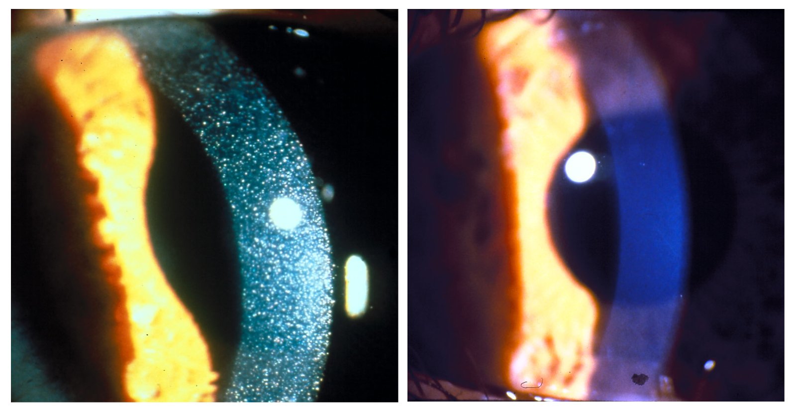
Abderhalden–Kaufmann–Lignac syndrome (AKL syndrome), also called nephropathic cystinosis, is an autosomal recessive renal disorder of childhood comprising cystinosis and renal rickets. Presentation Affected children are developmentally delayed with dwarfism, rickets and osteoporosis. Renal tubular disease is usually present causing aminoaciduria, glycosuria and hypokalemia. Cysteine deposition is most evident in the conjunctiva and cornea. Diagnosis Eponym It is named for Emil Abderhalden Emil Abderhalden (9 March 1877 – 5 August 1950) was a Swiss biochemist and physiologist. His main findings, though disputed already in the 1910s, were not finally rejected until the late 1990s. Whether his misleading findings were based on f ..., Eduard Kaufmann and George Lignac. See also * Cystinosin References External links Autosomal recessive disorders Syndromes affecting the kidneys {{Genetic-disorder-stub ... [...More Info...] [...Related Items...] OR: [Wikipedia] [Google] [Baidu] |
Autosomal Recessive
In genetics, dominance is the phenomenon of one variant (allele) of a gene on a chromosome masking or overriding the effect of a different variant of the same gene on the other copy of the chromosome. The first variant is termed dominant and the second recessive. This state of having two different variants of the same gene on each chromosome is originally caused by a mutation in one of the genes, either new (''de novo'') or inherited. The terms autosomal dominant or autosomal recessive are used to describe gene variants on non-sex chromosomes ( autosomes) and their associated traits, while those on sex chromosomes (allosomes) are termed X-linked dominant, X-linked recessive or Y-linked; these have an inheritance and presentation pattern that depends on the sex of both the parent and the child (see Sex linkage). Since there is only one copy of the Y chromosome, Y-linked traits cannot be dominant or recessive. Additionally, there are other forms of dominance such as incomplete d ... [...More Info...] [...Related Items...] OR: [Wikipedia] [Google] [Baidu] |
Cysteine
Cysteine (symbol Cys or C; ) is a semiessential proteinogenic amino acid with the formula . The thiol side chain in cysteine often participates in enzymatic reactions as a nucleophile. When present as a deprotonated catalytic residue, sometimes the symbol Cyz is used. The deprotonated form can generally be described by the symbol Cym as well. The thiol is susceptible to oxidation to give the disulfide derivative cystine, which serves an important structural role in many proteins. In this case, the symbol Cyx is sometimes used. When used as a food additive, it has the E number E920. Cysteine is encoded by the codons UGU and UGC. The sulfur-containing amino acids cysteine and methionine are more easily oxidized than the other amino acids. Structure Like other amino acids (not as a residue of a protein), cysteine exists as a zwitterion. Cysteine has chirality in the older / notation based on homology to - and -glyceraldehyde. In the newer ''R''/''S'' system of designating chi ... [...More Info...] [...Related Items...] OR: [Wikipedia] [Google] [Baidu] |
Cystinosin
''CTNS may also refer to the Center for Theology and the Natural Sciences.'' ''CTNS'' is the gene that encodes the protein cystinosin in humans. Cystinosin is a lysosomal seven-transmembrane protein that functions as an active transporter for the export of cystine molecules out of the lysosome. Mutations in ''CTNS'' are responsible for cystinosis, an autosomal recessive lysosomal storage disease. Gene ''The CTNS'' gene is located on the p arm of human chromosome 17, at position 13.2. It spans base pairs 3,636,468 and 3,661,542, and comprises 12 exons. In 1995, the gene was localized to the short arm of chromosome 17. An international collaborative effort finally succeeded in isolating ''CTNS'' by positional cloning in 1998. The CTNSN323K, CTNSK280R, and CTNSN288K mutations completely stop the movement of CySS out of the lysosome via cystinosin. /sup> interestingly, CTNSN323K and CTNSK280R are related to juvenile nephropathic cystinosis while CTNSN288K mutations are found ... [...More Info...] [...Related Items...] OR: [Wikipedia] [Google] [Baidu] |
George Lignac
George Otto Emil Lignac (30 August 1891 – 5 September 1954) was a Dutch pathologist-anatomist. Lignac was born in Passoeroean, Java, Dutch East Indies, where his father worked as a civil servant. He studied medicine at Leiden and then returned to the Dutch East Indies and was a teacher at the STOVIA (''School tot Opleiding van Indische Artsen'' or "school for the training of Indian physicians") in Batavia. He returned to the Netherlands and was appointed professor of pathology, general diseases, pathological anatomy, and juridical medicine in Leiden in 1934. He published work on skin pigmentation, cysteine metabolism and the carcinogenic nature of benzol and many more. The Abderhalden–Kaufmann–Lignac syndrome is named for Emil Abderhalden, Eduard Kaufmann Eduard Kaufmann (24 March 1860, Bonn – 15 December 1931, Göttingen) was a German physician. The disease Abderhalden–Kaufmann–Lignac syndrome is named for him. Career Kaufmann studied in Bonn and Berlin, ... [...More Info...] [...Related Items...] OR: [Wikipedia] [Google] [Baidu] |
Eduard Kaufmann
Eduard Kaufmann (24 March 1860, Bonn – 15 December 1931, Göttingen) was a German physician. The disease Abderhalden–Kaufmann–Lignac syndrome is named for him. Career Kaufmann studied in Bonn and Berlin, and earned his doctorate from the University of Bonn in 1884. He was appointed privatdozent for anatomical pathology in Breslau three years later, and assisted at Emil Ponfick’s institute. In 1896 Kaufmann became prosector at Allerheiligenhospital in Breslau and professor in 1897. In 1898 he became professor of pathology and anatomical pathology and head of the Pathological Anatomical Institute of the University of Basel. Kaufmann moved in 1907 to Göttingen and finally withdrew from active work in 1927. Kaufmann undertook the first study of cartilage changes in achondroplasia. His textbook on pathological anatomy, "''Lehrbuch der speciellen pathologischen Anatomie''", has been tractitioners He was the author of significant works involving chondrodystrophy and on ... [...More Info...] [...Related Items...] OR: [Wikipedia] [Google] [Baidu] |
Emil Abderhalden
Emil Abderhalden (9 March 1877 – 5 August 1950) was a Swiss biochemist and physiologist. His main findings, though disputed already in the 1910s, were not finally rejected until the late 1990s. Whether his misleading findings were based on fraud or simply the result of a lack of scientific rigor remains unclear. Abderhalden's drying pistol, used in chemistry, was first described by one of his students in a textbook Abderhalden edited. Biography Emil Abderhalden was born in Oberuzwil in the Canton of St. Gallen in Switzerland. He moved to Basel to study at the University of Basel. During his time in Basel, he joined the rowing club and was founder member of FC Basel. Eleven men attended the meeting of founding Fussball Club Basel on 15 November 1893. Abderhalden played his first game for the club in the home game in the Stadion Schützenmatte on 22 September 1894 as Basel won 2–0 against FC Gymnasia. Abderhalden left the club in January 1895. Abderhalden studied medicine a ... [...More Info...] [...Related Items...] OR: [Wikipedia] [Google] [Baidu] |
Cornea
The cornea is the transparent front part of the eye that covers the iris, pupil, and anterior chamber. Along with the anterior chamber and lens, the cornea refracts light, accounting for approximately two-thirds of the eye's total optical power. In humans, the refractive power of the cornea is approximately 43 dioptres. The cornea can be reshaped by surgical procedures such as LASIK. While the cornea contributes most of the eye's focusing power, its focus is fixed. Accommodation (the refocusing of light to better view near objects) is accomplished by changing the geometry of the lens. Medical terms related to the cornea often start with the prefix "'' kerat-''" from the Greek word κέρας, ''horn''. Structure The cornea has unmyelinated nerve endings sensitive to touch, temperature and chemicals; a touch of the cornea causes an involuntary reflex to close the eyelid. Because transparency is of prime importance, the healthy cornea does not have or need blood vessels with ... [...More Info...] [...Related Items...] OR: [Wikipedia] [Google] [Baidu] |
Conjunctiva
The conjunctiva is a thin mucous membrane that lines the inside of the eyelids and covers the sclera (the white of the eye). It is composed of non-keratinized, stratified squamous epithelium with goblet cells, stratified columnar epithelium and stratified cuboidal epithelium (depending on the zone). The conjunctiva is highly vascularised, with many microvessels easily accessible for imaging studies. Structure The conjunctiva is typically divided into three parts: Blood supply Blood to the bulbar conjunctiva is primarily derived from the ophthalmic artery. The blood supply to the palpebral conjunctiva (the eyelid) is derived from the external carotid artery. However, the circulations of the bulbar conjunctiva and palpebral conjunctiva are linked, so both bulbar conjunctival and palpebral conjunctival vessels are supplied by both the ophthalmic artery and the external carotid artery, to varying extents. Nerve supply Sensory innervation of the conjunctiva is divided into ... [...More Info...] [...Related Items...] OR: [Wikipedia] [Google] [Baidu] |
Hypokalemia
Hypokalemia is a low level of potassium (K+) in the blood serum. Mild low potassium does not typically cause symptoms. Symptoms may include feeling tired, leg cramps, weakness, and constipation. Low potassium also increases the risk of an abnormal heart rhythm, which is often too slow and can cause cardiac arrest. Causes of hypokalemia include vomiting, diarrhea, medications like furosemide and steroids, dialysis, diabetes insipidus, hyperaldosteronism, hypomagnesemia, and not enough intake in the diet. Normal potassium levels are between 3.5 and 5.0 mmol/L (3.5 and 5.0 mEq/L) with levels below 3.5 mmol/L defined as hypokalemia. It is classified as severe when levels are less than 2.5 mmol/L. Low levels may also be suspected based on an electrocardiogram (ECG). Hyperkalemia is a high level of potassium in the blood serum. The speed at which potassium should be replaced depends on whether or not there are symptoms or abnormalities on an electrocardiogram. Potas ... [...More Info...] [...Related Items...] OR: [Wikipedia] [Google] [Baidu] |
Cystinosis
Cystinosis is a lysosomal storage disease characterized by the abnormal accumulation of cystine, the oxidized dimer of the amino acid cysteine. It is a genetic disorder that follows an autosomal recessive inheritance pattern. It is a rare autosomal recessive disorder resulting from accumulation of free cystine in lysosomes, eventually leading to intracellular crystal formation throughout the body. Cystinosis is the most common cause of Fanconi syndrome in the pediatric age group. Fanconi syndrome occurs when the function of cells in renal tubules is impaired, leading to abnormal amounts of carbohydrates and amino acids in the urine, excessive urination, and low blood levels of potassium and phosphates. Cystinosis was the first documented genetic disease belonging to the group of lysosomal storage disease disorders.Nesterova G, Gahl WA. Cystinosis: the evolution of a treatable disease. Pediatr Nephrol 2012;28:51–9. Cystinosis is caused by mutations in the '' CTNS'' gene that code ... [...More Info...] [...Related Items...] OR: [Wikipedia] [Google] [Baidu] |
Glycosuria
Glycosuria is the excretion of glucose into the urine. Ordinarily, urine contains no glucose because the kidneys are able to reabsorb all of the filtered glucose from the tubular fluid back into the bloodstream. Glycosuria is nearly always caused by elevated blood glucose levels, most commonly due to untreated diabetes mellitus. Rarely, glycosuria is due to an intrinsic problem with glucose reabsorption within the kidneys (such as Fanconi syndrome), producing a condition termed ''renal glycosuria''. Glycosuria leads to excessive water loss into the urine with resultant dehydration, a process called osmotic diuresis. Alimentary glycosuria is a temporary condition, when a high amount of carbohydrate is taken, it is rapidly absorbed in some cases where a part of the stomach is surgically removed, the excessive glucose appears in urine producing glycosuria. Follow-up In a patient with glucosuria, diabetes is confirmed by measuring fasting or random plasma glucose and glycated hemog ... [...More Info...] [...Related Items...] OR: [Wikipedia] [Google] [Baidu] |


