|
Auditory Ossicles
The ossicles (also called auditory ossicles) are three bones in either middle ear that are among the smallest bones in the human body. They serve to transmit sounds from the air to the fluid-filled labyrinth (inner ear), labyrinth (cochlea). The absence of the auditory ossicles would constitute a moderate-to-severe hearing loss. The term "ossicle" literally means "tiny bone". Though the term may refer to any small bone throughout the body, it typically refers to the malleus, incus, and stapes (hammer, anvil, and stirrup) of the middle ear. Structure The ossicles are, in order from the eardrum to the inner ear (from superficial to deep): the malleus, incus, and stapes, terms that in Latin are translated as "the hammer, anvil, and stirrup". * The malleus ( la, "hammer") articulates with the incus through the incudomalleolar joint and is attached to the tympanic membrane (eardrum), from which vibrational sound pressure motion is passed. * The incus ( la, "anvil") is connected to ... [...More Info...] [...Related Items...] OR: [Wikipedia] [Google] [Baidu] |
Malleus
The malleus, or hammer, is a hammer-shaped small bone or ossicle of the middle ear. It connects with the incus, and is attached to the inner surface of the eardrum. The word is Latin for 'hammer' or 'mallet'. It transmits the sound vibrations from the eardrum to the ''incus'' (anvil). Structure The malleus is a bone situated in the middle ear. It is the first of the three ossicles, and attached to the tympanic membrane. The head of the malleus is the large protruding section, which attaches to the incus. The head connects to the neck of malleus. The bone continues as the handle (or manubrium) of malleus, which connects to the tympanic membrane. Between the neck and handle of the malleus, lateral and anterior processes emerge from the bone. The bone is oriented so that the head is superior and the handle is inferior. Development Embryologically, the malleus is derived from the first pharyngeal arch along with the ''incus''. It grows from Meckel's cartilage. Function The malleu ... [...More Info...] [...Related Items...] OR: [Wikipedia] [Google] [Baidu] |
Tympanic Membrane
In the anatomy of humans and various other tetrapods, the eardrum, also called the tympanic membrane or myringa, is a thin, cone-shaped membrane that separates the external ear from the middle ear. Its function is to transmit sound from the air to the ossicles inside the middle ear, and then to the oval window in the fluid-filled cochlea. Hence, it ultimately converts and amplifies vibration in the air to vibration in cochlear fluid. The malleus bone bridges the gap between the eardrum and the other ossicles. Rupture or perforation of the eardrum can lead to conductive hearing loss. Collapse or retraction of the eardrum can cause conductive hearing loss or cholesteatoma. Structure Orientation and relations The tympanic membrane is oriented obliquely in the anteroposterior, mediolateral, and superoinferior planes. Consequently, its superoposterior end lies lateral to its anteroinferior end. Anatomically, it relates superiorly to the middle cranial fossa, posteriorly to the o ... [...More Info...] [...Related Items...] OR: [Wikipedia] [Google] [Baidu] |
Angular Bone
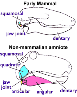
The angular is a large bone in the lower jaw (mandible) of amphibians and reptiles (birds included), which is connected to all other lower jaw bones: the dentary (which is the entire lower jaw in mammals), the splenial, the suprangular, and the articular. It is homologous to the tympanic bone in mammals, due to the incorporation of several jaw bones into the mammalian middle ear early in mammal evolution. In therapsids (mammal ancestors and their kin), the lower jaw is made up of the dentary (the mandible in mammals) and a group of smaller "postdentary" bones near the jaw joint. As the dentary increased in size over million of years, two of these postdentary bones, the articular and angular, became increasingly reduced and the dentary eventually made direct contact with the upper jaw. These postdentary bones, even before their articular function was lost, probably transmitted sound vibrations to the stapes and, in some therapsids, a bent plate that might have supported a membrane ... [...More Info...] [...Related Items...] OR: [Wikipedia] [Google] [Baidu] |
Articular
The articular bone is part of the lower jaw of most vertebrates, including most jawed fish, amphibians, birds and various kinds of reptiles, as well as ancestral mammals. Anatomy In most vertebrates, the articular bone is connected to two other lower jaw bones, the suprangular and the angular. Developmentally, it originates from the embryonic mandibular cartilage. The most caudal portion of the mandibular cartilage ossifies to form the articular bone, while the remainder of the mandibular cartilage either remains cartilaginous or disappears. In snakes In snakes, the articular, surangular, and prearticular bones have fused to form the compound bone. The mandible is suspended from the quadrate bone and articulates at this compound bone. Function In amphibians and reptiles In most tetrapods, the articular bone forms the lower portion of the jaw joint. The upper jaw articulates at the quadrate bone. In mammals In mammals, the articular bone evolves to form the malle ... [...More Info...] [...Related Items...] OR: [Wikipedia] [Google] [Baidu] |
Quadrate Bone
The quadrate bone is a skull bone in most tetrapods, including amphibians, sauropsids (reptiles, birds), and early synapsids. In most tetrapods, the quadrate bone connects to the quadratojugal and squamosal bones in the skull, and forms upper part of the jaw joint. The lower jaw articulates at the articular bone, located at the rear end of the lower jaw. The quadrate bone forms the lower jaw articulation in all classes except mammals. Evolutionarily, it is derived from the hindmost part of the primitive cartilaginous upper jaw. Function in reptiles In certain extinct reptiles, the variation and stability of the morphology of the quadrate bone has helped paleontologists in the species-level taxonomy and identification of mosasaur squamates and spinosaurine dinosaurs. In some lizards and dinosaurs, the quadrate is articulated at both ends and movable. In snakes, the quadrate bone has become elongated and very mobile, and contributes greatly to their ability to swallow very ... [...More Info...] [...Related Items...] OR: [Wikipedia] [Google] [Baidu] |
Columella (auditory System)
In the auditory system, the columella contributes to hearing in amphibians, reptiles and birds. The columella form thin, bony structures in the interior of the skull and serve the purpose of transmitting sounds from the eardrum. It is an evolutionary homolog of the stapes, one of the auditory ossicles in mammals. In many species, the extracolumella is a cartilaginous structure that grows in association with the columella. During development, the columella is derived from the dorsal end of the hyoid arch. Evolution The evolution of the columella is closely related to the evolution of the jaw joint. It is an ancestral homolog of the stapes, and is derived from the hyomandibular bone of fishes. As the columella is derived from the hyomandibula, many of its functional relationships remain the same. The columella resides in the air-filled tympanic cavity of the middle ear. The footplate, or proximal end of the columella, rests in the oval window. Sound is conducted through the ov ... [...More Info...] [...Related Items...] OR: [Wikipedia] [Google] [Baidu] |
Meckel's Cartilage
In humans, the cartilaginous bar of the mandibular arch is formed by what are known as Meckel's cartilages (right and left) also known as Meckelian cartilages; above this the incus and malleus are developed. Meckel's cartilage arises from the first pharyngeal arch. The dorsal end of each cartilage is connected with the ear-capsule and is ossified to form the malleus; the ventral ends meet each other in the region of the symphysis menti, and are usually regarded as undergoing ossification to form that portion of the mandible which contains the incisor teeth. The intervening part of the cartilage disappears; the portion immediately adjacent to the malleus is replaced by fibrous membrane, which constitutes the sphenomandibular ligament, while from the connective tissue covering the remainder of the cartilage the greater part of the mandible is ossified. Johann Friedrich Meckel, the Younger discovered this cartilage in 1820. Evolution Meckel's cartilage is a piece of cartilage from ... [...More Info...] [...Related Items...] OR: [Wikipedia] [Google] [Baidu] |
Cartilage
Cartilage is a resilient and smooth type of connective tissue. In tetrapods, it covers and protects the ends of long bones at the joints as articular cartilage, and is a structural component of many body parts including the rib cage, the neck and the bronchial tubes, and the intervertebral discs. In other taxa, such as chondrichthyans, but also in cyclostomes, it may constitute a much greater proportion of the skeleton. It is not as hard and rigid as bone, but it is much stiffer and much less flexible than muscle. The matrix of cartilage is made up of glycosaminoglycans, proteoglycans, collagen fibers and, sometimes, elastin. Because of its rigidity, cartilage often serves the purpose of holding tubes open in the body. Examples include the rings of the trachea, such as the cricoid cartilage and carina. Cartilage is composed of specialized cells called chondrocytes that produce a large amount of collagenous extracellular matrix, abundant ground substance that is rich in pro ... [...More Info...] [...Related Items...] OR: [Wikipedia] [Google] [Baidu] |
Dentary
In anatomy, the mandible, lower jaw or jawbone is the largest, strongest and lowest bone in the human facial skeleton. It forms the lower jaw and holds the lower tooth, teeth in place. The mandible sits beneath the maxilla. It is the only movable bone of the skull (discounting the ossicles of the middle ear). It is connected to the temporal bones by the temporomandibular joints. The bone is formed prenatal development, in the fetus from a fusion of the left and right mandibular prominences, and the point where these sides join, the mandibular symphysis, is still visible as a faint ridge in the midline. Like other symphyses in the body, this is a midline articulation where the bones are joined by fibrocartilage, but this articulation fuses together in early childhood.Illustrated Anatomy of the Head and Neck, Fehrenbach and Herring, Elsevier, 2012, p. 59 The word "mandible" derives from the Latin word ''mandibula'', "jawbone" (literally "one used for chewing"), from ''wikt:mandere ... [...More Info...] [...Related Items...] OR: [Wikipedia] [Google] [Baidu] |
Inner Ear
The inner ear (internal ear, auris interna) is the innermost part of the vertebrate ear. In vertebrates, the inner ear is mainly responsible for sound detection and balance. In mammals, it consists of the bony labyrinth, a hollow cavity in the temporal bone of the skull with a system of passages comprising two main functional parts: * The cochlea, dedicated to hearing; converting sound pressure patterns from the outer ear into electrochemical impulses which are passed on to the brain via the auditory nerve. * The vestibular system, dedicated to balance The inner ear is found in all vertebrates, with substantial variations in form and function. The inner ear is innervated by the eighth cranial nerve in all vertebrates. Structure The labyrinth can be divided by layer or by region. Bony and membranous labyrinths The bony labyrinth, or osseous labyrinth, is the network of passages with bony walls lined with periosteum. The three major parts of the bony labyrinth are the vestib ... [...More Info...] [...Related Items...] OR: [Wikipedia] [Google] [Baidu] |
Vestibule Of The Ear
The vestibule is the central part of the bony labyrinth in the inner ear, and is situated medial to the eardrum, behind the cochlea, and in front of the three semicircular canals. The name comes from the Latin ', literally an entrance hall. Structure The vestibule is somewhat oval in shape, but flattened transversely; it measures about 5 mm from front to back, the same from top to bottom, and about 3 mm across. In its lateral or tympanic wall is the oval window, closed, in the fresh state, by the base of the stapes and annular ligament. On its medial wall, at the forepart, is a small circular depression, the recessus sphæricus, which is perforated, at its anterior and inferior part, by several minute holes (macula cribrosa media) for the passage of filaments of the acoustic nerve to the saccule; and behind this depression is an oblique ridge, the crista vestibuli, the anterior end of which is named the pyramid of the vestibule. This ridge bifurcates below to enclose ... [...More Info...] [...Related Items...] OR: [Wikipedia] [Google] [Baidu] |
Oval Window
The oval window (or ''fenestra vestibuli'' or ''fenestra ovalis'') is a membrane-covered opening from the middle ear to the cochlea of the inner ear. Vibrations that contact the tympanic membrane travel through the three ossicles and into the inner ear. The oval window is the intersection of the middle ear with the inner ear and is directly contacted by the ''stapes''; by the time vibrations reach the oval window, they have been reduced in amplitude and increased in force due to the lever action of the ossicle bones. This is not an amplification function, as often incorrectly reported. Rather, it is an impedance-matching function, allowing sound to be transferred from air (outer ear) to liquid (cochlea). It is a reniform (kidney-shaped) opening leading from the tympanic cavity into the vestibule of the internal ear; its long diameter is horizontal and its convex border is upward. It is occupied by the base of the ''stapes'', the circumference of which is fixed by the annular l ... [...More Info...] [...Related Items...] OR: [Wikipedia] [Google] [Baidu] |


