|
Anterior Intercondyloid Fossa
The intercondylar area is the separation between the medial and lateral condyle on the upper extremity of the tibia. The anterior and posterior cruciate ligaments and the menisci attach to the intercondylar area. The intercondyloid eminence is composed of the medial and lateral intercondylar tubercles, and divides the intercondylar area into an anterior and a posterior area. Structure Anterior area The anterior intercondylar area (or anterior intercondyloid fossa) is an area on the tibia, a bone in the lower leg. Together with the posterior intercondylar area it makes up the intercondylar area. The intercondylar area is the separation between the medial and lateral condyle located toward the proximal portion of the tibia. The intercondylar eminence composed of the medial and lateral intercondylar tubercle divides the intercondylar area into anterior and posterior part. The anterior intercondylar area is the location where the anterior cruciate ligament attaches to the t ... [...More Info...] [...Related Items...] OR: [Wikipedia] [Google] [Baidu] |
Tibia
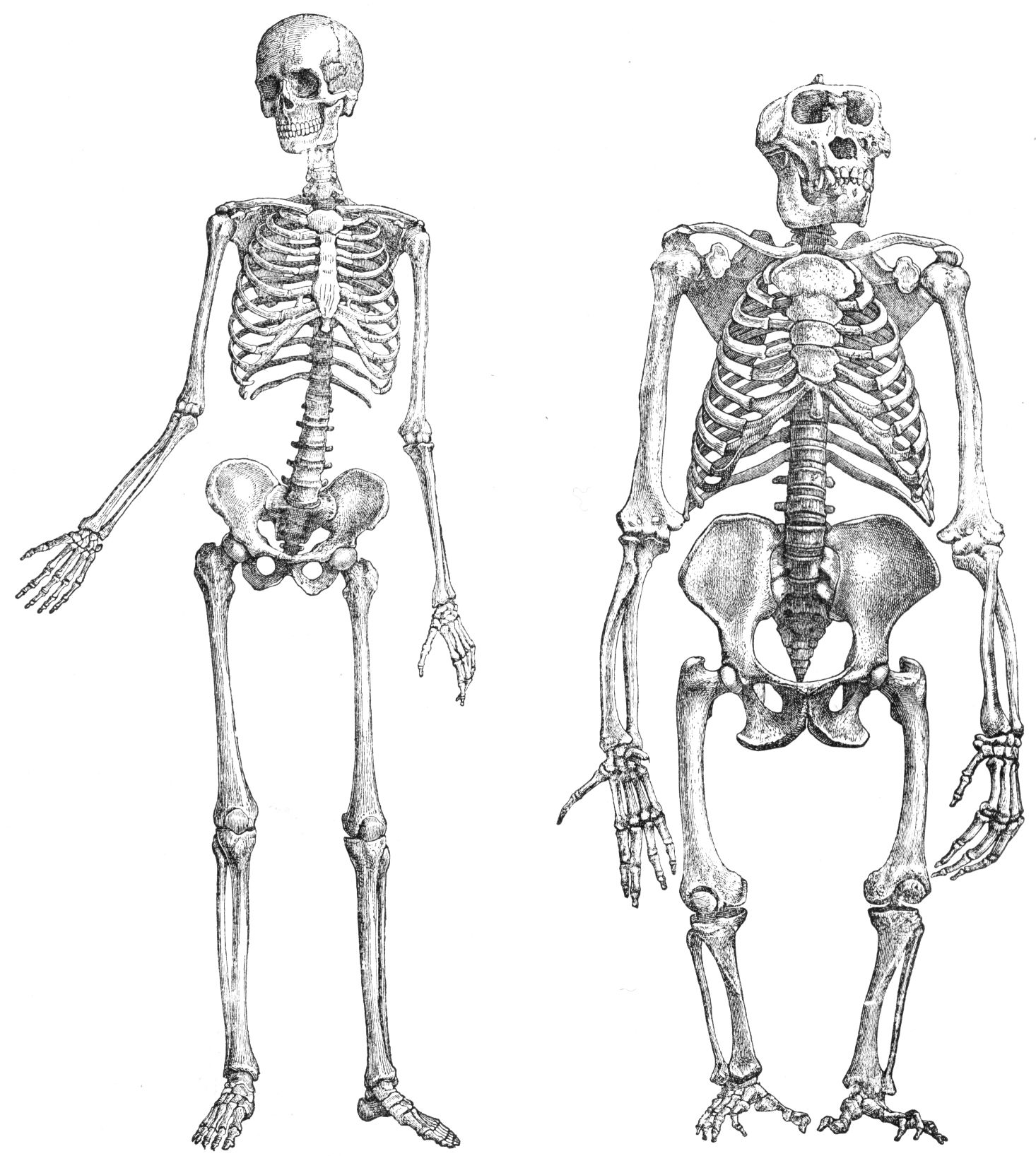
The tibia (; ), also known as the shinbone or shankbone, is the larger, stronger, and anterior (frontal) of the two bones in the leg below the knee in vertebrates (the other being the fibula, behind and to the outside of the tibia); it connects the knee with the ankle. The tibia is found on the medial side of the leg next to the fibula and closer to the median plane. The tibia is connected to the fibula by the interosseous membrane of leg, forming a type of fibrous joint called a syndesmosis with very little movement. The tibia is named for the flute ''tibia''. It is the second largest bone in the human body, after the femur. The leg bones are the strongest long bones as they support the rest of the body. Structure In human anatomy, the tibia is the second largest bone next to the femur. As in other vertebrates the tibia is one of two bones in the lower leg, the other being the fibula, and is a component of the knee and ankle joints. The ossification or formation of the bone ... [...More Info...] [...Related Items...] OR: [Wikipedia] [Google] [Baidu] |
Intercondylar Eminence
The intercondylar area is the separation between the medial and lateral condyle on the upper extremity of the tibia. The anterior and posterior cruciate ligaments and the menisci attach to the intercondylar area. The intercondyloid eminence is composed of the medial and lateral intercondylar tubercles, and divides the intercondylar area into an anterior and a posterior area. Structure Anterior area The anterior intercondylar area (or anterior intercondyloid fossa) is an area on the tibia, a bone in the lower leg. Together with the posterior intercondylar area it makes up the intercondylar area. The intercondylar area is the separation between the medial and lateral condyle located toward the proximal portion of the tibia. The intercondylar eminence composed of the medial and lateral intercondylar tubercle divides the intercondylar area into anterior and posterior part. The anterior intercondylar area is the location where the anterior cruciate ligament attaches to the t ... [...More Info...] [...Related Items...] OR: [Wikipedia] [Google] [Baidu] |
Knee
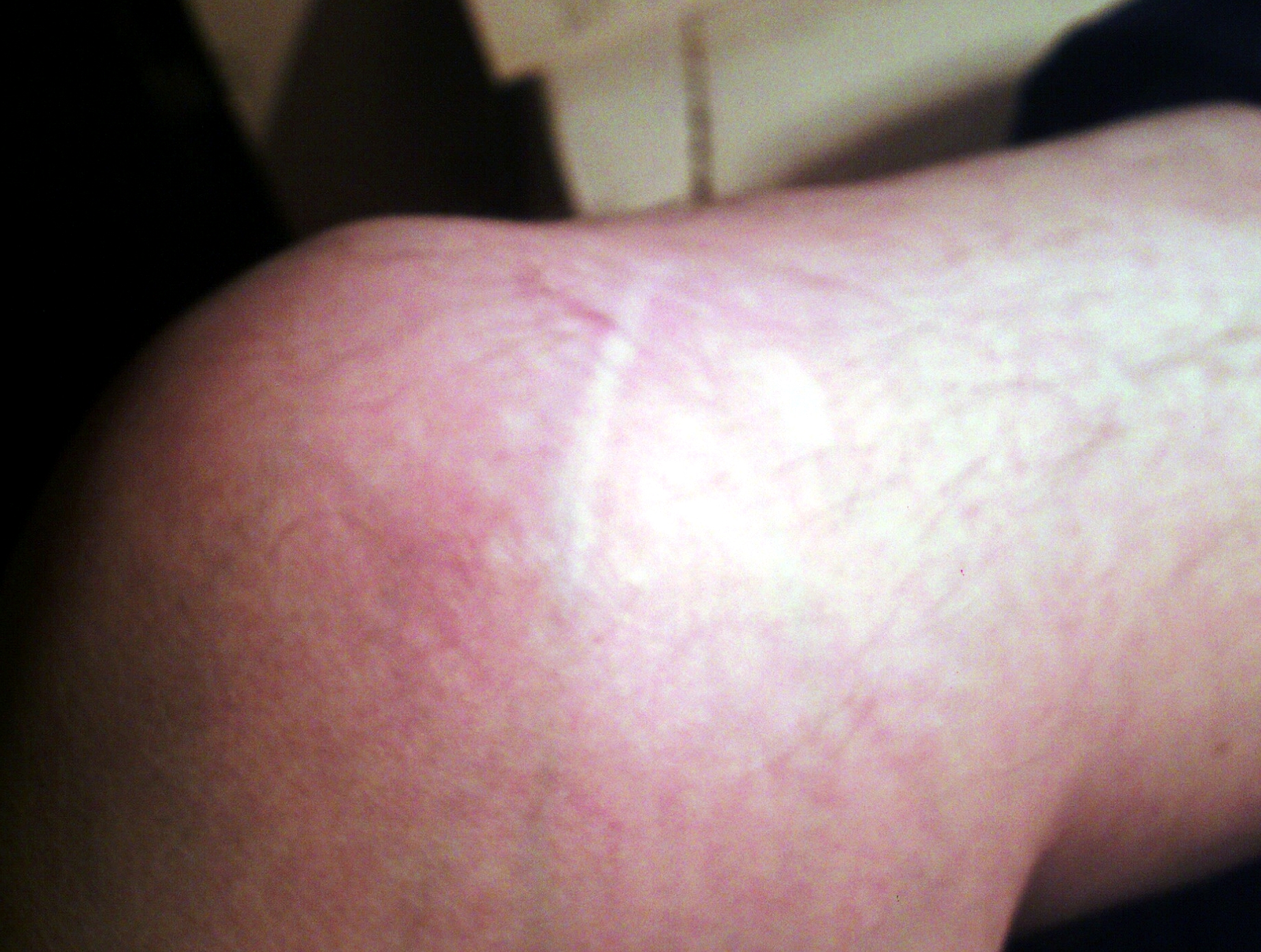
In humans and other primates, the knee joins the thigh with the leg and consists of two joints: one between the femur and tibia (tibiofemoral joint), and one between the femur and patella (patellofemoral joint). It is the largest joint in the human body. The knee is a modified hinge joint, which permits flexion and extension as well as slight internal and external rotation. The knee is vulnerable to injury and to the development of osteoarthritis. It is often termed a ''compound joint'' having tibiofemoral and patellofemoral components. (The fibular collateral ligament is often considered with tibiofemoral components.) Structure The knee is a modified hinge joint, a type of synovial joint, which is composed of three functional compartments: the patellofemoral articulation, consisting of the patella, or "kneecap", and the patellar groove on the front of the femur through which it slides; and the medial and lateral tibiofemoral articulations linking the femur, or thigh bone ... [...More Info...] [...Related Items...] OR: [Wikipedia] [Google] [Baidu] |
Medial Condyle Of Tibia
The medial condyle is the medial (or inner) portion of the upper extremity of tibia. It is the site of insertion for the semimembranosus muscle. See also * Lateral condyle of tibia * Medial collateral ligament The medial collateral ligament (MCL), or tibial collateral ligament (TCL), is one of the four major ligaments of the knee. It is on the medial (inner) side of the knee joint in humans and other primates. Its primary function is to resist out ... Additional images File:Gray258.png, Bones of the right leg. Anterior surface. File:Gray259.png, Bones of the right leg. Posterior surface. File:Slide2bib.JPG, Right knee in extension. Deep dissection. Posterior view. File:Slide2cocc.JPG, Right knee in extension. Deep dissection. Posterior view. References External links * * * () Bones of the lower limb Tibia {{musculoskeletal-stub ... [...More Info...] [...Related Items...] OR: [Wikipedia] [Google] [Baidu] |
Tubercle
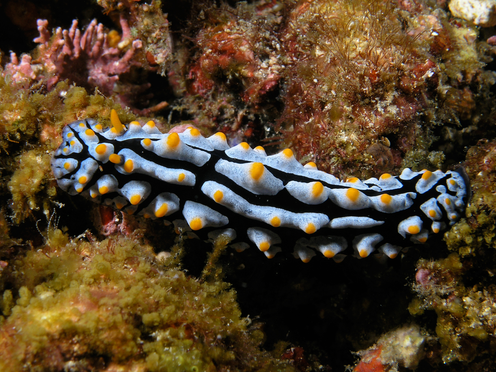
In anatomy, a tubercle (literally 'small tuber', Latin for 'lump') is any round nodule, small eminence, or warty outgrowth found on external or internal organs of a plant or an animal. In plants A tubercle is generally a wart-like projection, but it has slightly different meaning depending on which family of plants or animals it is used to refer to. In the case of certain orchids and cacti, it denotes a round nodule, small eminence, or warty outgrowth found on the lip. They are also known as podaria (singular ''podarium''). When referring to some members of the pea family, it is used to refer to the wart-like excrescences that are found on the roots. In fungi In mycology, a tubercle is used to refer to a mass of hyphae from which a mushroom is made. In animals When it is used in relation to certain dorid nudibranchs such as '' Peltodoris nobilis'', it means the nodules on the dorsum of the animal. The tubercles in nudibranchs can present themselves in different ways: e ... [...More Info...] [...Related Items...] OR: [Wikipedia] [Google] [Baidu] |
Fossa (anatomy)
In anatomy, a fossa (; plural ''fossae'' ( or ); from Latin ''fossa'', "ditch" or "trench") is a depression or hollow, usually in a bone, such as the hypophyseal fossa (the depression in the sphenoid bone).Venieratos D, Anagnostopoulou S, Garidou A., A new morphometric method for the sella turcica and the hypophyseal fossa and its clinical relevance.;Folia Morphol (Warsz). 2005 Nov;64(4):240-7. Some examples include: In the Skull: * Cranial fossa ** Anterior cranial fossa ** Middle cranial fossa *** Interpeduncular fossa ** Posterior cranial fossa * Hypophyseal fossa * Temporal bone fossa ** Mandibular fossa ** Jugular fossa * Infratemporal fossa * Pterygopalatine fossa * Pterygoid fossa * Lacrimal fossa ** Fossa for lacrimal gland ** Fossa for lacrimal sac * Mandibular fossa * Scaphoid fossa * Jugular fossa * Condyloid fossa * Rhomboid fossa In the Mandible: * Retromolar fossa In the Torso: * Fossa ovalis (heart) * Infraclavicular fossa *Pyriform fossa * Substernal fossa * ... [...More Info...] [...Related Items...] OR: [Wikipedia] [Google] [Baidu] |
Lateral Intercondylar Tubercle
The intercondylar area is the separation between the medial and lateral condyle on the upper extremity of the tibia. The anterior and posterior cruciate ligaments and the menisci attach to the intercondylar area. The intercondyloid eminence is composed of the medial and lateral intercondylar tubercles, and divides the intercondylar area into an anterior and a posterior area. Structure Anterior area The anterior intercondylar area (or anterior intercondyloid fossa) is an area on the tibia, a bone in the lower leg. Together with the posterior intercondylar area it makes up the intercondylar area. The intercondylar area is the separation between the medial and lateral condyle located toward the proximal portion of the tibia. The intercondylar eminence composed of the medial and lateral intercondylar tubercle divides the intercondylar area into anterior and posterior part. The anterior intercondylar area is the location where the anterior cruciate ligament attaches to the t ... [...More Info...] [...Related Items...] OR: [Wikipedia] [Google] [Baidu] |
Medial Intercondylar Tubercle
The intercondylar area is the separation between the medial and lateral condyle on the upper extremity of the tibia. The anterior and posterior cruciate ligaments and the menisci attach to the intercondylar area. The intercondyloid eminence is composed of the medial and lateral intercondylar tubercles, and divides the intercondylar area into an anterior and a posterior area. Structure Anterior area The anterior intercondylar area (or anterior intercondyloid fossa) is an area on the tibia, a bone in the lower leg. Together with the posterior intercondylar area it makes up the intercondylar area. The intercondylar area is the separation between the medial and lateral condyle located toward the proximal portion of the tibia. The intercondylar eminence composed of the medial and lateral intercondylar tubercle divides the intercondylar area into anterior and posterior part. The anterior intercondylar area is the location where the anterior cruciate ligament attaches to the t ... [...More Info...] [...Related Items...] OR: [Wikipedia] [Google] [Baidu] |
Posterior Intercondylar Area
The intercondylar area is the separation between the medial and lateral condyle on the upper extremity of the tibia. The anterior and posterior cruciate ligaments and the menisci attach to the intercondylar area. The intercondyloid eminence is composed of the medial and lateral intercondylar tubercles, and divides the intercondylar area into an anterior and a posterior area. Structure Anterior area The anterior intercondylar area (or anterior intercondyloid fossa) is an area on the tibia, a bone in the lower leg. Together with the posterior intercondylar area it makes up the intercondylar area. The intercondylar area is the separation between the medial and lateral condyle located toward the proximal portion of the tibia. The intercondylar eminence composed of the medial and lateral intercondylar tubercle divides the intercondylar area into anterior and posterior part. The anterior intercondylar area is the location where the anterior cruciate ligament attaches to the t ... [...More Info...] [...Related Items...] OR: [Wikipedia] [Google] [Baidu] |
Medial Condyle Of Tibia
The medial condyle is the medial (or inner) portion of the upper extremity of tibia. It is the site of insertion for the semimembranosus muscle. See also * Lateral condyle of tibia * Medial collateral ligament The medial collateral ligament (MCL), or tibial collateral ligament (TCL), is one of the four major ligaments of the knee. It is on the medial (inner) side of the knee joint in humans and other primates. Its primary function is to resist out ... Additional images File:Gray258.png, Bones of the right leg. Anterior surface. File:Gray259.png, Bones of the right leg. Posterior surface. File:Slide2bib.JPG, Right knee in extension. Deep dissection. Posterior view. File:Slide2cocc.JPG, Right knee in extension. Deep dissection. Posterior view. References External links * * * () Bones of the lower limb Tibia {{musculoskeletal-stub ... [...More Info...] [...Related Items...] OR: [Wikipedia] [Google] [Baidu] |
Lower Leg
The human leg, in the general word sense, is the entire lower limb of the human body, including the foot, thigh or sometimes even the hip or gluteal region. However, the definition in human anatomy refers only to the section of the lower limb extending from the knee to the ankle, also known as the crus or, especially in non-technical use, the shank. Legs are used for standing, and all forms of locomotion including recreational such as dancing, and constitute a significant portion of a person's mass. Female legs generally have greater hip anteversion and tibiofemoral angles, but shorter femur and tibial lengths than those in males. Structure In human anatomy, the lower leg is the part of the lower limb that lies between the knee and the ankle. Anatomists restrict the term ''leg'' to this use, rather than to the entire lower limb. The thigh is between the hip and knee and makes up the rest of the lower limb. The term ''lower limb'' or ''lower extremity'' is commonly used to descri ... [...More Info...] [...Related Items...] OR: [Wikipedia] [Google] [Baidu] |
Meniscus (anatomy)
A meniscus is a crescent-shaped fibrocartilaginous anatomical structure that, in contrast to an articular disc, only partly divides a joint cavity.Platzer (2004), p 208 In humans they are present in the knee, wrist, acromioclavicular, sternoclavicular, and temporomandibular joints; in other animals they may be present in other joints. Generally, the term "meniscus" is used to refer to the cartilage of the knee, either to the lateral or medial meniscus. Both are cartilaginous tissues that provide structural integrity to the knee when it undergoes tension and torsion. The menisci are also known as "semi-lunar" cartilages, referring to their half-moon, crescent shape. The term "meniscus" is from the Ancient Greek word (), meaning "crescent". Structure The menisci of the knee are two pads of fibrocartilaginous tissue which serve to disperse friction in the knee joint between the lower leg (tibia) and the thigh (femur). They are concave on the top and flat on the bottom, articula ... [...More Info...] [...Related Items...] OR: [Wikipedia] [Google] [Baidu] |


