|
Angiofibroma Of Soft Tissue
Angiofibroma of soft tissue (AFST), also termed angiofibroma, not otherwise specified, is a recently recognized and rare disorder that was classified in the category of benign fibroblastic and myofibroblastic tumors by the World Health Organization in 2020. An AFST tumor is a neoplasm (i.e. growth of tissue that is not coordinated with the normal surrounding tissue and persists in growing even if the original trigger for growth is removed) that was first described by A. Mariño-Enríquez and C.D. Fletcher in 2012. AFST tumors typically occur in a leg but can occur in other locations; they develop in older children and adults including elderly individuals. AFSTs are slow-growing, often painless tumors composed primarily of spindle-shaped cells and a prominent vascular network. The spindle-shaped cells are benign tumor cells that in almost all cases have chromosome abnormalities that are thought to contribute to their abnormal development and/or growth. AFST tumors are commonly trea ... [...More Info...] [...Related Items...] OR: [Wikipedia] [Google] [Baidu] |
Dermatology
Dermatology is the branch of medicine dealing with the skin.''Random House Webster's Unabridged Dictionary.'' Random House, Inc. 2001. Page 537. . It is a speciality with both medical and surgical aspects. A dermatologist is a specialist medical doctor who manages diseases related to skin, hair, nails, and some cosmetic problems. Etymology Attested in English in 1819, the word "dermatology" derives from the Greek δέρματος (''dermatos''), genitive of δέρμα (''derma''), "skin" (itself from δέρω ''dero'', "to flay") and -λογία '' -logia''. Neo-Latin ''dermatologia'' was coined in 1630, an anatomical term with various French and German uses attested from the 1730s. History In 1708, the first great school of dermatology became a reality at the famous Hôpital Saint-Louis in Paris, and the first textbooks (Willan's, 1798–1808) and atlases ( Alibert's, 1806–1816) appeared in print around the same time.Freedberg, et al. (2003). ''Fitzpatrick's Dermatology in ... [...More Info...] [...Related Items...] OR: [Wikipedia] [Google] [Baidu] |
Histopathological
Histopathology (compound of three Greek words: ''histos'' "tissue", πάθος ''pathos'' "suffering", and -λογία ''-logia'' "study of") refers to the microscopic examination of tissue in order to study the manifestations of disease. Specifically, in clinical medicine, histopathology refers to the examination of a biopsy or surgical specimen by a pathologist, after the specimen has been processed and histological sections have been placed onto glass slides. In contrast, cytopathology examines free cells or tissue micro-fragments (as "cell blocks"). Collection of tissues Histopathological examination of tissues starts with surgery, biopsy, or autopsy. The tissue is removed from the body or plant, and then, often following expert dissection in the fresh state, placed in a fixative which stabilizes the tissues to prevent decay. The most common fixative is 10% neutral buffered formalin (corresponding to 3.7% w/v formaldehyde in neutral buffered water, such as phosphate buff ... [...More Info...] [...Related Items...] OR: [Wikipedia] [Google] [Baidu] |
Cytokeratin
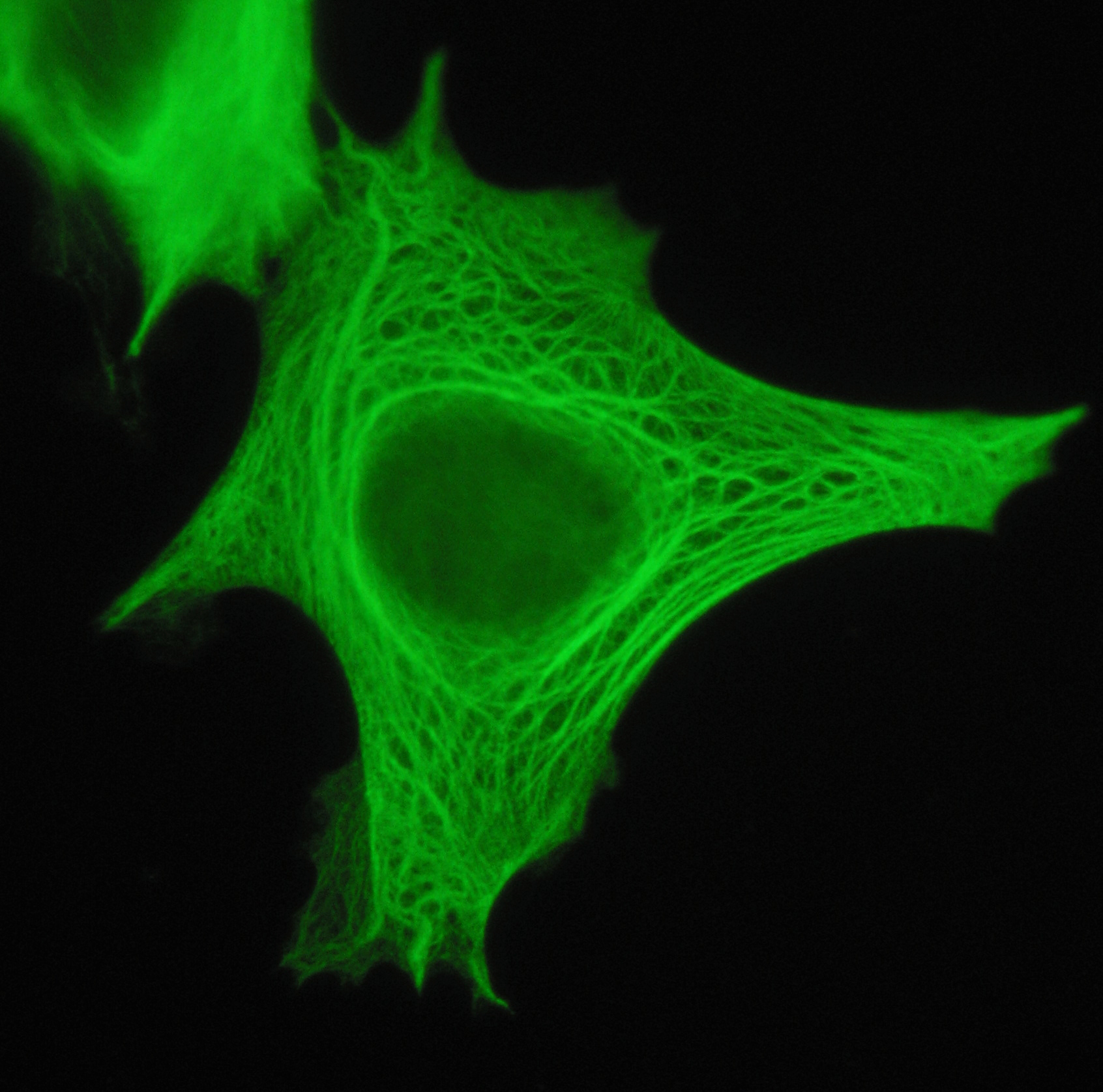
Cytokeratins are keratin proteins found in the intracytoplasmic cytoskeleton of epithelial tissue. They are an important component of intermediate filaments, which help cells resist mechanical stress. Expression of these cytokeratins within epithelial cells is largely specific to particular organs or tissues. Thus they are used clinically to identify the cell of origin of various human tumors. Naming The term ''cytokeratin'' began to be used in the late 1970s, when the protein subunits of keratin intermediate filaments inside cells were first being identified and characterized. In 2006 a new systematic nomenclature for mammalian keratins was created, and the proteins previously called ''cytokeratins'' are simply called ''keratins'' (human epithelial category). For example, cytokeratin-4 (CK-4) has been renamed keratin-4 (K4). However, they are still commonly referred to as cytokeratins in clinical practice. Types There are two categories of cytokeratins: the acidic type I cyt ... [...More Info...] [...Related Items...] OR: [Wikipedia] [Google] [Baidu] |
S100 Protein
The S100 proteins are a family of low molecular-weight proteins found in vertebrates characterized by two calcium-binding sites that have helix-loop-helix ("EF-hand-type") conformation. At least 21 different S100 proteins are known. They are encoded by a family of genes whose symbols use the ''S100'' prefix, for example, ''S100A1'', ''S100A2'', ''S100A3''. They are also considered as damage-associated molecular pattern molecules (DAMPs), and knockdown of aryl hydrocarbon receptor downregulates the expression of S100 proteins in THP-1 cells. Structure Most S100 proteins consist of two identical polypeptides (homodimeric), which are held together by noncovalent bonds. They are structurally similar to calmodulin. They differ from calmodulin, though, on the other features. For instance, their expression pattern is cell-specific, i.e. they are expressed in particular cell types. Their expression depends on environmental factors. In contrast, calmodulin is a ubiquitous and universa ... [...More Info...] [...Related Items...] OR: [Wikipedia] [Google] [Baidu] |
STAT6
Signal transducer and activator of transcription 6 (STAT6) is a transcription factor that belongs to the Signal Transducer and Activator of Transcription (STAT) family of proteins. The proteins of STAT family transmit signals from a receptor complex to the nucleus and activate gene expression. Similarly as other STAT family proteins, STAT6 is also activated by growth factors and cytokines. STAT6 is mainly activated by cytokines interleukin-4 and interleukin-13. Molecular biology In the human genome, STAT6 protein is encoded by the STAT6 gene, located on the chromosome 12q13.3-q14.1. The gene encompasses over 19 kb and consists of 23 exons. STAT6 shares structural similarity with the other STAT proteins and is composed of the N-terminal domain, DNA binding domain, SH3- like domain, SH2 domain and transactivation domain (TAD). STAT proteins are activated by the Janus family (JAKs) tyrosine kinases in response to cytokine exposure. STAT6 is activated by cytokines interleukin-4 ... [...More Info...] [...Related Items...] OR: [Wikipedia] [Google] [Baidu] |
CD34
CD34 is a transmembrane phosphoglycoprotein protein encoded by the CD34 gene in humans, mice, rats and other species. CD34 derives its name from the cluster of differentiation protocol that identifies cell surface antigens. CD34 was first described on hematopoietic stem cells independently by Civin et al. and Tindle et al. as a cell surface glycoprotein and functions as a cell-cell adhesion factor. It may also mediate the attachment of hematopoietic stem cells to bone marrow extracellular matrix or directly to stromal cells. Clinically, it is associated with the selection and enrichment of hematopoietic stem cells for bone marrow transplants. Due to these historical and clinical associations, CD34 expression is almost ubiquitously related to hematopoietic cells; however, it is actually found on many other cell types as well. Function The CD34 protein is a member of a family of single-pass transmembrane sialomucin proteins that show expression on early haematopoietic and vascul ... [...More Info...] [...Related Items...] OR: [Wikipedia] [Google] [Baidu] |
ACTA2
ACTA2 (actin alpha 2) is an actin protein with several aliases including alpha-actin, alpha-actin-2, aortic smooth muscle or alpha smooth muscle actin (α-SMA, SMactin, alpha-SM-actin, ASMA). Actins are a family of globular multi-functional proteins that form microfilaments. ACTA2 is one of 6 different actin isoforms and is involved in the contractile apparatus of smooth muscle. ACTA2 (as with all the actins) is extremely highly conserved and found in nearly all mammals. In humans, ACTA2 is encoded by the ''ACTA2'' gene located on 10q22-q24. Mutations in this gene cause a variety of vascular diseases, such as thoracic aortic disease, coronary artery disease, stroke, Moyamoya disease, and multisystemic smooth muscle dysfunction syndrome. ACTA2 (commonly referred to as alpha-smooth muscle actin or α-SMA) is often used as a marker of myofibroblast A myofibroblast is a cell phenotype that was first described as being in a state between a fibroblast and a smooth muscle cell. ... [...More Info...] [...Related Items...] OR: [Wikipedia] [Google] [Baidu] |
Desmin
Desmin is a protein that in humans is encoded by the ''DES'' gene. Desmin is a muscle-specific, type III intermediate filament that integrates the sarcolemma, Z disk, and nuclear membrane in sarcomeres and regulates sarcomere architecture. Structure Desmin is a 53.5 kD protein composed of 470 amino acids, encoded by the human ''DES'' gene located on the long arm of chromosome 2. There are three major domains to the desmin protein: a conserved alpha helix rod, a variable non alpha helix head, and a carboxy-terminal tail. Desmin, as all intermediate filaments, shows no polarity when assembled. The rod domain consists of 308 amino acids with parallel alpha helical coiled coil dimers and three linkers to disrupt it. The rod domain connects to the head domain. The head domain 84 amino acids with many arginine, serine, and aromatic residues is important in filament assembly and dimer-dimer interactions. The tail domain is responsible for the integration of filaments and interaction ... [...More Info...] [...Related Items...] OR: [Wikipedia] [Google] [Baidu] |
MUC1
Mucin short variant S1, also called polymorphic epithelial mucin (PEM) or epithelial membrane antigen (EMA), is a mucin encoded by the ''MUC1'' gene in humans. Mucin short variant S1 is a glycoprotein with extensive O-linked glycosylation of its extracellular domain. Mucins line the apical surface of epithelial cells in the lungs, stomach, intestines, eyes and several other organs. Mucins protect the body from infection by pathogen binding to oligosaccharides in the extracellular domain, preventing the pathogen from reaching the cell surface. Overexpression of MUC1 is often associated with colon, breast, ovarian, lung and pancreatic cancers. Joyce Taylor-Papadimitriou identified and characterised the antigen during her work with breast and ovarian tumors. Structure MUC1 is a member of the mucin family and encodes a membrane bound, glycosylated phosphoprotein. MUC1 has a core protein mass of 120-225 kDa which increases to 250-500 kDa with glycosylation. It extends 200-500 n ... [...More Info...] [...Related Items...] OR: [Wikipedia] [Google] [Baidu] |
NCOA2
NCOA may refer to: *National Change Of Address database (see United States Postal Service) *National Chamber Orchestra of Armenia *National Council on Aging * The Non-commissioned officer, Noncomissioned Officer Academy in the United States Air Force {{disambig ... [...More Info...] [...Related Items...] OR: [Wikipedia] [Google] [Baidu] |
CD163
CD163 (Cluster of Differentiation 163) is a protein that in humans is encoded by the CD163 gene. CD163 is the high affinity scavenger receptor for the hemoglobin-haptoglobin complex and in the absence of haptoglobin - with lower affinity - for hemoglobin alone. It also is a marker of cells from the monocyte/macrophage lineage. CD163 functions as innate immune sensor for gram-positive and gram-negative bacteria. The receptor was discovered in 1987. Structure The molecular size is 130 kDa. The receptor belongs to the scavenger receptor cysteine rich family type B and consists of a 1048 amino acid residues extracellular domain, a single transmembrane segment and a cytoplasmic tail with several splice variants. Clinical significance A soluble form of the receptor exists in plasma, and cerebrospinal fluid., commonly denoted sCD163. It is generated by ectodomain shedding of the membrane bound receptor, which may represent a form of modulation of CD163 function. sCD163 shedding oc ... [...More Info...] [...Related Items...] OR: [Wikipedia] [Google] [Baidu] |
Estrogen Receptor
Estrogen receptors (ERs) are a group of proteins found inside cells. They are receptors that are activated by the hormone estrogen ( 17β-estradiol). Two classes of ER exist: nuclear estrogen receptors (ERα and ERβ), which are members of the nuclear receptor family of intracellular receptors, and membrane estrogen receptors (mERs) (GPER (GPR30), ER-X, and Gq-mER), which are mostly G protein-coupled receptors. This article refers to the former (ER). Once activated by estrogen, the ER is able to translocate into the nucleus and bind to DNA to regulate the activity of different genes (i.e. it is a DNA-binding transcription factor). However, it also has additional functions independent of DNA binding. As hormone receptors for sex steroids (steroid hormone receptors), ERs, androgen receptors (ARs), and progesterone receptors (PRs) are important in sexual maturation and gestation. Proteomics There are two different forms of the estrogen receptor, usually referred to as α a ... [...More Info...] [...Related Items...] OR: [Wikipedia] [Google] [Baidu] |


