|
Amethicium
''Amethicium'' is a fungal genus in the family Phanerochaetaceae. A monotypic genus, it contains the single species ''Amethicium rimosum'', a crust fungus first reported from Tanzania in 1983. ''Amethicium'' is primarily characterized by its purple fruit body and a dimitic hyphal system (two types of hyphae: generative and skeletal). The felt-like tissue layer covering the substrate (the subiculum) comprises a thin layer of densely intertwined skeletal hyphae. Taxonomy ''Amethicium rimosum'' was described scientifically by Swedish mycologist Kurt Hjortstam in 1983, based on collections made by Leif Ryvarden a decade earlier. The type locality was on the southern slopes of Mount Kilimanjaro in Tanzania, at an elevation between . There have been a few species formerly classified in the ''Amethicium'' that have since been transferred to other genera. ''Amethicium chrysocreas'' (Berk. & M.A. Curtis) Sheng H. Wu 1990, and ''Amethicium leoninum'' (Burds. & Nakasone) Sheng H. Wu 1990 ... [...More Info...] [...Related Items...] OR: [Wikipedia] [Google] [Baidu] |
Cericium
''Cericium'' is a fungal genus in the family Cystostereaceae. It is a monotypic genus with the single species ''Cericium luteoincrustatum'' , a crust fungus. This species was originally described in 1986 by Kurt Hjortstam and Leif Ryvarden, who called it ''Amethicium luteoincrustatum''. They placed it in the genus ''Amethicium'' based on microscopic similarities with the African species '' Amethicium rimosum''. Description ''Cericium luteoincrustatum'' is a waxy and brittle crust fungus with a fruit body that is about 0.2–1 mm thick. Its surface is either smooth or somewhat tuberculate with a pale yellow colour. It has a dimitic hyphal system, without clamp connections in the generative hyphae. The hyphae in the subiculum are arboriform (tree-like); these are binding-type hyphae. Cystidia feature yellowish incrustations at their bases. The spores produced by the fungus are smooth and thin-walled with an ellipsoid shape, and measure about 5 by 3 μm. Habitat and dist ... [...More Info...] [...Related Items...] OR: [Wikipedia] [Google] [Baidu] |
Crustodontia
''Crustodontia'' is a fungal genus of uncertain familial placement in the order Polyporales. The genus was circumscribed in 2005 to contain the crust fungus ''Crustodontia chrysocreas''. This species was originally described as ''Corticium chrysocreas'' by Miles Berkeley and Moses Ashley Curtis in 1873. Their description was as follows: "Subiculum bright yellow, thin; hymenium immarginate pallid, or yellow tinged with tawny." ''Crustodontia'' has a monomitic hyphal system, meaning it contains only generative hyphae, and these hyphae have clamp connections. ''Crustodontia chrysocreas'' has a pantropical distribution. It has been widely collected, including the United States, Costa Rica, Caribbean Islands, Venezuela, equatorial Africa, Sri Lanka, China, Taiwan, Japan, Hawaii, Brunei, western Australia, and New Zealand. It is rare in Europe; its northernmost recorded collection location (51.7°N) is in Belarus. The fungus causes a white rot in the woody debris of live oak and othe ... [...More Info...] [...Related Items...] OR: [Wikipedia] [Google] [Baidu] |
Phanerochaetaceae
The Phanerochaetaceae are a family of mostly crust fungi in the order Polyporales. Taxonomy Phanerochaetaceae was first conceived by Swedish mycologist John Eriksson in 1958 as the subfamily Phanerochaetoideae of the Corticiaceae. It was later published validly by Erast Parmasto in 1986, and raised to familial status by Swiss mycologist Walter Jülich in 1982. The type genus is ''Phanerochaete''. In 2007, Karl-Henrik Larsson proposed using the name Phanerochaetaceae to refer to the clade of crust fungi clustered near ''Phanerochaete''. In 2013, a more extensive molecular analysis showed that the Phanerochaetaceae were a subclade of the large phlebioid clade, which also contains members of the families Meruliaceae and Irpicaceae. The generic limits of ''Phanerochaete'' were revised in 2015, and new genera were added in 2016. , Index Fungorum accepts 30 genera and 367 species in the family. Description Most Phanerochaetaceae species are crust-like. Their hyphal system is mo ... [...More Info...] [...Related Items...] OR: [Wikipedia] [Google] [Baidu] |
Hyphoderma
''Hyphoderma'' is a genus of crust fungi in the family Meruliaceae. It was circumscribed by German botanist Karl Friedrich Wilhelm Wallroth in 1833. Species , Index Fungorum ''Index Fungorum'' is an international project to index all formal names ( scientific names) in the fungus kingdom. the project is based at the Royal Botanic Gardens, Kew, one of three partners along with Landcare Research and the Institute of M ... accepts 102 species of ''Hyphoderma'': References Taxa described in 1833 Meruliaceae Polyporales genera Taxa named by Karl Friedrich Wilhelm Wallroth {{Polyporales-stub ... [...More Info...] [...Related Items...] OR: [Wikipedia] [Google] [Baidu] |
Hyaline
A hyaline substance is one with a glassy appearance. The word is derived from el, ὑάλινος, translit=hyálinos, lit=transparent, and el, ὕαλος, translit=hýalos, lit=crystal, glass, label=none. Histopathology Hyaline cartilage is named after its glassy appearance on fresh gross pathology. On light microscopy of H&E stained slides, the extracellular matrix of hyaline cartilage looks homogeneously pink, and the term "hyaline" is used to describe similarly homogeneously pink material besides the cartilage. Hyaline material is usually acellular and proteinaceous. For example, arterial hyaline is seen in aging, high blood pressure, diabetes mellitus and in association with some drugs (e.g. calcineurin inhibitors). It is bright pink with PAS staining. Ichthyology and entomology In ichthyology and entomology, ''hyaline'' denotes a colorless, transparent substance, such as unpigmented fins of fishes or clear insect wings. Resh, Vincent H. and R. T. Cardé, Eds. Encyclo ... [...More Info...] [...Related Items...] OR: [Wikipedia] [Google] [Baidu] |
Basidiospore
A basidiospore is a reproductive spore produced by Basidiomycete fungi, a grouping that includes mushrooms, shelf fungi, rusts, and smuts. Basidiospores typically each contain one haploid nucleus that is the product of meiosis, and they are produced by specialized fungal cells called basidia. Typically, four basidiospores develop on appendages from each basidium, of which two are of one strain and the other two of its opposite strain. In gills under a cap of one common species, there exist millions of basidia. Some gilled mushrooms in the order Agaricales have the ability to release billions of spores. The puffball fungus ''Calvatia gigantea'' has been calculated to produce about five trillion basidiospores. Most basidiospores are forcibly discharged, and are thus considered ballistospores. These spores serve as the main air dispersal units for the fungi. The spores are released during periods of high humidity and generally have a night-time or pre-dawn peak concentration in the ... [...More Info...] [...Related Items...] OR: [Wikipedia] [Google] [Baidu] |
Sterigmata
In biology, a sterigma (pl. sterigmata) is a small supporting structure. It commonly refers to an extension of the basidium (the spore-bearing cells) consisting of a basal filamentous part and a slender projection which carries a spore at the tip. The sterigmata are formed on the basidium as it develops and undergoes meiosis, to result in the production of (typically) four nuclei. The nuclei gradually migrate to the tips of the basidium, and one nucleus will migrate into each spore that develops at the tip of each sterigma. In less common usage, a sterigma is a structure within the posterior end of the genitalia of female Lepidoptera. It also refers to the stem-like structure, also called a "woody peg" at the base of the leaves of some, but not all conifers, specifically ''Picea'' and ''Tsuga ''Tsuga'' (, from Japanese (), the name of ''Tsuga sieboldii'') is a genus of conifers in the subfamily Abietoideae of Pinaceae, the pine family. The common name hemlock is derived ... [...More Info...] [...Related Items...] OR: [Wikipedia] [Google] [Baidu] |
Basidia
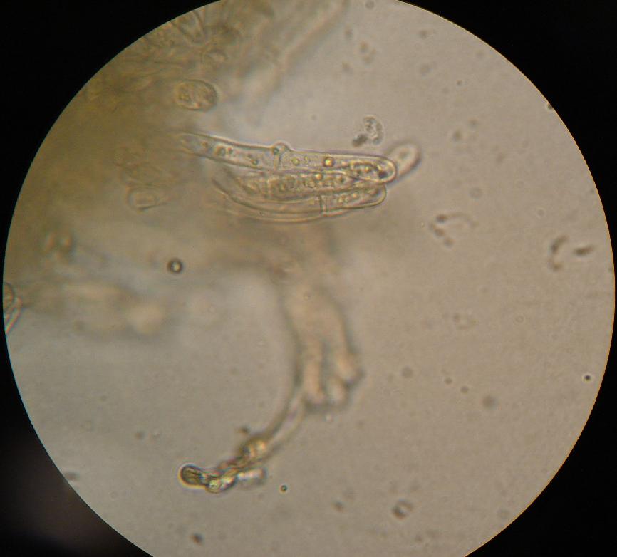
A basidium () is a microscopic sporangium (a spore-producing structure) found on the hymenophore of fruiting bodies of basidiomycete fungi which are also called tertiary mycelium, developed from secondary mycelium. Tertiary mycelium is highly-coiled secondary myceliuma dikaryon. The presence of basidia is one of the main characteristic features of the Basidiomycota. A basidium usually bears four sexual spores called basidiospores; occasionally the number may be two or even eight. In a typical basidium, each basidiospore is borne at the tip of a narrow prong or horn called a sterigma (), and is forcibly discharged upon maturity. The word ''basidium'' literally means "little pedestal", from the way in which the basidium supports the spores. However, some biologists suggest that the structure more closely resembles a club. An immature basidium is known as a basidiole. Structure Most basidiomycota have single celled basidia (holobasidia), but in some groups basidia can be multice ... [...More Info...] [...Related Items...] OR: [Wikipedia] [Google] [Baidu] |
Cystidia
A cystidium (plural cystidia) is a relatively large cell found on the sporocarp of a basidiomycete (for example, on the surface of a mushroom gill), often between clusters of basidia. Since cystidia have highly varied and distinct shapes that are often unique to a particular species or genus, they are a useful micromorphological characteristic in the identification of basidiomycetes. In general, the adaptive significance of cystidia is not well understood. Classification of cystidia By position Cystidia may occur on the edge of a lamella (or analogous hymenophoral structure) (cheilocystidia), on the face of a lamella (pleurocystidia), on the surface of the cap (dermatocystidia or pileocystidia), on the margin of the cap (circumcystidia) or on the stipe (caulocystidia). Especially the pleurocystidia and cheilocystidia are important for identification within many genera. Sometimes the cheilocystidia give the gill edge a distinct colour which is visible to the naked eye or wit ... [...More Info...] [...Related Items...] OR: [Wikipedia] [Google] [Baidu] |
Clamp Connection
A clamp connection is a hook-like structure formed by growing hyphal cells of certain fungi. It is a characteristic feature of Basidiomycetes fungi. It is created to ensure that each cell, or segment of hypha separated by septa (cross walls), receives a set of differing nuclei, which are obtained through mating of hyphae of differing sexual types. It is used to maintain genetic variation within the hypha much like the mechanisms found in crozier (hook) during sexual reproduction. Formation Clamp connections are formed by the terminal hypha during elongation. Before the clamp connection is formed this terminal segment contains two nuclei. Once the terminal segment is long enough it begins to form the clamp connection. At the same time, each nucleus undergoes mitotic division to produce two daughter nuclei. As the clamp continues to develop it uptakes one of the daughter (green circle) nuclei and separates it from its sister nucleus. While this is occurring the remaining nuclei ... [...More Info...] [...Related Items...] OR: [Wikipedia] [Google] [Baidu] |
Hymenium
The hymenium is the tissue layer on the hymenophore of a fungal fruiting body where the cells develop into basidia or asci, which produce spores. In some species all of the cells of the hymenium develop into basidia or asci, while in others some cells develop into sterile cells called cystidia (basidiomycetes) or paraphyses (ascomycetes). Cystidia are often important for microscopic identification. The subhymenium consists of the supportive hyphae from which the cells of the hymenium grow, beneath which is the hymenophoral trama, the hyphae that make up the mass of the hymenophore. The position of the hymenium is traditionally the first characteristic used in the classification and identification of mushrooms. Below are some examples of the diverse types which exist among the macroscopic Basidiomycota and Ascomycota. * In agarics, the hymenium is on the vertical faces of the gills. * In boletes and polypores, it is in a spongy mass of downward-pointing tubes. * In puffballs, ... [...More Info...] [...Related Items...] OR: [Wikipedia] [Google] [Baidu] |
Amyloid (mycology)
In mycology a tissue or feature is said to be amyloid if it has a positive amyloid reaction when subjected to a crude chemical test using iodine as an ingredient of either Melzer's reagent or Lugol's solution, producing a blue to blue-black staining. The term "amyloid" is derived from the Latin ''amyloideus'' ("starch-like"). It refers to the fact that starch gives a similar reaction, also called an amyloid reaction. The test can be on microscopic features, such as spore walls or hyphal walls, or the apical apparatus or entire ascus wall of an ascus, or be a macroscopic reaction on tissue where a drop of the reagent is applied. Negative reactions, called inamyloid or nonamyloid, are for structures that remain pale yellow-brown or clear. A reaction producing a deep reddish to reddish-brown staining is either termed a dextrinoid reaction (pseudoamyloid is a synonym) or a hemiamyloid reaction. Melzer's reagent reactions Hemiamyloidity Hemiamyloidity in mycology refers to a special ... [...More Info...] [...Related Items...] OR: [Wikipedia] [Google] [Baidu] |


