|
Ambient Cistern
The ambient cistern is a bilaterally paired (one on each side of the body) subarachnoid cistern situated at either lateral aspect of the mesencephalon (midbrain). Each ambient cistern has a supratentorial compartment and an infratentorial compartment. Each is continuous anteriorly with the interpeduncular cistern, and posteriorly with the quadrigeminal cistern. Anatomy Each ambient cistern is situated between (the parahippocampal gyrus and dentate gyrus of) the temporal lobe (laterally), and the lateral aspect of the posterior of the mesencephalon posterior to the cerebral peduncle (medially). Relations Each ambient cistern extends anterolaterally around the mesencephalon to become continuous rostrally/anteriorly with the interpeduncular cistern. Each ambient cistern is continuous dorsally/posteriorly with the quadrigeminal cistern; inversely, each ambient cistern is an anterolateral extension of the quadrigeminal cistern on either side (some sources define the quad ... [...More Info...] [...Related Items...] OR: [Wikipedia] [Google] [Baidu] |
Bilateral Symmetry
Symmetry in biology refers to the symmetry observed in organisms, including plants, animals, fungi, and bacteria. External symmetry can be easily seen by just looking at an organism. For example, take the face of a human being which has a plane of symmetry down its centre, or a pine cone with a clear symmetrical spiral pattern. Internal features can also show symmetry, for example the tubes in the human body (responsible for transporting gases, nutrients, and waste products) which are cylindrical and have several planes of symmetry. Biological symmetry can be thought of as a balanced distribution of duplicate body parts or shapes within the body of an organism. Importantly, unlike in mathematics, symmetry in biology is always approximate. For example, plant leaves – while considered symmetrical – rarely match up exactly when folded in half. Symmetry is one class of patterns in nature whereby there is near-repetition of the pattern element, either by reflection or rotation ... [...More Info...] [...Related Items...] OR: [Wikipedia] [Google] [Baidu] |
Subarachnoid Cisterns
The subarachnoid cisterns are spaces formed by openings in the subarachnoid space, an anatomic space in the meninges of the brain. The space is situated between the two meninges, the arachnoid mater and the pia mater. These cisterns are filled with cerebrospinal fluid. Structure Although the pia mater adheres to the surface of the brain, closely following the contours of its gyri and sulci, the arachnoid mater only covers its superficial surface, bridging across the gyri. This leaves wider spaces between the pia and arachnoid and the cavities are known as the subarachnoid cisterns. Although they are often described as distinct compartments, the subarachnoid cisterns are not truly anatomically distinct. Rather, these subarachnoid cisterns are separated from each other by a trabeculated porous wall with various-sized openings. Cisterns There are many cisterns in the brain with several large ones noted with their own name. At the base of the spinal cord is another subarachnoid ciste ... [...More Info...] [...Related Items...] OR: [Wikipedia] [Google] [Baidu] |
Midbrain
The midbrain or mesencephalon is the forward-most portion of the brainstem and is associated with vision, hearing, motor control, sleep and wakefulness, arousal (alertness), and temperature regulation. The name comes from the Greek ''mesos'', "middle", and ''enkephalos'', "brain". Structure The principal regions of the midbrain are the tectum, the cerebral aqueduct, tegmentum, and the cerebral peduncles. Rostrally the midbrain adjoins the diencephalon (thalamus, hypothalamus, etc.), while caudally it adjoins the hindbrain (pons, medulla and cerebellum). In the rostral direction, the midbrain noticeably splays laterally. Sectioning of the midbrain is usually performed axially, at one of two levels – that of the superior colliculi, or that of the inferior colliculi. One common technique for remembering the structures of the midbrain involves visualizing these cross-sections (especially at the level of the superior colliculi) as the upside-down face of a be ... [...More Info...] [...Related Items...] OR: [Wikipedia] [Google] [Baidu] |
Interpeduncular Cistern
The interpeduncular cistern of Sweeney is the subarachnoid cistern that encloses the cerebral peduncles and the structures contained in the interpeduncular fossa and contains the arterial circle of Willis as well as the oculomotor nerve The oculomotor nerve, also known as the third cranial nerve, cranial nerve III, or simply CN III, is a cranial nerve that enters the orbit through the superior orbital fissure and innervates extraocular muscles that enable most movements of ... (CN3). References Meninges {{neuroanatomy-stub ... [...More Info...] [...Related Items...] OR: [Wikipedia] [Google] [Baidu] |
Quadrigeminal Cistern
The quadrigeminal cistern (also cistern of great cerebral vein, vein of Galen cistern, superior cistern, Bichat's canal, or peripineal cistern) is a subarachnoid cistern situated between splenium of corpus callosum, and the superior surface of the cerebellum. It contains a part of the great cerebral vein, the posterior cerebral artery, quadrigeminal artery, glossopharyngeal nerve (CN IX), and the pineal gland. Structure The quadrigeminal cistern lies between the splenium of the corpus callosum (superiorly), the cerebellar vermis (inferiorly and posteriorly), and the tentorial margin. It is just superior to the tectum of the mesencephalon (midbrain). It lies medial to part of the medial occipital cortex. It is posterior to the brainstem and third ventricle; it extends between the layers of the tela choroidea of the third ventricle. The cistern may extend anterior-ward between the thalamus and corpus callosum to form the ''cistern of velum interpositum''. Content ... [...More Info...] [...Related Items...] OR: [Wikipedia] [Google] [Baidu] |
Parahippocampal Gyrus
The parahippocampal gyrus (or hippocampal gyrus') is a grey matter cortical region of the brain that surrounds the hippocampus and is part of the limbic system. The region plays an important role in memory encoding and retrieval. It has been involved in some cases of hippocampal sclerosis. Asymmetry has been observed in schizophrenia. Structure The anterior part of the gyrus includes the perirhinal and entorhinal cortices. The term parahippocampal cortex is used to refer to an area that encompasses both the posterior parahippocampal gyrus and the medial portion of the fusiform gyrus. Function Scene recognition The parahippocampal place area (PPA) is a sub-region of the parahippocampal cortex that lies medially in the inferior temporo-occipital cortex. PPA plays an important role in the encoding and recognition of environmental scenes (rather than faces). fMRI studies indicate that this region of the brain becomes highly active when human subjects view topographical scene sti ... [...More Info...] [...Related Items...] OR: [Wikipedia] [Google] [Baidu] |
Dentate Gyrus
The dentate gyrus (DG) is part of the hippocampal formation in the temporal lobe of the brain, which also includes the hippocampus and the subiculum. The dentate gyrus is part of the hippocampal trisynaptic circuit and is thought to contribute to the formation of new episodic memories, the spontaneous exploration of novel environments and other functions. It is notable as being one of a select few brain structures known to have significant rates of adult neurogenesis in many species of mammals, from rodents to primates. Other sites of adult neurogenesis include the subventricular zone, the striatum and the cerebellum. However, whether significant neurogenesis exists in the adult human dentate gyrus has been a matter of debate. 2019 evidence has shown that adult neurogenesis does take place in the subventricular zone and in the subgranular zone of the dentate gyrus. Structure The dentate gyrus, like the hippocampus, consists of three distinct layers: an outer molecular layer ... [...More Info...] [...Related Items...] OR: [Wikipedia] [Google] [Baidu] |
Temporal Lobe
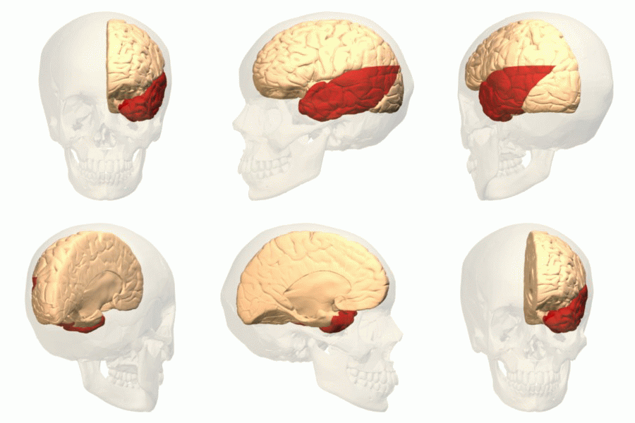
The temporal lobe is one of the four Lobes of the brain, major lobes of the cerebral cortex in the brain of mammals. The temporal lobe is located beneath the lateral fissure on both cerebral hemispheres of the mammalian brain. The temporal lobe is involved in processing sensory input into derived meanings for the appropriate retention of visual memory, language comprehension, and emotion association. ''Temporal'' refers to the head's Temple (anatomy), temples. Structure The Temple (anatomy)#Etymology, temporal Lobe (anatomy), lobe consists of structures that are vital for declarative or long-term memory. Declarative memory, Declarative (denotative) or Explicit memory, explicit memory is conscious memory divided into semantic memory (facts) and episodic memory (events). Medial temporal lobe structures that are critical for long-term memory include the hippocampus, along with the surrounding Hippocampal formation, hippocampal region consisting of the Perirhinal cortex, perirhinal, ... [...More Info...] [...Related Items...] OR: [Wikipedia] [Google] [Baidu] |
Cerebral Peduncle
The cerebral peduncles are the two stalks that attach the cerebrum to the brainstem. They are structures at the front of the midbrain which arise from the ventral pons and contain the large ascending (sensory) and descending (motor) nerve tracts that run to and from the cerebrum from the pons. Mainly, the three common areas that give rise to the cerebral peduncles are the cerebral cortex, the spinal cord and the cerebellum. The region includes the tegmentum, crus cerebri and pretectum. By this definition, the cerebral peduncles are also known as the basis pedunculi, while the large ventral bundle of efferent fibers is referred to as the cerebral crus or the pes pedunculi. The cerebral peduncles are located on either side of the midbrain and are the frontmost part of the midbrain, and act as the connectors between the rest of the midbrain and the thalamic nuclei and thus the cerebrum. As a whole, the cerebral peduncles assist in refining motor movements, learning new motor ski ... [...More Info...] [...Related Items...] OR: [Wikipedia] [Google] [Baidu] |
Trochlear Nerve
The trochlear nerve (), ( lit. ''pulley-like'' nerve) also known as the fourth cranial nerve, cranial nerve IV, or CN IV, is a cranial nerve that innervates just one muscle: the superior oblique muscle of the eye, which operates through the pulley-like trochlea. CN IV is a motor nerve only (a somatic efferent nerve), unlike most other CNs. The trochlear nerve is unique among the cranial nerves in several respects: * It is the ''smallest'' nerve in terms of the number of axons it contains. * It has the greatest intracranial length. * It is the only cranial nerve that exits from the dorsal (rear) aspect of the brainstem. * It innervates a muscle, the superior oblique muscle, on the opposite side (contralateral) from its nucleus. The trochlear nerve decussates within the brainstem before emerging on the contralateral side of the brainstem (at the level of the inferior colliculus). An injury to the trochlear nucleus in the brainstem will result in an contralateral superior obliqu ... [...More Info...] [...Related Items...] OR: [Wikipedia] [Google] [Baidu] |
Basal Vein
The basal vein is a vein in the brain. It is formed at the anterior perforated substance by the union of * (a) a ''small anterior cerebral vein'' which accompanies the anterior cerebral artery and supplies the medial surface of the frontal lobe by the fronto-basal vein. * (b) the ''deep middle cerebral vein'' (''deep Sylvian vein''), which receives tributaries from the insula and neighboring gyri, and runs in the lower part of the lateral cerebral fissure, and * (c) the ''inferior striate veins'', which leave the corpus striatum through the anterior perforated substance. The basal vein passes backward around the cerebral peduncle, and ends in the great cerebral vein; it receives tributaries from the interpeduncular fossa, the inferior horn of the lateral ventricle, the hippocampal gyrus, and the mid-brain The midbrain or mesencephalon is the forward-most portion of the brainstem and is associated with vision, hearing, motor control, sleep and wakefulness, arousal (alertness) ... [...More Info...] [...Related Items...] OR: [Wikipedia] [Google] [Baidu] |
Posterior Cerebral Artery
The posterior cerebral artery (PCA) is one of a pair of cerebral arteries that supply oxygenated blood to the occipital lobe, part of the back of the human brain. The two arteries originate from the distal end of the basilar artery, where it bifurcates into the left and right posterior cerebral arteries. These anastomose with the middle cerebral arteries and internal carotid arteries via the posterior communicating arteries. Structure The posterior cerebral artery is subdivided into 4 sections P1: pre-communicating segment * Originated at the termination of the basilar artery * May give rise to the artery of Percheron if present P2: post-communicating segment * From the PCOM around the midbrain * Terminates as it enters the quadrigeminal ganglion * Gives rise to the choroidal branches (medial and lateral posterior choroidal arteries) P3: quadrigeminal segment * Courses posteromedially through the quadrigeminal cistern * Terminates as it enters the sulk of the occipital ... [...More Info...] [...Related Items...] OR: [Wikipedia] [Google] [Baidu] |


