|
Cerebral Peduncle
The cerebral peduncles are the two stalks that attach the cerebrum to the brainstem. They are structures at the front of the midbrain which arise from the ventral pons and contain the large ascending (sensory) and descending (motor) nerve tracts that run to and from the cerebrum from the pons. Mainly, the three common areas that give rise to the cerebral peduncles are the cerebral cortex, the spinal cord and the cerebellum. The region includes the tegmentum, crus cerebri and pretectum. By this definition, the cerebral peduncles are also known as the basis pedunculi, while the large ventral bundle of efferent fibers is referred to as the cerebral crus or the pes pedunculi. The cerebral peduncles are located on either side of the midbrain and are the frontmost part of the midbrain, and act as the connectors between the rest of the midbrain and the thalamic nuclei and thus the cerebrum. As a whole, the cerebral peduncles assist in refining motor movements, learning new motor ... [...More Info...] [...Related Items...] OR: [Wikipedia] [Google] [Baidu] |
Superior Colliculus
In neuroanatomy, the superior colliculus () is a structure lying on the roof of the mammalian midbrain. In non-mammalian vertebrates, the homologous structure is known as the optic tectum, or optic lobe. The adjective form '' tectal'' is commonly used for both structures. In mammals, the superior colliculus forms a major component of the midbrain. It is a paired structure and together with the paired inferior colliculi forms the corpora quadrigemina. The superior colliculus is a layered structure, with a pattern that is similar to all mammals. The layers can be grouped into the superficial layers ( stratum opticum and above) and the deeper remaining layers. Neurons in the superficial layers receive direct input from the retina and respond almost exclusively to visual stimuli. Many neurons in the deeper layers also respond to other modalities, and some respond to stimuli in multiple modalities. The deeper layers also contain a population of motor-related neurons, capable of ac ... [...More Info...] [...Related Items...] OR: [Wikipedia] [Google] [Baidu] |
Thalamic Nuclei
This traditional list does not accord strictly with human thalamic anatomy. Nuclear groups of the thalamus include: *anterior nuclear group ** anteroventral nucleus ** anterodorsal nucleus ** anteromedial nucleus **superficial ("lateral dorsal") *medial nuclear group (or dorsomedial nucleus) ** parvocellular part ** magnocellular part * midline nuclear group or paramedian ** paratenial nucleus ** paraventricular nucleus of thalamus ** reuniens nucleus ** rhomboidal nucleus *Intralaminar nuclear group (Intralaminar nuclei) **anterior (rostral) group *** paracentral nucleus *** central lateral nucleus *** central medial nucleus **posterior (caudal) intralaminar group *** centromedian nucleus ***parafascicular nucleus *lateral nuclear group in fact a false entity replaced by **posterior region *** pulvinar ****anterior pulvinar nucleus ****lateral pulvinar nucleus ****medial pulvinar nucleus ****inferior pulvinar nucleus ***lateral posterior nucleus belongs to pulvinar ***(later ... [...More Info...] [...Related Items...] OR: [Wikipedia] [Google] [Baidu] |
Neuroscience Information Framework
The Neuroscience Information Framework is a repository of global neuroscience web resources, including experimental, clinical, and translational neuroscience databases, knowledge bases, atlases, and genetic/ genomic resources and provides many authoritative links throughout the neuroscience portal of Wikipedia. Description The Neuroscience Information Framework (NIF) is an initiative of the NIH Blueprint for Neuroscience Research, which was established in 2004 by the National Institutes of Health. Development of the NIF started in 2008, when the University of California, San Diego School of Medicine obtained an NIH contract to create and maintain "a dynamic inventory of web-based neurosciences data, resources, and tools that scientists and students can access via any computer connected to the Internet". The project is headed by Maryann Martone, co-director of the National Center for Microscopy and Imaging Research (NCMIR), part of the multi-disciplinary Center for Research in B ... [...More Info...] [...Related Items...] OR: [Wikipedia] [Google] [Baidu] |
List Of Regions In The Human Brain
The human brain anatomical regions are ordered following standard neuroanatomy hierarchies. Functional, connective, and developmental regions are listed in parentheses where appropriate. Hindbrain (rhombencephalon) Myelencephalon *Medulla oblongata ** Medullary pyramids ** Arcuate nucleus ** Olivary body *** Inferior olivary nucleus ** Rostral ventrolateral medulla ** Caudal ventrolateral medulla **Solitary nucleus (Nucleus of the solitary tract) ** Respiratory center- Respiratory groups *** Dorsal respiratory group *** Ventral respiratory group or Apneustic centre **** Pre-Bötzinger complex **** Botzinger complex **** Retrotrapezoid nucleus **** Nucleus retrofacialis **** Nucleus retroambiguus **** Nucleus para-ambiguus ** Paramedian reticular nucleus ** Gigantocellular reticular nucleus ** Parafacial zone ** Cuneate nucleus **Gracile nucleus **Perihypoglossal nuclei ***Intercalated nucleus *** Prepositus nucleus ***Sublingual nucleus ** Area postrema **Medullary ... [...More Info...] [...Related Items...] OR: [Wikipedia] [Google] [Baidu] |
Trochlear Nerve
The trochlear nerve (), ( lit. ''pulley-like'' nerve) also known as the fourth cranial nerve, cranial nerve IV, or CN IV, is a cranial nerve that innervates just one muscle: the superior oblique muscle of the eye, which operates through the pulley-like trochlea. CN IV is a motor nerve only (a somatic efferent nerve), unlike most other CNs. The trochlear nerve is unique among the cranial nerves in several respects: * It is the ''smallest'' nerve in terms of the number of axons it contains. * It has the greatest intracranial length. * It is the only cranial nerve that exits from the dorsal (rear) aspect of the brainstem. * It innervates a muscle, the superior oblique muscle, on the opposite side (contralateral) from its nucleus. The trochlear nerve decussates within the brainstem before emerging on the contralateral side of the brainstem (at the level of the inferior colliculus). An injury to the trochlear nucleus in the brainstem will result in an contralateral superior ... [...More Info...] [...Related Items...] OR: [Wikipedia] [Google] [Baidu] |
Interpeduncular Fossa
The interpeduncular fossa is a deep depression of the ventral surface of the midbrain between the two crura cerebri. It has been found in humans and macaques, but not in rats or mice, showing that this is a relatively new evolutionary region. Anatomy The interpeduncular fossa is a somewhat rhomboid-shaped area of the base of the brain. Features The lateral wall of the interpeduncular fossa bears a groove - the oculomotor sulcus - from which rootlets of the oculomotor nerve emerge from the substance of the brainstem and aggregate into a single fascicle. Anatomical relations The ventral tegmental area lies at the depth of the interpeduncular fossa. Boundaries The interpeduncular fossa is in front by the optic chiasma, behind by the antero-superior surface of the pons, antero-laterally by the converging optic tracts, and postero-laterally by the diverging cerebral peduncles. The floor of interpeduncular fossa, from behind forward, are the posterior perforated substance ... [...More Info...] [...Related Items...] OR: [Wikipedia] [Google] [Baidu] |
Internal Capsule
The internal capsule is a white matter structure situated in the inferomedial part of each cerebral hemisphere of the brain. It carries information past the basal ganglia, separating the caudate nucleus and the thalamus from the putamen and the globus pallidus. The internal capsule contains both ascending and descending axons, going to and coming from the cerebral cortex. It also separates the caudate nucleus and the putamen in the dorsal striatum, a brain region involved in motor and reward pathways. The corticospinal tract constitutes a large part of the internal capsule, carrying motor information from the primary motor cortex to the lower motor neurons in the spinal cord. Above the basal ganglia the corticospinal tract is a part of the corona radiata. Below the basal ganglia the tract is called cerebral crus (a part of the cerebral peduncle) and below the pons it is referred to as the corticospinal tract. Structure The internal capsule consists of three parts and is V-s ... [...More Info...] [...Related Items...] OR: [Wikipedia] [Google] [Baidu] |
Proprioception
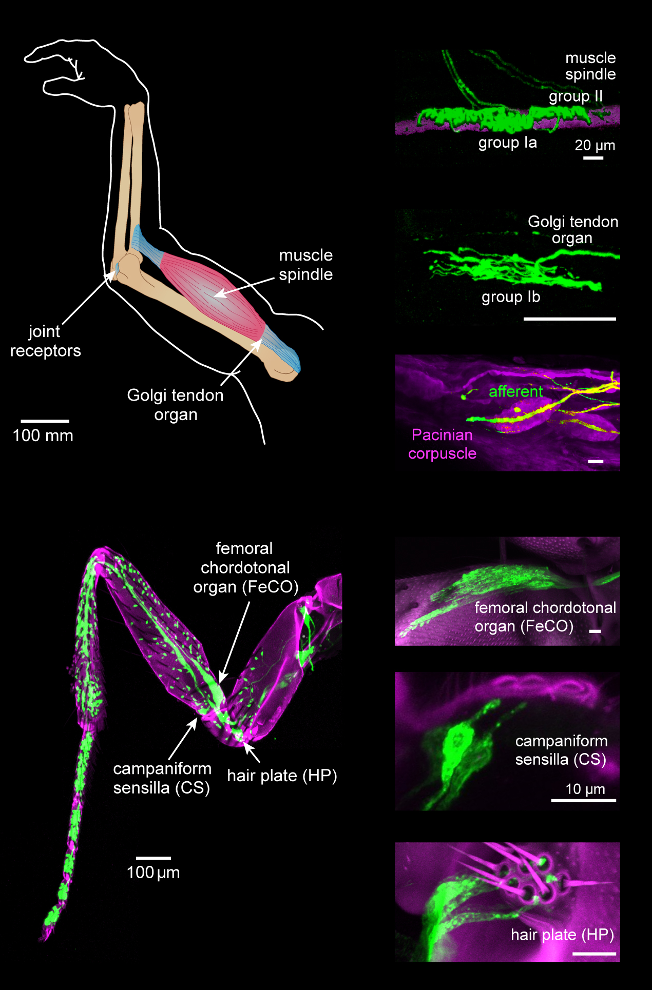
Proprioception ( ), also referred to as kinaesthesia (or kinesthesia), is the sense of self-movement, force, and body position. It is sometimes described as the "sixth sense". Proprioception is mediated by proprioceptors, mechanosensory neurons located within muscles, tendons, and joints. Most animals possess multiple subtypes of proprioceptors, which detect distinct kinematic parameters, such as joint position, movement, and load. Although all mobile animals possess proprioceptors, the structure of the sensory organs can vary across species. Proprioceptive signals are transmitted to the central nervous system, where they are integrated with information from other sensory systems, such as the visual system and the vestibular system, to create an overall representation of body position, movement, and acceleration. In many animals, sensory feedback from proprioceptors is essential for stabilizing body posture and coordinating body movement. System overview In vertebrates, limb ... [...More Info...] [...Related Items...] OR: [Wikipedia] [Google] [Baidu] |
Corticobulbar Tract
In neuroanatomy, the corticobulbar (or corticonuclear) tract is a two-neuron white matter motor pathway connecting the motor cortex in the cerebral cortex to the medullary pyramids, which are part of the brainstem's medulla oblongata (also called "bulbar") region, and are primarily involved in carrying the motor function of the non-oculomotor cranial nerves. The corticobulbar tract is one of the pyramidal tracts, the other being the corticospinal tract. Structure The corticobulbar tract originates in the primary motor cortex of the frontal lobe, just superior to the lateral fissure and rostral to the central sulcus in the precentral gyrus (Brodmann area 4). The tract descends through the corona radiata and genu of the internal capsule with a few fibers in the posterior limb of the internal capsule, as it passes from the cortex down to the midbrain. In the midbrain, the internal capsule becomes the cerebral peduncles. The white matter is located in the ventral portion of the ... [...More Info...] [...Related Items...] OR: [Wikipedia] [Google] [Baidu] |
Corticopontine Tract
Corticopontine fibers are projections from the cerebral cortex to the pontine nuclei Pontine may refer to: * Having to do with the pons, a structure located in the brain stem (from ''pons'', "bridge") * Pontine Marshes, a region of Italy near Rome * Pontine Islands The Pontine Islands (, also ; it, Isole Ponziane ) are an a .... Depending upon the lobe of origin, they can be classified as frontopontine fibers, parietopontine fibers, temporopontine fibers or occipitopontine fibers.http://braininfo.rprc.washington.edu/AncilDefinition.aspx?ID=1322&questID=1322 References External links Cortex->Pons->Cerebellum: * https://www.csuchico.edu/~pmccaffrey/syllabi/CMSD%20320/362unit7.html * Cerebral white matter {{neuroanatomy-stub ... [...More Info...] [...Related Items...] OR: [Wikipedia] [Google] [Baidu] |
Corticospinal Tract
The corticospinal tract is a white matter motor pathway starting at the cerebral cortex that terminates on lower motor neurons and interneurons in the spinal cord, controlling movements of the limbs and trunk. There are more than one million neurons in the corticospinal tract, and they become myelinated usually in the first two years of life. The corticospinal tract is one of the pyramidal tracts, the other being the corticobulbar tract. Anatomy The corticospinal tract originates in several parts of the brain, including not just the motor areas, but also the primary somatosensory cortex and premotor areas. Most of the neurons originate in the primary motor cortex (precentral gyrus, Brodmann area 4) or the premotor frontal areas.Purves, D. et al. (2012). Neuroscience: Fifth edition. Sunderland, MA: Sinauer Associates, Inc.Kolb, B. & Whishaw, I. Q. (2014). An introduction to brain and behavior: Fourth edition. New York, NY: Worth Publishers. About 30% of corticospinal neurons origi ... [...More Info...] [...Related Items...] OR: [Wikipedia] [Google] [Baidu] |
Proprioceptive
Proprioception ( ), also referred to as kinaesthesia (or kinesthesia), is the sense of self-movement, force, and body position. It is sometimes described as the "sixth sense". Proprioception is mediated by proprioceptors, mechanosensory neurons located within muscles, tendons, and joints. Most animals possess multiple subtypes of proprioceptors, which detect distinct kinematic parameters, such as joint position, movement, and load. Although all mobile animals possess proprioceptors, the structure of the sensory organs can vary across species. Proprioceptive signals are transmitted to the central nervous system, where they are integrated with information from other sensory systems, such as the visual system and the vestibular system, to create an overall representation of body position, movement, and acceleration. In many animals, sensory feedback from proprioceptors is essential for stabilizing body posture and coordinating body movement. System overview In vertebrates, l ... [...More Info...] [...Related Items...] OR: [Wikipedia] [Google] [Baidu] |


