|
Alpha-granules
Alpha granules, (α-granules) also known as platelet alpha-granules are a cellular component of platelets. Platelets contain different types of granules that perform different functions, and include alpha granules, dense granules, and lysosomes. Of these, alpha granules are the most common, making up between 50% to 80% of the secretory granules. Alpha granules contain several growth factors. Contents Contents include insulin-like growth factor 1, platelet-derived growth factors, TGF beta, platelet factor 4 (which is a heparin-binding chemokine) and other clotting proteins (such as thrombospondin, fibronectin, factor V, and von Willebrand factor). The alpha granules express the adhesion molecule P-selectin and CD63. These are transferred to the membrane after synthesis. The other type of granules within platelets are called dense granules. Clinical significance A deficiency of alpha granules is known as gray platelet syndrome Gray platelet syndrome (GPS), or platelet alpha-gra ... [...More Info...] [...Related Items...] OR: [Wikipedia] [Google] [Baidu] |
Gray Platelet Syndrome
Gray platelet syndrome (GPS), or platelet alpha-granule deficiency, is a rare congenital autosomal recessive bleeding disorder caused by a reduction or absence of alpha-granules in blood platelets, and the release of proteins normally contained in these granules into the marrow, causing myelofibrosis. The name derives from the initial observation of gray appearance of platelets with a paucity of granules on blood films from a patient with a lifelong bleeding disorder. Signs and symptoms Signs of GPS typically arise at birth or in childhood, these signs and symptoms include thrombocytopenia, bruising susceptibility, and epistaxis. Typically, the observed low platelet count in individuals is progressive, this can result in fatal hemorrhages later in life. Additionally, females who are affected may experience irregular menstrual cycles and heavy menstrual bleeding. Another common effect of GPS is myelofibrosis, where scar tissue builds up in the bone marrow causing it to be unable to ... [...More Info...] [...Related Items...] OR: [Wikipedia] [Google] [Baidu] |
Platelet Factor 4
Platelet factor 4 (PF4) is a small cytokine belonging to the CXC chemokine family that is also known as chemokine (C-X-C motif) ligand 4 (CXCL4) . This chemokine is released from alpha-granules of activated platelets during platelet aggregation, and promotes blood coagulation by moderating the effects of heparin-like molecules. Due to these roles, it is predicted to play a role in wound repair and inflammation. It is usually found in a complex with proteoglycan. Genomics The gene for human PF4 is located on human chromosome 4. Function Platelet factor-4 is a 70-amino acid protein that is released from the alpha-granules of activated platelets and binds with high affinity to heparin. Its major physiologic role appears to be neutralization of heparin-like molecules on the endothelial surface of blood vessels, thereby inhibiting local antithrombin activity and promoting coagulation. As a strong chemoattractant for neutrophils and fibroblasts, PF4 probably has a role in infla ... [...More Info...] [...Related Items...] OR: [Wikipedia] [Google] [Baidu] |
Clotting
Coagulation, also known as clotting, is the process by which blood changes from a liquid to a gel, forming a blood clot. It potentially results in hemostasis, the cessation of blood loss from a damaged vessel, followed by repair. The mechanism of coagulation involves activation, adhesion and aggregation of platelets, as well as deposition and maturation of fibrin. Coagulation begins almost instantly after an injury to the endothelium lining a blood vessel. Exposure of blood to the subendothelial space initiates two processes: changes in platelets, and the exposure of subendothelial tissue factor to plasma factor VII, which ultimately leads to cross-linked fibrin formation. Platelets immediately form a plug at the site of injury; this is called ''primary hemostasis. Secondary hemostasis'' occurs simultaneously: additional coagulation (clotting) factors beyond factor VII ( listed below) respond in a cascade to form fibrin strands, which strengthen the platelet plug. Disorders of ... [...More Info...] [...Related Items...] OR: [Wikipedia] [Google] [Baidu] |
CD63
CD63 antigen is a protein that, in humans, is encoded by the ''CD63'' gene. CD63 is mainly associated with membranes of intracellular vesicles, although cell surface expression may be induced. Function The protein encoded by this gene is a member of the transmembrane 4 superfamily, also known as the tetraspanin family. Most of these members are cell-surface proteins that are characterized by the presence of four hydrophobic domains. The proteins mediate signal transduction events that play a role in the regulation of cell development, activation, growth, and motility. This encoded protein is a cell surface glycoprotein that is known to complex with integrins. It may function as a blood platelet activation marker. Deficiency of this protein is associated with Hermansky-Pudlak Syndrome . Also this gene has been associated with tumor progression. The use of alternate polyadenylation sites has been found for this gene. Alternative splicing results in multiple transcript variants en ... [...More Info...] [...Related Items...] OR: [Wikipedia] [Google] [Baidu] |
P-selectin
P-selectin is a type-1 transmembrane protein that in humans is encoded by the SELP gene. P-selectin functions as a cell adhesion molecule (CAM) on the surfaces of activated endothelial cells, which line the inner surface of blood vessels, and activated platelets. In unactivated endothelial cells, it is stored in granules called Weibel-Palade bodies. In unactivated platelets P-selectin is stored in α-granules. Other names for P-selectin include CD62P, Granule Membrane Protein 140 (GMP-140), and Platelet Activation-Dependent Granule to External Membrane Protein (PADGEM). It was first identified in endothelial cells in 1989. Gene and regulation P-selectin is located on chromosome 1q21-q24, spans > 50 kb and contains 17 exons in humans. P-selectin is constitutively expressed in megakaryocytes (the precursor of platelets) and endothelial cells. P-selectin expression is induced by two distinct mechanisms. First, P-selectin is synthesized by megakaryocytes and endothelial cells ... [...More Info...] [...Related Items...] OR: [Wikipedia] [Google] [Baidu] |
Von Willebrand Factor
Von Willebrand factor (VWF) () is a blood glycoprotein involved in hemostasis, specifically, platelet adhesion. It is deficient and/or defective in von Willebrand disease and is involved in many other diseases, including thrombotic thrombocytopenic purpura, Heyde's syndrome, and possibly hemolytic–uremic syndrome. Increased plasma levels in many cardiovascular, neoplastic, metabolic (e.g. diabetes), and connective tissue diseases are presumed to arise from adverse changes to the endothelium, and may predict an increased risk of thrombosis. Biochemistry Synthesis VWF is a large multimeric glycoprotein present in blood plasma and produced constitutively as ultra-large VWF in endothelium (in the Weibel–Palade bodies), megakaryocytes (α-granules of platelets), and subendothelial connective tissue. Structure The basic VWF monomer is a 2050-amino acid protein. Every monomer contains a number of specific domains with a specific function; elements of note are: * the D'/D3 do ... [...More Info...] [...Related Items...] OR: [Wikipedia] [Google] [Baidu] |
Factor V
Factor V (pronounced factor five) is a protein of the coagulation system, rarely referred to as proaccelerin or labile factor. In contrast to most other coagulation factors, it is not enzymatically active but functions as a cofactor. Deficiency leads to predisposition for hemorrhage, while some mutations (most notably factor V Leiden) predispose for thrombosis. Genetics The gene for factor V is located on the first chromosome (1q24). It is genomically related to the family of multicopper oxidases, and is homologous to coagulation factor VIII. The gene spans 70 kb, consists of 25 exons, and the resulting protein has a relative molecular mass of approximately 330kDa. Structure Factor V protein consists of six domains: A1-A2-B-A3-C1-C2. The A domains are homologous to the A domains of the copper-binding protein ceruloplasmin, and form a triangular as in that protein. A copper ion is bound in the A1-A3 interface, and A3 interacts with the plasma. The C domains belong to the p ... [...More Info...] [...Related Items...] OR: [Wikipedia] [Google] [Baidu] |
Fibronectin
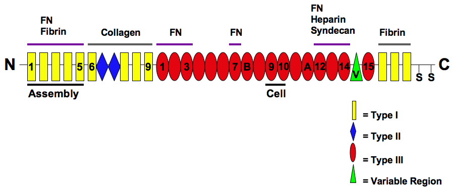
Fibronectin is a high- molecular weight (~500-~600 kDa) glycoprotein of the extracellular matrix that binds to membrane-spanning receptor proteins called integrins. Fibronectin also binds to other extracellular matrix proteins such as collagen, fibrin, and heparan sulfate proteoglycans (e.g. syndecans). Fibronectin exists as a protein dimer, consisting of two nearly identical monomers linked by a pair of disulfide bonds. The fibronectin protein is produced from a single gene, but alternative splicing of its pre-mRNA leads to the creation of several isoforms. Two types of fibronectin are present in vertebrates: * soluble plasma fibronectin (formerly called "cold-insoluble globulin", or CIg) is a major protein component of blood plasma (300 μg/ml) and is produced in the liver by hepatocytes. * insoluble cellular fibronectin is a major component of the extracellular matrix. It is secreted by various cells, primarily fibroblasts, as a soluble protein dimer and is then ass ... [...More Info...] [...Related Items...] OR: [Wikipedia] [Google] [Baidu] |
Thrombospondin
Thrombospondins (TSPs) are a family of secreted glycoproteins with antiangiogenic functions. Due to their dynamic role within the extracellular matrix they are considered matricellular proteins. The first member of the family, thrombospondin 1 (THBS1), was discovered in 1971 by Nancy L. Baenziger. Types The thrombospondins are a family of multifunctional proteins. The family consists of thrombospondins 1-5 and can be divided into 2 subgroups: A, which contains TSP-1 and TSP-2, and B, which contains TSP-3, TSP-4 and TSP-5 (also designated cartilage oligomeric protein or COMP). TSP-1 and TSP-2 are homotrimers, consisting of three identical subunits, whereas TSP-3, TSP-4 and TSP-5 are homopentamers. TSP-1 and TSP-2 are produced by immature astrocytes during brain development, which promotes the development of new synapses. Thrombospondin 1 Thrombospondin 1 (TSP-1) is encoded by THBS1. It was first isolated from platelets that had been stimulated with thrombin, and so was design ... [...More Info...] [...Related Items...] OR: [Wikipedia] [Google] [Baidu] |
Platelet
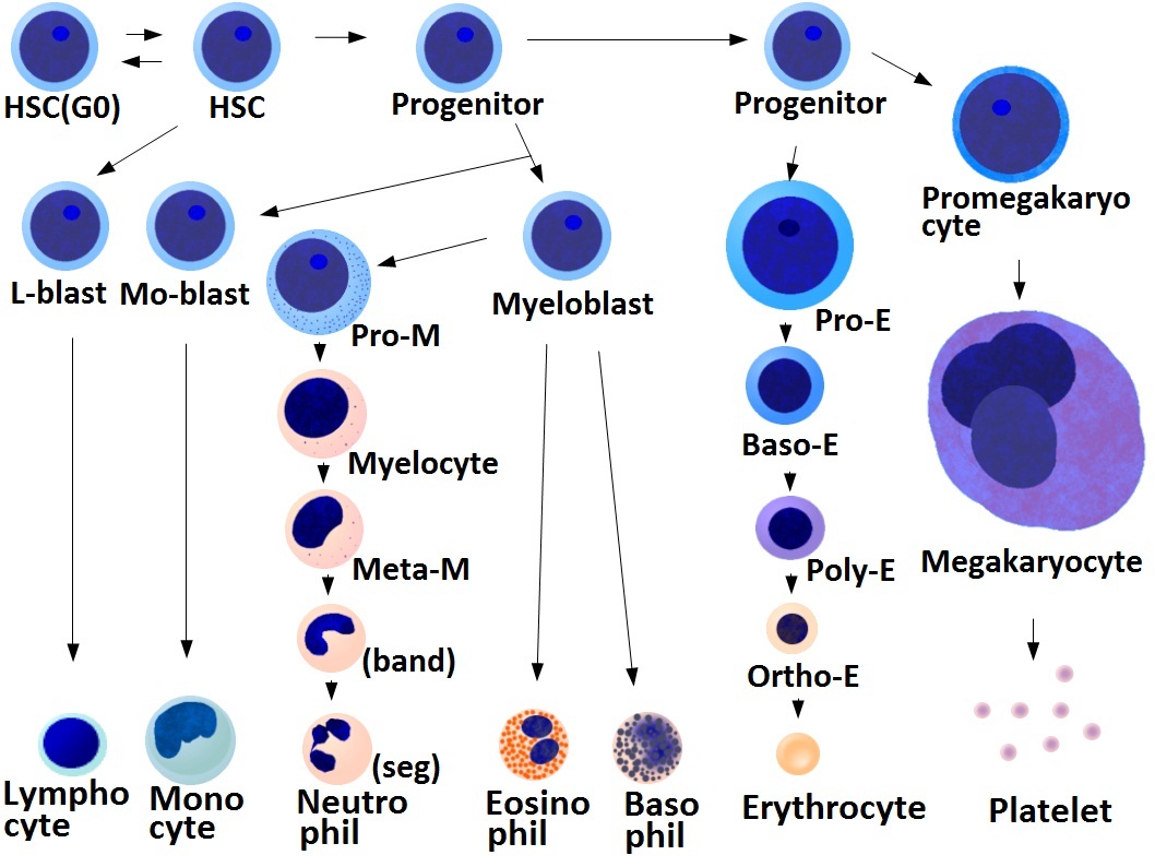
Platelets, also called thrombocytes (from Greek θρόμβος, "clot" and κύτος, "cell"), are a component of blood whose function (along with the coagulation factors) is to react to bleeding from blood vessel injury by clumping, thereby initiating a blood clot. Platelets have no cell nucleus; they are fragments of cytoplasm that are derived from the megakaryocytes of the bone marrow or lung, which then enter the circulation. Platelets are found only in mammals, whereas in other vertebrates (e.g. birds, amphibians), thrombocytes circulate as intact mononuclear cells. One major function of platelets is to contribute to hemostasis: the process of stopping bleeding at the site of interrupted endothelium. They gather at the site and, unless the interruption is physically too large, they plug the hole. First, platelets attach to substances outside the interrupted endothelium: ''adhesion''. Second, they change shape, turn on receptors and secrete chemical messengers: ''activatio ... [...More Info...] [...Related Items...] OR: [Wikipedia] [Google] [Baidu] |
Platelets
Platelets, also called thrombocytes (from Greek θρόμβος, "clot" and κύτος, "cell"), are a component of blood whose function (along with the coagulation factors) is to react to bleeding from blood vessel injury by clumping, thereby initiating a blood clot. Platelets have no cell nucleus; they are fragments of cytoplasm that are derived from the megakaryocytes of the bone marrow or lung, which then enter the circulation. Platelets are found only in mammals, whereas in other vertebrates (e.g. birds, amphibians), thrombocytes circulate as intact mononuclear cells. One major function of platelets is to contribute to hemostasis: the process of stopping bleeding at the site of interrupted endothelium. They gather at the site and, unless the interruption is physically too large, they plug the hole. First, platelets attach to substances outside the interrupted endothelium: ''adhesion''. Second, they change shape, turn on receptors and secrete chemical messengers: ''activatio ... [...More Info...] [...Related Items...] OR: [Wikipedia] [Google] [Baidu] |
TGF Beta
Transforming growth factor beta (TGF-β) is a multifunctional cytokine belonging to the transforming growth factor superfamily that includes three different mammalian isoforms (TGF-β 1 to 3, HGNC symbols TGFB1, TGFB2, TGFB3) and many other signaling proteins. TGFB proteins are produced by all white blood cell lineages. Activated TGF-β complexes with other factors to form a serine/threonine kinase complex that binds to TGF-β receptors. TGF-β receptors are composed of both type 1 and type 2 receptor subunits. After the binding of TGF-β, the type 2 receptor kinase phosphorylates and activates the type 1 receptor kinase that activates a signaling cascade. This leads to the activation of different downstream substrates and regulatory proteins, inducing transcription of different target genes that function in differentiation, chemotaxis, proliferation, and activation of many immune cells. TGF-β is secreted by many cell types, including macrophages, in a latent form in which it ... [...More Info...] [...Related Items...] OR: [Wikipedia] [Google] [Baidu] |


