|
AIFM1
Apoptosis-inducing factor 1, mitochondrial is a protein that in humans is encoded by the ''AIFM1'' gene on the X chromosome. This protein localizes to the mitochondria, as well as the nucleus, where it carries out nuclear fragmentation as part of caspase-independent apoptosis. Structure AIFM1 is expressed as a 613- residue precursor protein that containing a mitochondrial targeting sequence (MTS) at its N-terminal and two nuclear leading sequences (NLS). Once imported into the mitochondria, the first 54 residues of the N-terminal are cleaved to produce the mature protein, which inserts into the inner mitochondrial membrane. The mature protein incorporates the FAD cofactor and folds into three structural domains: the FAD-binding domain, the NAD-binding domain, and the C-terminal. While the C-terminal is responsible for the proapoptotic activity of AIFM1, the FAD-binding and NAD-binding domains share the classical Rossmann topology with other flavoproteins and the NAD(P)H depe ... [...More Info...] [...Related Items...] OR: [Wikipedia] [Google] [Baidu] |
HSPA1A
Heat shock 70 kDa protein 1, also termed Hsp72, is a protein that in humans is encoded by the ''HSPA1A'' gene. As a member of the heat shock protein 70 family and a chaperone protein, it facilitates the proper folding of newly translated and misfolded proteins, as well as stabilize or degrade mutant proteins. In addition, Hsp72 also facilitates DNA repair. Its functions contribute to biological processes including signal transduction, apoptosis, protein homeostasis, and cell growth and differentiation. It has been associated with an extensive number of cancers, neurodegenerative diseases, cell senescence and aging, and inflammatory diseases such as Diabetes mellitus type 2 and rheumatoid arthritis. Structure This intronless gene encodes a 70kDa heat shock protein which is a member of the heat shock protein 70 (Hsp70) family. As a Hsp70 protein, it has a C-terminal protein substrate-binding domain and an N-terminal ATP-binding domain. The substrate-binding domain consists ... [...More Info...] [...Related Items...] OR: [Wikipedia] [Google] [Baidu] |
Protein
Proteins are large biomolecules and macromolecules that comprise one or more long chains of amino acid residues. Proteins perform a vast array of functions within organisms, including catalysing metabolic reactions, DNA replication, responding to stimuli, providing structure to cells and organisms, and transporting molecules from one location to another. Proteins differ from one another primarily in their sequence of amino acids, which is dictated by the nucleotide sequence of their genes, and which usually results in protein folding into a specific 3D structure that determines its activity. A linear chain of amino acid residues is called a polypeptide. A protein contains at least one long polypeptide. Short polypeptides, containing less than 20–30 residues, are rarely considered to be proteins and are commonly called peptides. The individual amino acid residues are bonded together by peptide bonds and adjacent amino acid residues. The sequence of amino acid resid ... [...More Info...] [...Related Items...] OR: [Wikipedia] [Google] [Baidu] |
Isoform
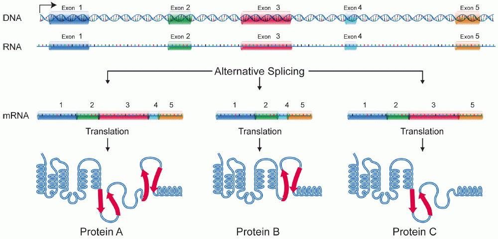
A protein isoform, or "protein variant", is a member of a set of highly similar proteins that originate from a single gene or gene family and are the result of genetic differences. While many perform the same or similar biological roles, some isoforms have unique functions. A set of protein isoforms may be formed from alternative splicings, variable promoter usage, or other post-transcriptional modifications of a single gene; post-translational modifications are generally not considered. (For that, see Proteoforms.) Through RNA splicing mechanisms, mRNA has the ability to select different protein-coding segments (exons) of a gene, or even different parts of exons from RNA to form different mRNA sequences. Each unique sequence produces a specific form of a protein. The discovery of isoforms could explain the discrepancy between the small number of protein coding regions genes revealed by the human genome project and the large diversity of proteins seen in an organism: differ ... [...More Info...] [...Related Items...] OR: [Wikipedia] [Google] [Baidu] |
Ataxia
Ataxia is a neurological sign consisting of lack of voluntary coordination of muscle movements that can include gait abnormality, speech changes, and abnormalities in eye movements. Ataxia is a clinical manifestation indicating dysfunction of the parts of the nervous system that coordinate movement, such as the cerebellum. Ataxia can be limited to one side of the body, which is referred to as hemiataxia. Several possible causes exist for these patterns of neurological dysfunction. Dystaxia is a mild degree of ataxia. Friedreich's ataxia has gait abnormality as the most commonly presented symptom. The word is from Greek α- negative prefix+ -τάξις rder= "lack of order". Types Cerebellar The term cerebellar ataxia is used to indicate ataxia due to dysfunction of the cerebellum. The cerebellum is responsible for integrating a significant amount of neural information that is used to coordinate smoothly ongoing movements and to participate in motor planning. Although ... [...More Info...] [...Related Items...] OR: [Wikipedia] [Google] [Baidu] |
Muscle Atrophy
Muscle atrophy is the loss of skeletal muscle mass. It can be caused by immobility, aging, malnutrition, medications, or a wide range of injuries or diseases that impact the musculoskeletal or nervous system. Muscle atrophy leads to muscle weakness and causes disability. Disuse causes rapid muscle atrophy and often occurs during injury or illness that requires immobilization of a limb or bed rest. Depending on the duration of disuse and the health of the individual, this may be fully reversed with activity. Malnutrition first causes fat loss but may progress to muscle atrophy in prolonged starvation and can be reversed with nutritional therapy. In contrast, cachexia is a wasting syndrome caused by an underlying disease such as cancer that causes dramatic muscle atrophy and cannot be completely reversed with nutritional therapy. Sarcopenia is age-related muscle atrophy and can be slowed by exercise. Finally, diseases of the muscles such as muscular dystrophy or myopathies can caus ... [...More Info...] [...Related Items...] OR: [Wikipedia] [Google] [Baidu] |
Oxidative Phosphorylation
Oxidative phosphorylation (UK , US ) or electron transport-linked phosphorylation or terminal oxidation is the metabolic pathway in which cells use enzymes to oxidize nutrients, thereby releasing chemical energy in order to produce adenosine triphosphate (ATP). In eukaryotes, this takes place inside mitochondria. Almost all aerobic organisms carry out oxidative phosphorylation. This pathway is so pervasive because it releases more energy than alternative fermentation processes such as anaerobic glycolysis. The energy stored in the chemical bonds of glucose is released by the cell in the citric acid cycle producing carbon dioxide, and the energetic electron donors NADH and FADH. Oxidative phosphorylation uses these molecules and O2 to produce ATP, which is used throughout the cell whenever energy is needed. During oxidative phosphorylation, electrons are transferred from the electron donors to a series of electron acceptors in a series of redox reactions ending in oxygen, ... [...More Info...] [...Related Items...] OR: [Wikipedia] [Google] [Baidu] |
Redox
Redox (reduction–oxidation, , ) is a type of chemical reaction in which the oxidation states of substrate (chemistry), substrate change. Oxidation is the loss of Electron, electrons or an increase in the oxidation state, while reduction is the gain of electrons or a decrease in the oxidation state. There are two classes of redox reactions: * ''Electron-transfer'' – Only one (usually) electron flows from the reducing agent to the oxidant. This type of redox reaction is often discussed in terms of redox couples and electrode potentials. * ''Atom transfer'' – An atom transfers from one substrate to another. For example, in the rusting of iron, the oxidation state of iron atoms increases as the iron converts to an oxide, and simultaneously the oxidation state of oxygen decreases as it accepts electrons released by the iron. Although oxidation reactions are commonly associated with the formation of oxides, other chemical species can serve the same function. In hydrogen ... [...More Info...] [...Related Items...] OR: [Wikipedia] [Google] [Baidu] |
Reductase
A reductase is an enzyme that catalyzes a reduction reaction. Examples * 5α-Reductase * 5β-Reductase * Dihydrofolate reductase * HMG-CoA reductase * Methemoglobin reductase * Ribonucleotide reductase * Thioredoxin reductase * ''E. coli'' nitroreductase * Methylenetetrahydrofolate reductase See also * Oxidase * Oxidoreductase In biochemistry, an oxidoreductase is an enzyme that catalyzes the transfer of electrons from one molecule, the reductant, also called the electron donor, to another, the oxidant, also called the electron acceptor. This group of enzymes usually ... References Oxidoreductases {{Enzyme-stub ... [...More Info...] [...Related Items...] OR: [Wikipedia] [Google] [Baidu] |
Cathepsin
Cathepsins (Ancient Greek ''kata-'' "down" and ''hepsein'' "boil"; abbreviated CTS) are proteases ( enzymes that degrade proteins) found in all animals as well as other organisms. There are approximately a dozen members of this family, which are distinguished by their structure, catalytic mechanism, and which proteins they cleave. Most of the members become activated at the low pH found in lysosomes. Thus, the activity of this family lies almost entirely within those organelles. There are, however, exceptions such as cathepsin K, which works extracellularly after secretion by osteoclasts in bone resorption. Cathepsins have a vital role in mammalian cellular turnover. Classification * Cathepsin A (serine protease Serine proteases (or serine endopeptidases) are enzymes that cleave peptide bonds in proteins. Serine serves as the nucleophilic amino acid at the (enzyme's) active site. They are found ubiquitously in both eukaryotes and prokaryotes. S ...) * Cathepsin B ... [...More Info...] [...Related Items...] OR: [Wikipedia] [Google] [Baidu] |
Calpain
A calpain (; , ) is a protein belonging to the family of calcium-dependent, non-lysosomal cysteine proteases (proteolytic enzymes) expressed ubiquitously in mammals and many other organisms. Calpains constitute the C2 family of protease clan CA in the MEROPS database. The calpain proteolytic system includes the calpain proteases, the small regulatory subunit CAPNS1, also known as CAPN4, and the endogenous calpain-specific inhibitor, calpastatin. Discovery The history of calpain's discovery originates in 1964, when calcium-dependent proteolytic activities caused by a "calcium-activated neutral protease" (CANP) were detected in brain, lens of the eye and other tissues. In the late 1960s the enzymes were isolated and characterised independently in both rat brain and skeletal muscle. These activities were caused by an intracellular cysteine protease not associated with the lysosome and having an optimum activity at neutral pH, which clearly distinguished it from the cathepsin fa ... [...More Info...] [...Related Items...] OR: [Wikipedia] [Google] [Baidu] |


