|
ε-opioid Receptor
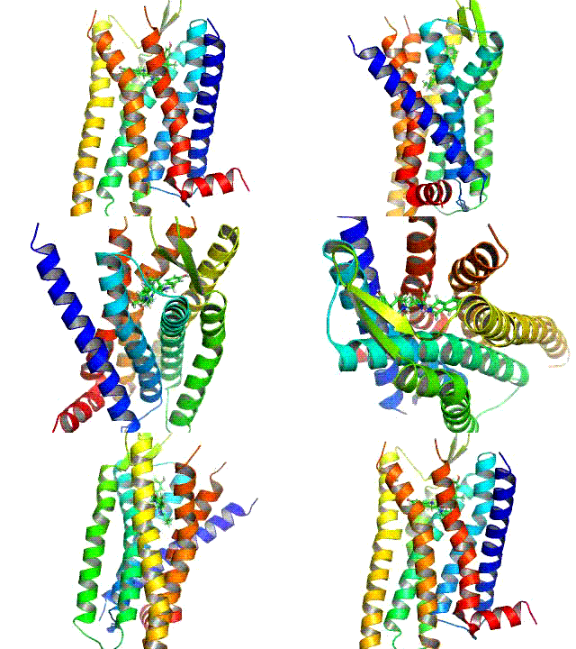
Opioid receptors are a group of inhibitory G protein-coupled receptors with opioids as ligands. The endogenous opioids are dynorphins, enkephalins, endorphins, endomorphins and nociceptin. The opioid receptors are ~40% identical to somatostatin receptors (SSTRs). Opioid receptors are distributed widely in the brain, in the spinal cord, on peripheral neurons, and digestive tract. Discovery By the mid-1960s, it had become apparent from pharmacologic studies that opioids were likely to exert their actions at specific receptor sites, and that there were likely to be multiple such sites. Early studies had indicated that opiates appeared to accumulate in the brain. The receptors were first identified as specific molecules through the use of binding studies, in which opiates that had been labeled with radioisotopes were found to bind to brain membrane homogenates. The first such study was published in 1971, using 3H-levorphanol. In 1973, Candace Pert and Solomon H. Snyder publishe ... [...More Info...] [...Related Items...] OR: [Wikipedia] [Google] [Baidu] |
Levorphanol
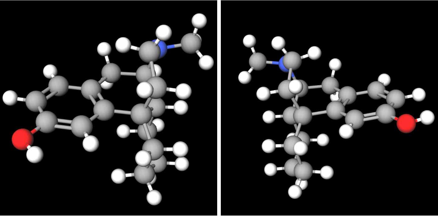
Levorphanol (brand name Levo-Dromoran) is an opioid medication used to treat moderate to severe pain. It is the levorotatory enantiomer of the compound racemorphan. Its dextrorotatory counterpart is dextrorphan. It was first described in Germany in 1946. The drug has been in medical use in the United States since 1953. Pharmacology Levorphanol acts predominantly as an agonist of the μ-opioid receptor (MOR), but is also an agonist of the δ-opioid receptor (DOR), κ-opioid receptor (KOR), and the nociceptin receptor (NOP), as well as an NMDA receptor antagonist and a serotonin-norepinephrine reuptake inhibitor (SNRI). Levorphanol, similarly to certain other opioids, also acts as a glycine receptor antagonist and GABA receptor antagonist at very high concentrations. As per the World Health Organization, levorphanol is a step 3 opioid and is considered eight times more potent than morphine at the MOR (2 mg levorphanol is equivalent to 15 mg morphine). Relative t ... [...More Info...] [...Related Items...] OR: [Wikipedia] [Google] [Baidu] |
Sensory Neurons
Sensory neurons, also known as afferent neurons, are neurons in the nervous system, that convert a specific type of stimulus, via their receptors, into action potentials or graded receptor potentials. This process is called sensory transduction. The cell bodies of the sensory neurons are located in the dorsal root ganglia of the spinal cord. The sensory information travels on the afferent nerve fibers in a sensory nerve, to the brain via the spinal cord. Spinal nerves transmit external sensations via sensory nerves to the brain through the spinal cord. The stimulus can come from exteroreceptors outside the body, for example those that detect light and sound, or from interoreceptors inside the body, for example those that are responsive to blood pressure or the sense of body position. Types and function Sensory neurons in vertebrates are predominantly pseudounipolar or bipolar, and different types of sensory neurons have different sensory receptors that respond to di ... [...More Info...] [...Related Items...] OR: [Wikipedia] [Google] [Baidu] |
Cerebral Cortex
The cerebral cortex, also known as the cerebral mantle, is the outer layer of neural tissue of the cerebrum of the brain in humans and other mammals. It is the largest site of Neuron, neural integration in the central nervous system, and plays a key role in attention, perception, awareness, thought, memory, language, and consciousness. The six-layered neocortex makes up approximately 90% of the Cortex (anatomy), cortex, with the allocortex making up the remainder. The cortex is divided into left and right parts by the longitudinal fissure, which separates the two cerebral hemispheres that are joined beneath the cortex by the corpus callosum and other commissural fibers. In most mammals, apart from small mammals that have small brains, the cerebral cortex is folded, providing a greater surface area in the confined volume of the neurocranium, cranium. Apart from minimising brain and cranial volume, gyrification, cortical folding is crucial for the Neural circuit, brain circuitry ... [...More Info...] [...Related Items...] OR: [Wikipedia] [Google] [Baidu] |
Olfactory Bulbs
The olfactory bulb (Latin: ''bulbus olfactorius'') is a grey matter, neural structure of the vertebrate forebrain involved in olfaction, the sense of odor, smell. It sends olfactory information to be further processed in the amygdala, the orbitofrontal cortex (OFC) and the hippocampus where it plays a role in emotion, memory and learning. The bulb is divided into two distinct structures: the main olfactory bulb and the accessory olfactory bulb. The main olfactory bulb connects to the amygdala via the piriform cortex of the primary olfactory cortex and directly projects from the main olfactory bulb to specific amygdala areas. The accessory olfactory bulb resides on the dorsal-posterior region of the main olfactory bulb and forms a parallel pathway. Destruction of the olfactory bulb results in ipsilateral anosmia, while irritative lesions of the uncus can result in olfactory and gustatory hallucinations. Structure In most vertebrates, the olfactory bulb is the most Anatomical te ... [...More Info...] [...Related Items...] OR: [Wikipedia] [Google] [Baidu] |
Amygdala
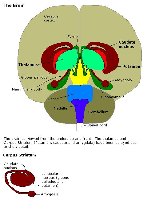
The amygdala (; : amygdalae or amygdalas; also '; Latin from Greek language, Greek, , ', 'almond', 'tonsil') is a paired nucleus (neuroanatomy), nuclear complex present in the Cerebral hemisphere, cerebral hemispheres of vertebrates. It is considered part of the limbic system. In Primate, primates, it is located lateral and medial, medially within the temporal lobes. It consists of many nuclei, each made up of further subnuclei. The subdivision most commonly made is into the Basolateral amygdala, basolateral, Central nucleus of the amygdala, central, cortical, and medial nuclei together with the intercalated cells of the amygdala, intercalated cell clusters. The amygdala has a primary role in the processing of memory, decision making, decision-making, and emotions, emotional responses (including fear, anxiety, and aggression). The amygdala was first identified and named by Karl Friedrich Burdach in 1822. Structure Thirteen Nucleus (neuroanatomy), nuclei have been identif ... [...More Info...] [...Related Items...] OR: [Wikipedia] [Google] [Baidu] |
Pons
The pons (from Latin , "bridge") is part of the brainstem that in humans and other mammals, lies inferior to the midbrain, superior to the medulla oblongata and anterior to the cerebellum. The pons is also called the pons Varolii ("bridge of Varolius"), after the Italian anatomist and surgeon Costanzo Varolio (1543–75). This region of the brainstem includes neural pathways and tracts that conduct signals from the brain down to the cerebellum and medulla, and tracts that carry the sensory signals up into the thalamus. Structure The pons in humans measures about in length. It is the part of the brainstem situated between the midbrain and the medulla oblongata. The horizontal ''medullopontine sulcus'' demarcates the boundary between the pons and medulla oblongata on the ventral aspect of the brainstem, and the roots of cranial nerves VI/VII/VIII emerge from the brainstem along this groove. The junction of pons, medulla oblongata, and cerebellum forms the cerebellopontine ... [...More Info...] [...Related Items...] OR: [Wikipedia] [Google] [Baidu] |
δ-opioid Receptor
The δ-opioid receptor, also known as delta opioid receptor or simply delta receptor, abbreviated DOR or DOP, is an inhibitory 7-transmembrane G-protein coupled receptor coupled to the G protein Gi alpha subunit, Gi/G0 and has enkephalins as its endogenous ligands. The regions of the brain where the δ-opioid receptor is largely expressed vary from species model to species model. In humans, the δ-opioid receptor is most heavily expressed in the basal ganglia and Neocortex, neocortical regions of the brain. Function The endogenous system of opioid receptors is well known for its analgesic potential; however, the exact role of δ-opioid receptor activation in pain modulation is largely up for debate. This also depends on the model at hand since receptor activity is known to change from species to species. Activation of delta receptors produces analgesia, perhaps as significant potentiators of μ-opioid receptor agonists. However, it seems like delta agonism provides heavy poten ... [...More Info...] [...Related Items...] OR: [Wikipedia] [Google] [Baidu] |
OGFr
Opioid growth factor receptor, also known as OGFr or the ζ-opioid receptor, is a protein which in humans is encoded by the ''OGFR'' gene. The protein encoded by this gene is a receptor for opioid growth factor (OGF), also known as et(5)enkephalin. The endogenous ligand is thus a known opioid peptide, and OGFr was originally discovered and named as a new opioid receptor zeta (ζ). However it was subsequently found that it shares little sequence similarity with the other opioid receptors, and has quite different function. Function The natural function of this receptor appears to be in regulation of tissue growth, and it has been shown to be important in embryonic development, wound repair, and certain forms of cancer. OGF is a negative regulator of cell proliferation and tissue organization in a variety of processes. The encoded unbound receptor for OGF has been localized to the outer nuclear envelope, where it binds OGF and is translocated into the nucleus. The coding sequen ... [...More Info...] [...Related Items...] OR: [Wikipedia] [Google] [Baidu] |
Opioid Antagonist
An opioid antagonist, or opioid receptor antagonist, is a receptor antagonist that acts on one or more of the opioid receptors. Naloxone and naltrexone are commonly used opioid antagonist drugs which are competitive antagonists that bind to the opioid receptors with higher affinity than agonists but do not activate the receptors. This effectively blocks the receptor, preventing the body from responding to opioids and endorphins. Some opioid antagonists are not pure antagonists but do produce some weak opioid partial agonist effects, and can produce analgesic effects when administered in high doses to opioid-naive individuals. Examples of such compounds include nalorphine and levallorphan. However, the analgesic effects from these specific drugs are limited and tend to be accompanied by dysphoria, most likely due to additional agonist action at the Îş-opioid receptor. As they induce opioid withdrawal effects in people who are taking, or have recently used, opioid full agoni ... [...More Info...] [...Related Items...] OR: [Wikipedia] [Google] [Baidu] |
Naloxone
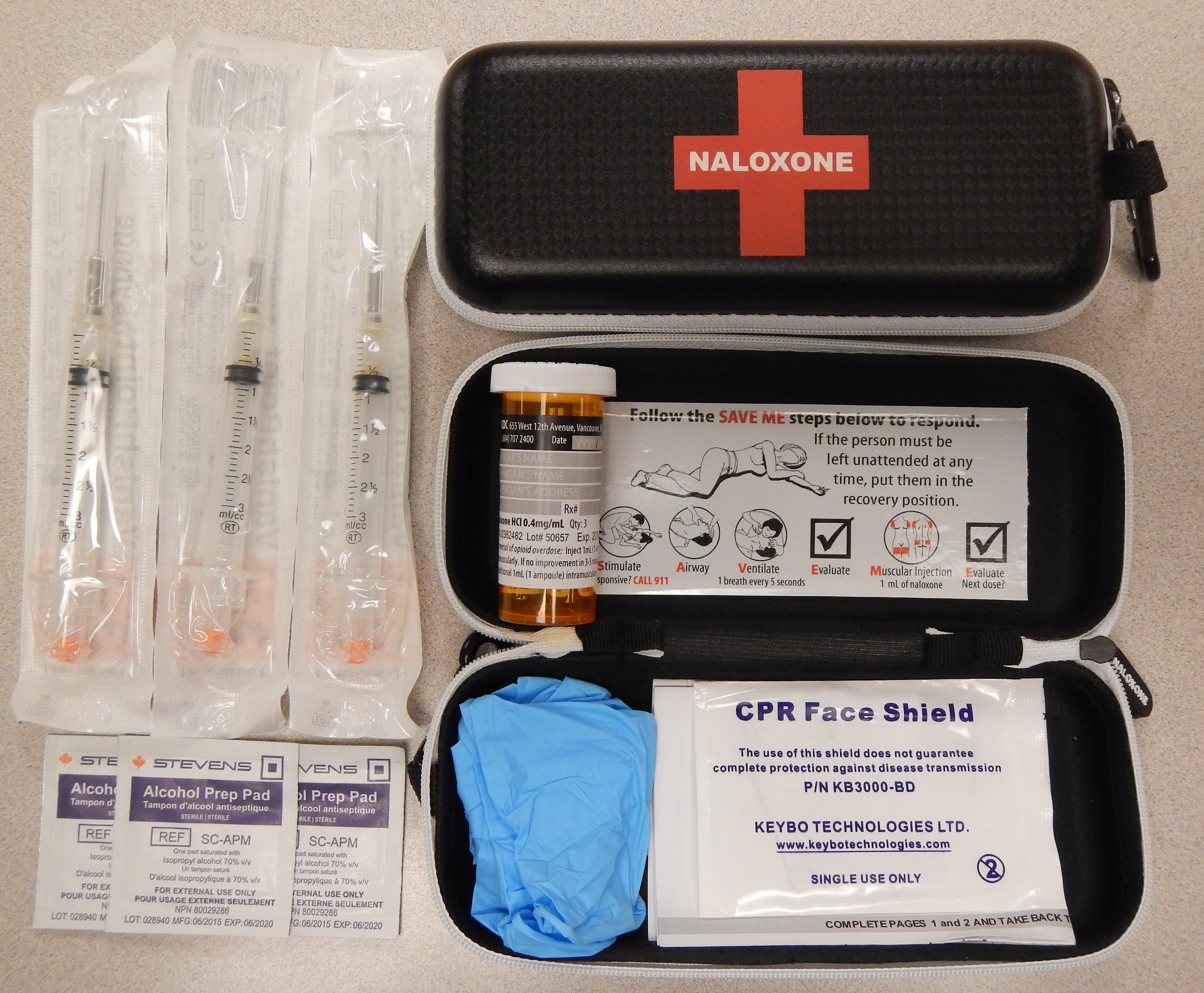
Naloxone, sold under the brand name Narcan among others, is an opioid antagonist, a medication used to reverse or reduce the effects of opioids. For example, it is used to restore breathing after an opioid overdose. Effects begin within two minutes when given intravenously, five minutes when injected into a muscle, and ten minutes as a nasal spray. Naloxone blocks the effects of opioids for 30 to 90 minutes. Administration to opioid-dependent individuals may cause symptoms of opioid withdrawal, including restlessness, agitation, nausea, vomiting, a fast heart rate, and sweating. To prevent this, small doses every few minutes can be given until the desired effect is reached. In those with previous heart disease or taking medications that negatively affect the heart, further heart problems have occurred. It appears to be safe in pregnancy, after having been given to a limited number of women. Naloxone is a non-selective and competitive opioid receptor antagonist. It revers ... [...More Info...] [...Related Items...] OR: [Wikipedia] [Google] [Baidu] |


