|
Olfactory Bulbs
The olfactory bulb (Latin: ''bulbus olfactorius'') is a neural structure of the vertebrate forebrain involved in olfaction, the sense of smell. It sends olfactory information to be further processed in the amygdala, the orbitofrontal cortex (OFC) and the hippocampus where it plays a role in emotion, memory and learning. The bulb is divided into two distinct structures: the main olfactory bulb and the accessory olfactory bulb. The main olfactory bulb connects to the amygdala via the piriform cortex of the primary olfactory cortex and directly projects from the main olfactory bulb to specific amygdala areas. The accessory olfactory bulb resides on the dorsal-posterior region of the main olfactory bulb and forms a parallel pathway. Destruction of the olfactory bulb results in ipsilateral anosmia, while irritative lesions of the uncus can result in olfactory and gustatory hallucinations. Structure In most vertebrates, the olfactory bulb is the most rostral (forward) part of the ... [...More Info...] [...Related Items...] OR: [Wikipedia] [Google] [Baidu] |
Vesalius
Andreas Vesalius (Latinized from Andries van Wezel) () was a 16th-century anatomist, physician, and author of one of the most influential books on human anatomy, ''De Humani Corporis Fabrica Libri Septem'' (''On the fabric of the human body'' ''in seven books''). Vesalius is often referred to as the founder of modern human anatomy. He was born in Brussels, which was then part of the Habsburg Netherlands. He was a professor at the University of Padua (1537–1542) and later became Imperial physician at the court of Emperor Charles V. ''Andreas Vesalius'' is the Latinized form of the Dutch name Andries van Wesel. It was a common practice among European scholars in his time to Latinize their names. His name is also given as ''Andrea Vesalius'', ''André Vésale'', ''Andrea Vesalio'', ''Andreas Vesal'', ''Andrés Vesalio'' and ''Andre Vesale''. Early life and education Vesalius was born as Andries van Wesel to his father Anders van Wesel and mother Isabel Crabbe on 31 December 151 ... [...More Info...] [...Related Items...] OR: [Wikipedia] [Google] [Baidu] |
Lesion
A lesion is any damage or abnormal change in the tissue of an organism, usually caused by disease or trauma. ''Lesion'' is derived from the Latin "injury". Lesions may occur in plants as well as animals. Types There is no designated classification or naming convention for lesions. Since lesions can occur anywhere in the body and the definition of a lesion is so broad, the varieties of lesions are virtually endless. Generally, lesions may be classified by their patterns, their sizes, their locations, or their causes. They can also be named after the person who discovered them. For example, Ghon lesions, which are found in the lungs of those with tuberculosis, are named after the lesion's discoverer, Anton Ghon. The characteristic skin lesions of a varicella zoster virus infection are called '' chickenpox''. Lesions of the teeth are usually called dental caries. Location Lesions are often classified by their tissue types or locations. For example, a "skin lesion" or a " b ... [...More Info...] [...Related Items...] OR: [Wikipedia] [Google] [Baidu] |
Afferent Nerve Fiber
Afferent nerve fibers are the axons (nerve fibers) carried by a sensory nerve that relay sensory information from sensory receptors to regions of the brain. Afferent projections ''arrive'' at a particular brain region. Efferent nerve fibers are carried by efferent nerves and ''exit'' a region to act on muscles and glands. In the peripheral nervous system afferent and efferent nerve fibers are part of the somatic nervous system and arise from outside of the spinal cord. Sensory nerves carry the afferent fibers to enter into the spinal cord, and motor nerves carry the efferent fibers out of the spinal cord to act on skeletal muscles. In the central nervous system non-motor efferents are carried in efferent nerves to act on glands. Structure Afferent neurons are pseudounipolar neurons that have a single process leaving the cell body dividing into two branches: the long one towards the sensory organ, and the short one toward the central nervous system (e.g. spinal co ... [...More Info...] [...Related Items...] OR: [Wikipedia] [Google] [Baidu] |
Neural Circuit
A neural circuit is a population of neurons interconnected by synapses to carry out a specific function when activated. Neural circuits interconnect to one another to form large scale brain networks. Biological neural networks have inspired the design of artificial neural networks, but artificial neural networks are usually not strict copies of their biological counterparts. Early study Early treatments of neural networks can be found in Herbert Spencer's ''Principles of Psychology'', 3rd edition (1872), Theodor Meynert's ''Psychiatry'' (1884), William James' ''Principles of Psychology'' (1890), and Sigmund Freud's Project for a Scientific Psychology (composed 1895). The first rule of neuronal learning was described by Hebb in 1949, in the Hebbian theory. Thus, Hebbian pairing of pre-synaptic and post-synaptic activity can substantially alter the dynamic characteristics of the synaptic connection and therefore either facilitate or inhibit signal transmission. In 1959, the ... [...More Info...] [...Related Items...] OR: [Wikipedia] [Google] [Baidu] |
Mitral Cell
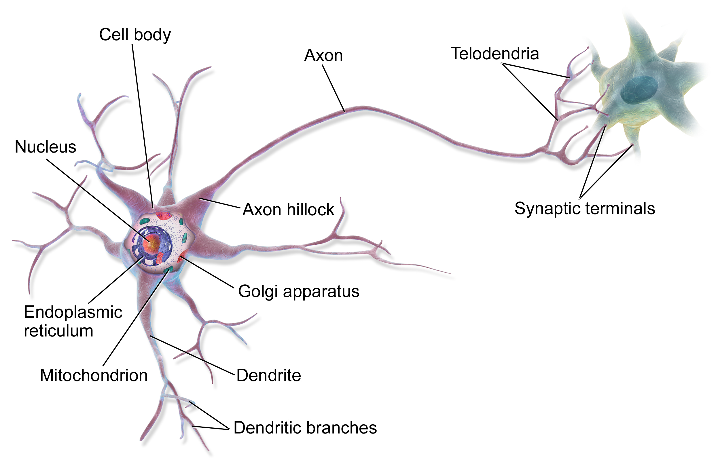
Mitral cells are neurons that are part of the olfactory system. They are located in the olfactory bulb in the mammalian central nervous system. They receive information from the axons of olfactory receptor neurons, forming synapses in neuropils called glomeruli. Axons of the mitral cells transfer information to a number of areas in the brain, including the piriform cortex, entorhinal cortex, and amygdala. Mitral cells receive excitatory input from olfactory sensory neurons and external tufted cells on their primary dendrites, whereas inhibitory input arises either from granule cells onto their lateral dendrites and soma or from periglomerular cells onto their dendritic tuft. Mitral cells together with tufted cells form an obligatory relay for all olfactory information entering from the olfactory nerve. Mitral cell output is not a passive reflection of their input from the olfactory nerve. In mice, each mitral cell sends a single primary dendrite into a glomerulus receiving input f ... [...More Info...] [...Related Items...] OR: [Wikipedia] [Google] [Baidu] |
Glomerulus (olfaction)
The glomerulus (plural glomeruli) is a spherical structure located in the olfactory bulb of the brain where synapses form between the terminals of the olfactory nerve and the dendrites of mitral, periglomerular and tufted cells. Each glomerulus is surrounded by a heterogeneous population of juxtaglomerular neurons (that include periglomerular, short axon, and external tufted cells) and glial cells. All glomeruli are located near the surface of the olfactory bulb. The olfactory bulb also includes a portion of the anterior olfactory nucleus, the cells of which contribute fibers to the olfactory tract. They are the initial sites for synaptic processing of odor information coming from the nose. A glomerulus is made up of a globular tangle of axons from the olfactory receptor neurons, and dendrites from the mitral and tufted cells, as well as, from cells that surround the glomerulus such as the external tufted cells, periglomerular cells, short axon cells, and astrocytes. In mamm ... [...More Info...] [...Related Items...] OR: [Wikipedia] [Google] [Baidu] |
Cytoarchitecture
Cytoarchitecture ( Greek '' κύτος''= "cell" + '' ἀρχιτεκτονική''= "architecture"), also known as cytoarchitectonics, is the study of the cellular composition of the central nervous system's tissues under the microscope. Cytoarchitectonics is one of the ways to parse the brain, by obtaining sections of the brain using a microtome and staining them with chemical agents which reveal where different neurons are located. The study of the parcellation of ''nerve fibers'' (primarily axons) into layers forms the subject of myeloarchitectonics ( History of the cerebral cytoarchitecture Defining cerebral cytoarchitecture began with the advent of —the science of slicin ...[...More Info...] [...Related Items...] OR: [Wikipedia] [Google] [Baidu] |
Olfactory Nerve
The olfactory nerve, also known as the first cranial nerve, cranial nerve I, or simply CN I, is a cranial nerve that contains sensory nerve fibers relating to the sense of smell. The afferent nerve fibers of the olfactory receptor neurons transmit nerve impulses about odors to the central nervous system ( olfaction). Derived from the embryonic nasal placode, the olfactory nerve is somewhat unusual among cranial nerves because it is capable of some regeneration if damaged. The olfactory nerve is sensory in nature and originates on the olfactory mucosa in the upper part of the nasal cavity. From the olfactory mucosa, the nerve (actually many small nerve fascicles) travels up through the cribriform plate of the ethmoid bone to reach the surface of the brain. Here the fascicles enter the olfactory bulb and synapse there; from the bulbs (one on each side) the olfactory information is transmitted into the brain via the olfactory tract. The fascicles of the olfactory nerve are no ... [...More Info...] [...Related Items...] OR: [Wikipedia] [Google] [Baidu] |
Olfactory Epithelium
The olfactory epithelium is a specialized epithelial tissue inside the nasal cavity that is involved in smell. In humans, it measures 9 cm2 and lies on the roof of the nasal cavity about 7 cm above and behind the nostrils. The olfactory epithelium is the part of the olfactory system directly responsible for detecting odors. Structure Olfactory epithelium consists of four distinct cell types: * Olfactory sensory neurons * Supporting cells * Basal cells * Brush cells Olfactory sensory neurons The olfactory receptor neurons are sensory neurons of the olfactory epithelium. They are bipolar neurons and their apical poles express odorant receptors on non-motile cilia at the ends of the dendritic knob, which extend out into the airspace to interact with odorants. Odorant receptors bind odorants in the airspace, which are made soluble by the serous secretions from olfactory glands located in the lamina propria of the mucosa.Ross, MH, ''Histology: A Text and Atlas'', 5th ... [...More Info...] [...Related Items...] OR: [Wikipedia] [Google] [Baidu] |
Ethmoid Bone
The ethmoid bone (; from grc, ἡθμός, hēthmós, sieve) is an unpaired bone in the skull that separates the nasal cavity from the brain. It is located at the roof of the nose, between the two orbits. The cubical bone is lightweight due to a spongy construction. The ethmoid bone is one of the bones that make up the orbit of the eye. Structure The ethmoid bone is an anterior cranial bone located between the eyes. It contributes to the medial wall of the orbit, the nasal cavity, and the nasal septum. The ethmoid has three parts: cribriform plate, ethmoidal labyrinth, and perpendicular plate. The cribriform plate forms the roof of the nasal cavity and also contributes to formation of the anterior cranial fossa, the ethmoidal labyrinth consists of a large mass on either side of the perpendicular plate, and the perpendicular plate forms the superior two-thirds of the nasal septum. Between the orbital plate and the nasal conchae are the ethmoidal sinuses or ethmoidal air cells, w ... [...More Info...] [...Related Items...] OR: [Wikipedia] [Google] [Baidu] |
Cribriform Plate
In mammalian anatomy, the cribriform plate (Latin for lit. ''sieve-shaped''), horizontal lamina or lamina cribrosa is part of the ethmoid bone. It is received into the ethmoidal notch of the frontal bone and roofs in the nasal cavities. It supports the olfactory bulb, and is perforated by olfactory foramina for the passage of the olfactory nerves to the roof of the nasal cavity to convey smell to the brain. The foramina at the medial part of the groove allow the passage of the nerves to the upper part of the nasal septum while the foramina at the lateral part transmit the nerves to the superior nasal concha. A fractured cribriform plate can result in olfactory dysfunction, septal hematoma, cerebrospinal fluid rhinorrhoea (CSF rhinorrhoea), and possibly infection which can lead to meningitis. CSF rhinorrhoea (clear fluid leaking from the nose) is very serious and considered a medical emergency. Aging can cause the openings in the cribriform plate to close, pinching olfactor ... [...More Info...] [...Related Items...] OR: [Wikipedia] [Google] [Baidu] |
Anatomical Terms Of Location
Standard anatomical terms of location are used to unambiguously describe the anatomy of animals, including humans. The terms, typically derived from Latin or Greek roots, describe something in its standard anatomical position. This position provides a definition of what is at the front ("anterior"), behind ("posterior") and so on. As part of defining and describing terms, the body is described through the use of anatomical planes and anatomical axes. The meaning of terms that are used can change depending on whether an organism is bipedal or quadrupedal. Additionally, for some animals such as invertebrates, some terms may not have any meaning at all; for example, an animal that is radially symmetrical will have no anterior surface, but can still have a description that a part is close to the middle ("proximal") or further from the middle ("distal"). International organisations have determined vocabularies that are often used as standard vocabularies for subdisciplines of ana ... [...More Info...] [...Related Items...] OR: [Wikipedia] [Google] [Baidu] |


