|
Zygomatic Process Of Frontal Bone
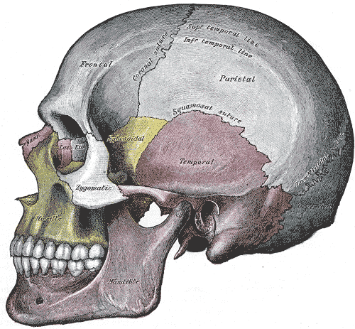
The zygomatic processes (aka. malar) are three processes (protrusions) from other bones of the skull which each articulate with the zygomatic bone. The three processes are: * Zygomatic process of frontal bone from the frontal bone * Zygomatic process of maxilla from the maxilla * Zygomatic process of temporal bone from the temporal bone The term ''zygomatic'' derives . The zygomatic process is occasionally referred to as the zygoma, but this term usually refers to the zygomatic bone or occasionally the zygomatic arch. Zygomatic process of frontal bone The supraorbital margin of the frontal bone ends laterally in its zygomatic process, which is strong and prominent, and articulates with the zygomatic bone. The zygomatic process of the frontal bone extends from the frontal bone laterally and inferiorly. Zygomatic process of maxilla The zygomatic process of the maxilla [...More Info...] [...Related Items...] OR: [Wikipedia] [Google] [Baidu] |
Infratemporal Fossa
The infratemporal fossa is an irregularly shaped cavity that is a part of the skull. It is situated below and medial to the zygomatic arch. It is not fully enclosed by bone in all directions. It contains superficial muscles, including the lower part of the temporalis muscle, the lateral pterygoid muscle, and the medial pterygoid muscle. It also contains important blood vessels such as the middle meningeal artery, the pterygoid plexus, and the retromandibular vein, and nerves such as the mandibular nerve (CN V3) and its branches. Structure Boundaries The boundaries of the infratemporal fossa occur: * ''anteriorly'', by the infratemporal surface of the maxilla, and the ridge which descends from its zygomatic process. This contains the alveolar canal. * ''posteriorly'', by the tympanic part of the temporal bone, and the spina angularis of the sphenoid. * ''superiorly'', by the greater wing of the sphenoid below the infratemporal crest, and by the under surface of the ... [...More Info...] [...Related Items...] OR: [Wikipedia] [Google] [Baidu] |
Tripod Fracture
The zygomaticomaxillary complex fracture, also known as a quadripod fracture, quadramalar fracture, and formerly referred to as a tripod fracture or trimalar fracture, has four components, three of which are directly related to connections between the zygoma and the face, and the fourth being the orbital floor. Its specific locations are the lateral orbital wall (at its superior junction with the zygomaticofrontal suture or its inferior junction with the zygomaticosphenoid suture at the sphenoid greater wing), separation of the maxilla and zygoma at the anterior maxilla (near the zygomaticomaxillary suture), the zygomatic arch, and the orbital floor near the infraorbital canal. Signs and symptoms On physical exam, the fracture appears as a loss of cheek projection with increased width of the face. In most cases, there is loss of sensation in the cheek and upper lip due to infraorbital nerve injury. Facial bruising, periorbital ecchymosis, soft tissue gas, swelling, trismus, al ... [...More Info...] [...Related Items...] OR: [Wikipedia] [Google] [Baidu] |
Zygomatic Arch
In anatomy, the zygomatic arch (colloquially known as the cheek bone), is a part of the skull formed by the zygomatic process of temporal bone, zygomatic process of the temporal bone (a bone extending forward from the side of the skull, over the opening of the ear) and the temporal Process (anatomy), process of the zygomatic bone (the side of the cheekbone), the two being united by an oblique Suture (anatomy), suture (the zygomaticotemporal suture); the tendon of the temporal muscle passes medial to (i.e. through the middle of) the arch, to gain insertion into the coronoid process of the mandible (jawbone). The jugal point is the point at the anterior (towards face) end of the upper border of the zygomatic arch where the Masseter muscle, masseteric and Maxilla, maxillary edges meet at an angle, and where it meets the process of the zygomatic bone. The arch is typical of ''Synapsida'' ("fused arch"), a clade of amniotes that includes mammals and their extinct relatives, such as ' ... [...More Info...] [...Related Items...] OR: [Wikipedia] [Google] [Baidu] |
Zygomatic Process Of The Temporal
The zygomatic processes (aka. malar) are three processes (protrusions) from other bones of the skull which each articulate with the zygomatic bone. The three processes are: * Zygomatic process of frontal bone from the frontal bone * Zygomatic process of maxilla from the maxilla * Zygomatic process of temporal bone from the temporal bone The term ''zygomatic'' derives . The zygomatic process is occasionally referred to as the zygoma, but this term usually refers to the zygomatic bone or occasionally the zygomatic arch. Zygomatic process of frontal bone The supraorbital margin of the frontal bone ends laterally in its zygomatic process, which is strong and prominent, and articulates with the zygomatic bone. The zygomatic process of the frontal bone extends from the frontal bone laterally and inferiorly. Zygomatic process of maxilla The zygomatic process of the maxilla [...More Info...] [...Related Items...] OR: [Wikipedia] [Google] [Baidu] |
Orbital Process
In the human skull, the zygomatic bone (from ), also called cheekbone or malar bone, is a paired irregular bone, situated at the upper and lateral part of the face and forming part of the lateral wall and floor of the orbit, of the temporal fossa and the infratemporal fossa. It presents a malar and a temporal surface; four processes (the frontosphenoidal, orbital, maxillary, and temporal), and four borders. Etymology The term ''zygomatic'' derives from the Ancient Greek , ''zygoma'', meaning "yoke". The zygomatic bone is occasionally referred to as the zygoma, but this term may also refer to the zygomatic arch. Structure Surfaces The ''malar surface'' is convex and perforated near its center by a small aperture, the zygomaticofacial foramen, for the passage of the zygomaticofacial nerve and vessels; below this foramen is a slight elevation, which gives origin to the zygomaticus muscle. The ''temporal surface'', directed posteriorly and medially, is concave, presenting ... [...More Info...] [...Related Items...] OR: [Wikipedia] [Google] [Baidu] |
Cranial Suture
In anatomy, fibrous joints are joints connected by fibrous tissue, consisting mainly of collagen. These are fixed joints where bones are united by a layer of white fibrous tissue of varying thickness. In the skull, the joints between the bones are called sutures. Such immovable joints are also referred to as synarthroses. Types Most fibrous joints are also called "fixed" or "immovable". These joints have no joint cavity and are connected via fibrous connective tissue. * Sutures: The skull bones are connected by fibrous joints called '' sutures''. In fetal skulls, the sutures are wide to allow slight movement during birth. They later become rigid ( synarthrodial). * Syndesmosis: Some of the long bones in the body such as the radius and ulna in the forearm are joined by a ''syndesmosis'' (along the interosseous membrane). Syndemoses are slightly moveable ( amphiarthrodial). The distal tibiofibular joint is another example. * A ''gomphosis'' is a joint between the root of a ... [...More Info...] [...Related Items...] OR: [Wikipedia] [Google] [Baidu] |
Articular Tubercle
The articular tubercle (eminentia articularis) is a bony eminence on the temporal bone in the skull. It is a rounded eminence of the anterior root of the posterior end of the outer surface of the squama temporalis. This tubercle forms the front boundary of the mandibular fossa, and in the fresh state is covered with cartilage. The mandibular condyle normally moves over the articular tubercle during physiologic maximal opening of the jaw. The articular tubercle is the site of attachment of the lateral ligament of the temporomandibular joint In anatomy, the temporomandibular joints (TMJ) are the two joints connecting the jawbone to the skull. It is a bilateral Synovial joint, synovial articulation between the temporal bone of the skull above and the condylar process of mandible be .... Earth's Lab- Retrieved 2018-08-30< ... [...More Info...] [...Related Items...] OR: [Wikipedia] [Google] [Baidu] |
External Auditory Meatus
The ear canal (external acoustic meatus, external auditory meatus, EAM) is a pathway running from the outer ear to the middle ear. The adult human ear canal extends from the auricle to the eardrum and is about in length and in diameter. Structure The human ear canal is divided into two parts. The elastic cartilage part forms the outer third of the canal; its anterior and lower wall are cartilaginous, whereas its superior and back wall are fibrous. The cartilage is the continuation of the cartilage framework of auricle. The cartilaginous portion of the ear canal contains small hairs and specialized sweat glands, called apocrine glands, which produce cerumen ( ear wax). The bony part forms the inner two thirds. The bony part is much shorter in children and is only a ring (''annulus tympanicus'') in the newborn. The layer of epithelium encompassing the bony portion of the ear canal is much thinner and therefore, more sensitive in comparison to the cartilaginous portion. Size a ... [...More Info...] [...Related Items...] OR: [Wikipedia] [Google] [Baidu] |
Masseter Muscle
In anatomy, the masseter is one of the muscles of mastication. Found only in mammals, it is particularly powerful in herbivores to facilitate chewing of plant matter. The most obvious muscle of mastication is the masseter muscle, since it is the most superficial and one of the strongest. Structure The masseter is a thick, somewhat quadrilateral muscle, consisting of three heads, superficial, deep and coronoid. The fibers of superficial and deep heads are continuous at their insertion. Superficial head The superficial head, the larger, arises by a thick, tendinous aponeurosis from the zygomatic process of the maxilla, the temporal process of the zygomatic bone and from the anterior two-thirds of the inferior border of the zygomatic arch. Its fibers pass inferior and posterior, to be inserted into the angle of the mandible and inferior half of the lateral surface of the ramus of the mandible. Deep head The deep head is much smaller, and more muscular in texture. It arises from t ... [...More Info...] [...Related Items...] OR: [Wikipedia] [Google] [Baidu] |
Temporal Fascia
The temporal fascia (or deep temporal fascia) is a fascia of the head that covers the temporalis muscle and structures situated superior to the zygomatic arch. The fascia is attached superiorly at the superior temporal line; inferiorly, it splits into two layers at the superior border of the zygomatic arch. The superficial layer then attaches to the lateral aspect of the superior border of the arch, and the deep layer to its medial aspect. The space between the two layers is occupied by adipose tissue and contains a branch of the superficial temporal artery, and the zygomaticotemporal nerve. Anatomy The temporal fascia is a strong fibrous investment. Structure Superiorly, it is a single layer, attached to the entire extent of the superior temporal line. Inferiorly, where it is fixed to the zygomatic arch, it consists of two layers, one of which is inserted into the lateral, and the other into the medial border of the arch. Contents A small quantity of fat, the orbital ... [...More Info...] [...Related Items...] OR: [Wikipedia] [Google] [Baidu] |
Squama Temporalis
The squamous part of temporal bone, or temporal squama, forms the front and upper part of the temporal bone, and is scale-like, thin, and translucent. Surfaces Its outer surface is smooth and convex; it affords attachment to the temporal muscle, and forms part of the temporal fossa; on its hinder part is a vertical groove for the middle temporal artery. A curved line, the ''temporal line'', or ''supramastoid crest'', runs backward and upward across its posterior part; it serves for the attachment of the temporal fascia, and limits the origin of the temporalis muscle. The boundary between the squamous part and the mastoid portion of the bone, as indicated by traces of the original suture, lies about 1 cm. below this line. Projecting from the lower part of the squamous part is a long, arched process, the '' zygomatic process''. This process is at first directed lateralward, its two surfaces looking upward and downward; it then appears as if twisted inward upon itself, a ... [...More Info...] [...Related Items...] OR: [Wikipedia] [Google] [Baidu] |


